. 2017 Apr;222(3):1495-1507.
doi: 10.1007/s00429-016-1289-7. Epub 2016 Aug 18.

Differential origin of the activation of dorsal and ventral dentate gyrus granule cells during paradoxical (REM) sleep in the rat

Affiliations
Free PMC article

Differential origin of the activation of dorsal and ventral dentate gyrus granule cells during paradoxical (REM) sleep in the rat

Francesca Billwiller et al. Brain Struct Funct. 2017 Apr.
Free PMC article

Abstract

We recently demonstrated that granule cells located in the dorsal dentate gyrus (dDG) are activated by neurons located in the lateral supramammillary nucleus (SumL) during paradoxical sleep (PS) hypersomnia. To determine whether these neurons are glutamatergic and/or GABAergic, we combined FOS immunostaining with in situ hybridization of vesicular glutamate transporter 2 (vGLUT2, a marker of glutamatergic neurons) or that of the vesicular GABA transporter (vGAT, a marker of GABAergic neurons) mRNA in rats displaying PS hypersomnia (PSR). We found that 84 and 76 % of the FOS+ SumL neurons in PSR rats expressed vGLUT2 and vGAT mRNA, respectively. Then, we examined vGLUT2 and FOS immunostaining in the dorsal and ventral DG of PSR rats with a neurochemical lesion of the Sum. In PSR-lesioned animals but not in sham animals, nearly all vGLUT2+ fibers and FOS+ neurons disappeared in the dDG, but not in the ventral DG (vDG). To identify the pathway (s) responsible (s) for the activation of the vDG during PS hypersomnia, we combined Fluorogold (FG) injection in the vDG of PSR rats with FOS staining. We found a large number of neurons FOS-FG+, specifically in the medial entorhinal cortex (ENTm). Altogether, our results suggest that SumL neurons with a unique dual glutamatergic and GABAergic phenotype are responsible for the activation of the dDG during PS hypersomnia, while vDG granule neurons are activated by ENTm cortical neurons. These results suggest differential mechanisms and functions for the activation of the dDG and the vDG granule cells during PS.

Keywords: Dentate gyrus; Entorhinal cortex; Hippocampus; REM sleep; Supramammillary nucleus.


Figures
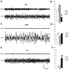
Fig. 1
Fig. 1
Polygraphic recordings and sleep quantities during the flowerpot paradoxical sleep (PS) deprivation and recovery protocol. (a, c, e) Representative electromyogram (EMG) and electroencephalogram (EEG) signals during waking (WK), slow wave sleep (SWS) and PS. (b, d, f) Quantities (%) of WK, SWS and PS in control (PSC, n=4), PS deprived rats (PSD, n=4) and rats with PS hypersomnia (PSR, n=12) during the 150 min before euthanasia. Values are mean±SEM. Significance: Kruskal-Wallis followed by Mann-Whitney. ** p<0.01 PSR vs PSD, ## p<0.01 PSR vs PSC, # p<0.05 PSR vs PSC or PSD vs PSC.
Fig. 2
Fig. 2
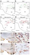
SumL neurons expressing FOS after PS recovery are glutamatergic and GABAergic. (a–c) Schematic distribution of singly FOS+ (black dots), singly vGLUT2+ (green dots) and FOS-vGLUT2 double-labelled (red squares) neurons on a coronal section in a representative animal for PSC (a), PSD (b) and PSR (c) conditions. (d) Schematic distribution of singly FOS+ (black dots), singly vGAT+ (green dots) and FOS-vGAT double-labelled (red squares) neurons on a coronal section in a representative PSR animal. (e) Photomicrographs showing FOS (brown nuclear staining) and vGLUT2 (blue diffuse cytoplasmic staining) double-stained neurons in the SumL of a PSR rat. (f) shows the area inside the rectangle in (e) at a higher magnification. Note the dense cluster of double-labelled neurons in the SumL (red arrowheads). The black and green arrowheads indicate the FOS+ and the vGLUT2+ singly labelled neurons, respectively. (g) Photomicrographs showing FOS (brown nuclear staining) and vGAT (blue diffuse cytoplasmic staining) double-stained neurons in the SumL of a PSR rat. (h) shows the area inside the rectangle in (f) at a higher magnification. Note the dense cluster of double-labelled neurons in the SumL (red arrowheads). The black arrowheads indicate the FOS+ singly labelled neurons. Abbreviations: cp: cerebral peduncle; LHA: lateral hypothalamic area; LM: lateral mammillary nucleus; MM: medial mammillary nucleus, medial part; MnM: medial mammillary nucleus, median part; PH: posterior hypothalamic nucleus; pm: principal mammillary tract; SumL: lateral supramammillary nucleus; SumM: medial supramammillary nucleus. Scale bars: 500 μm for (e, g) and 50 μm for (f, h).
Figure 3
Figure 3
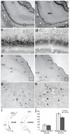
vGLUT2+ fibers and FOS+ neurons are absent in the dorsal DG (dDG) after lesion of the Sum. (a–d) Photomicrographs showing vGLUT2 immunostained sections at the level of the dDG in a PSR sham (a, c) and a PSR rat with a lesion of the Sum (b, d). (c) and (d) show the area inside the rectangle in (a) and (b) at a higher magnification. The Sum lesion causes a dramatic reduction in the number of vGLUT2+ fibers in the dDG. (e–h) Photomicrographs showing FOS immunostained sections at the level of the dDG in a PSR sham (e, g) and a PSR rat with a lesion of the Sum (f, h). (g) and (h) show the area inside the rectangle in (e) and (f) at a higher magnification. The Sum lesion causes a dramatic reduction in the number of FOS+ neurons in the dDG. (i) Illustration of a representative neurotoxic lesion of the Sum on a section immunostained with NeuN. (j) Mean±SEM optic density of vGLUT2 staining and number of FOS+ neurons in dDG in rats with a Sum lesion (n=4) relative to PSR sham animals (n=4). Significance: Kruskal-Wallis followed by Mann-Whitney. * p<0.05. Abbreviations: CA3: field CA3, Ammon’s Horn; DGlb: dentate gyrus, lateral blade; DGlb-mo: dentate gyrus, lateral blade-molecular layer; DGlb-sg: dentate gyrus, lateral blade-granule cell layer; DGmb: dentate gyrus, medial blade; DGmb-mo dentate gyrus, medial blade-molecular layer; DGmb-po: dentate gyrus, medial blade-polymorph layer; DGmb-sg: dentate gyrus, medial blade-granule cell layer; LM: lateral mammillary nucleus; MM: medial mammillary nucleus, medial part; MnM: medial mammillary nucleus, median part; pm: principal mammillary tract; SumL: lateral supramammillary nucleus; SumM: medial supramammillary nucleus; V3: third ventricle. Scale bars: 500 μm for (a, b, e, f, i); 50 μm for (c, d, g, h).
Figure 4
Figure 4
vGLUT2+ fibers and FOS+ neurons are present in the ventral DG (vDG) after lesion of the Sum. (a–d) Photomicrographs showing the vGLUT2 staining in a representative PSR sham (a, c) and a PSR rat with a Sum lesion (b, d). The Sum lesion does not induce a significant reduction in the number of vGLUT2+ fibers in the vDG. (c) and (d) show the area inside the rectangle in (a) and (b) at a higher magnification. (e–h) Photomicrographs showing FOS immunostained sections at the level of the vDG in a PSR sham (e, g) and a PSR rat with a lesion of the Sum (f, h). (g) and (h) show the area inside the rectangle in (e) and (f) at a higher magnification. The Sum lesion does not cause a reduction in the number of FOS+ neurons in the vDG. (i) Schematic distribution of FOS+ (black dots) neurons in the dDG and vDG on a coronal section in a sham (left) and a lesioned (right) rat. Note the strong decrease in the number of FOS+ neurons in the dDG but not the vDG. (j) Mean±SEM optic density of vGLUT2 staining and number of FOS neurons in vDG in rats with a Sum lesion (n=4) relative to PSR sham animals (n=4). Abbreviations: CA3: field CA3, Ammon’s Horn; dDG: dorsal dentate gyrus; DGlb: dentate gyrus, lateral blade; DGlb-mo: dentate gyrus, lateral blade-molecular layer; DGlb-sg: dentate gyrus, lateral blade-granule cell layer; DGmb: dentate gyrus, medial blade; DGmb-mo: dentate gyrus, medial blade-molecular layer; DGmb-po: dentate gyrus, medial blade-polymorph layer; DGmb-sg: dentate gyrus, medial blade-granule cell layer; vDG: ventral dentate gyrus. Scale bars: 500 μm for (a, b, e, f); 50 μm for (c, d, g, h).
Figure 5
Figure 5
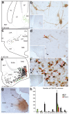
The FOS+ neurons projecting to the vDG in PSR animals are mostly located in the medial entorhinal cortex. (a, c, e) Schematic distribution of singly FOS+ (black dots), singly FG+ (green dots) and FOS-FG double-labelled (red squares) neurons in the medial septum (a), the Sum (c) and the entorhinal cortex (e) in a representative PSR animal. (b, d, f) Photomicrographs showing FOS (black nuclear staining) and FG (brown cytoplasmatic staining) double-stained neurons in the medial septum (b), the Sum (d) and the entorhinal cortex (f) of a PSR rat. Note the dense cluster of double-labelled neurons in the medial entorhinal cortex (red arrowheads). The black and green arrowheads indicate the FOS+ and FG+ singly labelled neurons, respectively. (g) Photomicrograph showing a representative FG injection in the vDG. (h) Mean±SEM number of FOS/FG+ cells contained in the structures projecting to the vDG. Abbreviations: BST: bed nuclei stria terminalis; cp: cerebral peduncle; DGlb: dentate gyrus, lateral blade; DGlb-mo: dentate gyrus, lateral blade-molecular layer; DGlb-sg: dentate gyrus, lateral blade-granule cell layer; DGmb: dentate gyrus, medial blade; DGmb-mo: dentate gyrus, medial blade-molecular layer; DGmb-po: dentate gyrus, medial blade-polymorph layer; DGmb-sg: dentate gyrus, medial blade-granule cell layer; ECT: ectorhinal area; ENTl: entorhinal area, lateral part; ENTm: entorhinal area, medial part; IPN: interpeduncular nucleus; LHA: lateral hypothalamic area; LM: lateral mammillary nucleus; LS: lateral septal nucleus; MS: medial septal nucleus; MM: medial mammillary nucleus, medial part; MnM: medial mammillary nucleus, median part; NBD: diagonal band nucleus; PAR: parasubiculum; PH: posterior hypothalamic nucleus; pm: principal mammillary tract; SF: septofimbrial nucleus; SUBv: subiculum, ventral part; SumL: lateral supramammillary nucleus; SumM: medial supramammillary nucleus; VL: lateral ventricle. Scale bars: 500 μm for (b, d, f, g); 50 μm for the insets.

References

    1. Boyce R, Glasgow SD, Williams S, Adamantidis A. Causal evidence for the role of REM sleep theta rhythm in contextual memory consolidation. Science. 2016;352:812–816. doi: 10.1126/science.aad5252. - DOI - PubMed
    1. Denny CA, et al. Hippocampal memory traces are differentially modulated by experience, time, and adult neurogenesis. Neuron. 2014;83:189–201. doi: 10.1016/j.neuron.2014.05.018. - DOI - PMC - PubMed
    1. Gandrillon O, Solari F, Legrand C, Jurdic P, Samarut J. A rapid and convenient method to prepare DIG-labelled RNA probes for use in non-radioactive in situ hybridization. Mol Cell Probes. 1996;10:51–55. doi: 10.1006/mcpr.1996.0007. - DOI - PubMed
    1. Jouvet M, Michel F, Courjon J. Sur un stade d’activité électrique cérébrale rapide au cours du sommeil physiologique CR Seances. Soc Biol. 1959;153:1024–1028.
    1. Kheirbek MA, et al. Differential control of learning and anxiety along the dorsoventral axis of the dentate gyrus. Neuron. 2013;77:955–968. doi: 10.1016/j.neuron.2012.12.038. - DOI - PMC - PubMed

Publication types

MeSH terms

Substances