. 2016 Oct 7;291(41):21596-21606.
doi: 10.1074/jbc.M116.749184. Epub 2016 Aug 18.

A Diverse Set of Single-domain Antibodies (VHHs) against the Anthrax Toxin Lethal and Edema Factors Provides a Basis for Construction of a Bispecific Agent That Protects against Anthrax Infection

Affiliations

A Diverse Set of Single-domain Antibodies (VHHs) against the Anthrax Toxin Lethal and Edema Factors Provides a Basis for Construction of a Bispecific Agent That Protects against Anthrax Infection

Catherine E Vrentas et al. J Biol Chem. .

Abstract

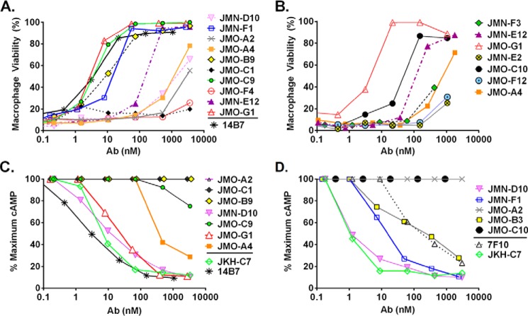
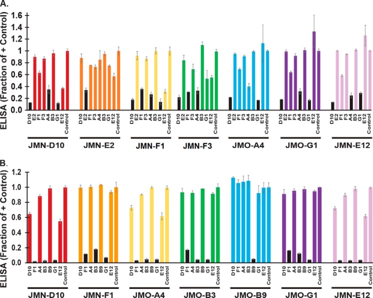
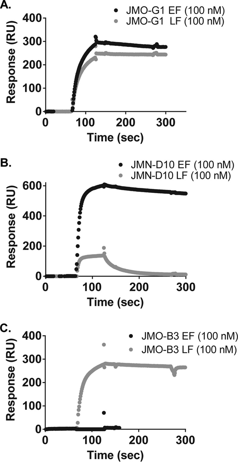
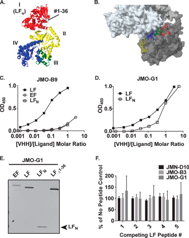
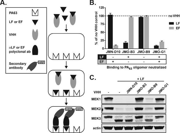
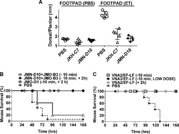
Infection with Bacillus anthracis, the causative agent of anthrax, can lead to persistence of lethal secreted toxins in the bloodstream, even after antibiotic treatment. VHH single-domain antibodies have been demonstrated to neutralize diverse bacterial toxins both in vitro and in vivo, with protein properties such as small size and high stability that make them attractive therapeutic candidates. Recently, we reported on VHHs with in vivo activity against the protective antigen component of the anthrax toxins. Here, we characterized a new set of 15 VHHs against the anthrax toxins that act by binding to the edema factor (EF) and/or lethal factor (LF) components. Six of these VHHs are cross-reactive against both EF and LF and recognize the N-terminal domain (LFN, EFN) of their target(s) with subnanomolar affinity. The cross-reactive VHHs block binding of EF/LF to the protective antigen C-terminal binding interface, preventing toxin entry into the cell. Another VHH appears to recognize the LF C-terminal domain and exhibits a kinetic effect on substrate cleavage by LF. A subset of the VHHs neutralized against EF and/or LF in murine macrophage assays, and the neutralizing VHHs that were tested improved survival of mice in a spore model of anthrax infection. Finally, a bispecific VNA (VHH-based neutralizing agent) consisting of two linked toxin-neutralizing VHHs, JMN-D10 and JMO-G1, was fully protective against lethal anthrax spore infection in mice as a single dose. This set of VHHs should facilitate development of new therapeutic VNAs and/or diagnostic agents for anthrax.

Keywords: EF; LF; VHH; VNA; anthrax toxin; antibody; epitope mapping; microbial pathogenesis; phage display; toxin.


Figures

FIGURE 1.
FIGURE 1.
LF and EF neutralization by VHHs. Representative LF (A and B) and EF (C and D) neutralization experiments measuring toxicity of LT or ET for macrophages in the presence of various doses of antibody are shown. Assays were performed using 250 ng/ml concentrations of each toxin component. In addition to testing the new EF and LF binding VHHs (Table 1), 7F10 (12), JKH-C7 (3), and 14B7 (24) are previously characterized neutralizing mAbs and VHHs included here as positive controls. Percent survival of macrophages is calculated relative to medium-treated cells.
FIGURE 2.
FIGURE 2.
EF/LF epitope mapping of VHHs. Sandwich ELISAs, as shown graphically in supplemental Fig. S3, were performed with a subset of anti-EF VHHs (A) and anti-LF VHHs (B) to investigate epitope binding competition. Briefly, HRP-labeled EF (A) or LF (B) was preincubated with a blocking VHH or PBS control for 1 h followed by application to an ELISA plate coated with a test VHH. Test VHHs are identified below each cluster of bars, and blocking agents are identified under each individual bar. ELISA values are expressed as a fraction of the signal in the PBS control wells, with smaller bars indicating increased competition between the blocking and the test VHHs. Pairs in which the binding of EF/LF to the test VHH was reduced ∼3-fold or more by the blocking VHH are indicated by black bars. Bars represent the average of three technical replicates ± S.D. Results were effectively duplicated within the experiment, as each VHH was tested as both a blocking and test agent with each other VHH; additionally, a matching pattern of competition was observed for two independent experiments.
FIGURE 3.
FIGURE 3.
SPR binding curves for selected EF/LF binding VHHs. Binding of JMO-G1 (A), JMN-D10 (B), and JMO-B3 (C) to EF (black curves) and LF (gray curves) were assessed by SPR. Association and dissociation phases of curves are depicted for chips coupled to EF or LF and assessed with ∼100 nm VHH protein in the flow chamber. Representative curves are shown; triplicate curves were utilized to generate the mean binding parameters ± S.E. displayed in Table 2.
FIGURE 4.
FIGURE 4.
Additional epitope mapping of VHH binding to LF. A, crystal structure of LF (PDB 1J7N; Ref. 29) colored by domain. The LFN domain is colored in red, and residues 1–36 are colored in gray. B, location of LF residues represented by peptides in supplemental Table S1, as depicted on the co-crystal structure of PA (white) with LF (gray) (PDB 3KWV). Yellow, residues #179–184; blue, residues #231–236; green, residues #227–231; red, residues #136–143. C and D, ELISA curves assessing direct binding of HRP-labeled VHHs to plates coated with LF, EF, or LFN. Panel C depicts binding of JMO-B9, and panel D depicts binding of JMO-G1. Note that molar ratios of [VHH]/[antigen] are calculated as based on the total concentration of VHH in the reaction; however, only a fraction of the VHH was HRP labeled. As depicted here in a representative experiment, JMO-B9 failed to effectively bind to LFN in four independent experiments. E, boiled LF, EF, LFN, and LF(Δ1–36) proteins were blotted and probed with E-tagged JMO-G1 as indicated using an anti-E tag goat antibody and an IR dye-coupled anti-goat antibody for detection. The same pattern was observed for three other members of the EF1/LF1 family in three sets of replicate blots. In a control experiment (data not shown), no reactivity was observed in a parallel blot in which the VHH primary antibody incubation was omitted. F, results of peptide competition assay for binding of HRP-VHHs to LF. ELISA signals for JMN-D10, JMO-B3, and JMO-G1 in the presence of peptides 1–5 (supplemental Table S1) are expressed as a percentage of controls in which HRP-VHHs were incubated in the absence of competing peptides. Data are the mean ± S.D. of six replicates across two independent experiments. Continued increases in the concentration of competing peptides did not block binding in two additional experiments conducted with JMN-D10 and JMO-B3 (data not shown).
FIGURE 5.
FIGURE 5.
VHH neutralization of LF and EF binding to PA63 oligomer. A, schematic diagram illustrating ELISA methods used in B. B, LF and EF were preincubated with JMN-D10, JMO-B3, and JMO-G1 at a molar ratio of 1:5 (LF or EF to VHH). The ability of LF and EF preincubated with VHH antibodies to bind to PA63 oligomer was assessed by ELISA and compared with LF and EF binding to PA63 oligomer in the absence of any VHH. Data are the mean ± S.E. of eight technical replicates from three independent experiments. The table (bottom) summarizes the ability of each VHH to neutralize LF or EF binding to PA63 oligomer. C, RAW264.7 cells were treated for 1 h with PA (1 μg/ml) and with either vehicle, VHHs, LF (1 μg/ml), or LF (1 μg/ml) preincubated for 1 h with VHHs (1.75 μg/ml) as in B, at a molar ratio of 1:5 (LF to VHH). Western blotting of cell lysates was performed to assess cleavage of MEK1, -2, and -3 by LF. The MEK1 and MEK2 antibodies recognize N-terminal epitopes that are degraded upon cleavage, and the MEK3 antibody recognizes an N-terminal epitope that is not degraded after cleavage. The larger MEK3 band is the full-length protein, and the smaller band is the N-terminal cleavage product. Actin is a control to demonstrate equal protein loading. Data are representative of three independent experiments.
FIGURE 6.
FIGURE 6.
Therapeutic efficacy of VHHs in mice. A, footpad edema model. BALB/cJ mice (n = 5/group) were injected with PBS, JKH-C7, or JMN-D10 (25 μg/100 μl, i.v.) 10–20 min before footpad administration of ET (0.2 μg/20 μl) or PBS (20 μl). Each circle represents the average of three dorsal/plantar measurements for a single footpad/mouse at 21 h post-toxin administration. The p value comparing the ET footpad measurements of both antibody pretreated groups to the PBS group is <0.0001 using a standard unpaired t test. B and C, spore challenge model. C57BL/6J mice were challenged with a lethal dose of 5 × 10⁷ spores (SC, 400 μl). In B, mice received PBS (n = 16) or VHH treatments, either as a single administration (16 μg/VHH, SC, 10 min pre-spore infection, n = 5) or as two administrations (16 μg, SC, 10 min pre-spore infection followed by 32 μg, SC, 2 h post-spore infection, n = 5). JMO-G1, targeting both LF and EF, was injected alone (n = 9), whereas JMN-D10 (targeting EF) and JMO-B3 (targeting LF) were administered to mice as a mixture (n = 10). Results shown are from three studies. In C, VNA2/EF-LF was administered at either 16 μg, SC, 10 min pre-spore infection, or 2 h post-spore infection. In one group (LOW DOSE), the VNA was administered at 4 μg, SC, only 10 min pre-spore infection. Control mice received PBS at 10 min pre-infection (n = 5/treatment group).

References

    1. Moayeri M., Leppla S. H., Vrentas C., Pomerantsev A. P., and Liu S. (2015) Anthrax pathogenesis. Annu. Rev. Microbiol. 69, 185–208 - PubMed
    1. Chen Z., Moayeri M., and Purcell R. (2011) Monoclonal antibody therapies against anthrax. Toxins 3, 1004–1019 - PMC - PubMed
    1. Moayeri M., Leysath C. E., Tremblay J. M., Vrentas C., Crown D., Leppla S. H., and Shoemaker C. B. (2015) A heterodimer of a VHH (variable domains of camelid heavy chain-only) antibody that inhibits anthrax toxin cell binding linked to a VHH antibody that blocks oligomer formation is highly protective in an anthrax spore challenge model. J. Biol. Chem. 290, 6584–6595 - PMC - PubMed
    1. Muyldermans S. (2013) Nanobodies: natural single-domain antibodies. Annu. Rev. Biochem. 82, 775–797 - PubMed
    1. Hassanzadeh-Ghassabeh G., Devoogdt N., De Pauw P., Vincke C., and Muyldermans S. (2013) Nanobodies and their potential applications. Nanomedicine 8, 1013–1026 - PubMed

MeSH terms