Review
. 2016 Nov;38(11):1117-1122.
doi: 10.1002/bies.201600116. Epub 2016 Aug 19.

R.I.P. to the PIP: PCNA-binding motif no longer considered specific: PIP motifs and other related sequences are not distinct entities and can bind multiple proteins involved in genome maintenance

Affiliations
Review

R.I.P. to the PIP: PCNA-binding motif no longer considered specific: PIP motifs and other related sequences are not distinct entities and can bind multiple proteins involved in genome maintenance

Elizabeth M Boehm et al. Bioessays. 2016 Nov.

Abstract

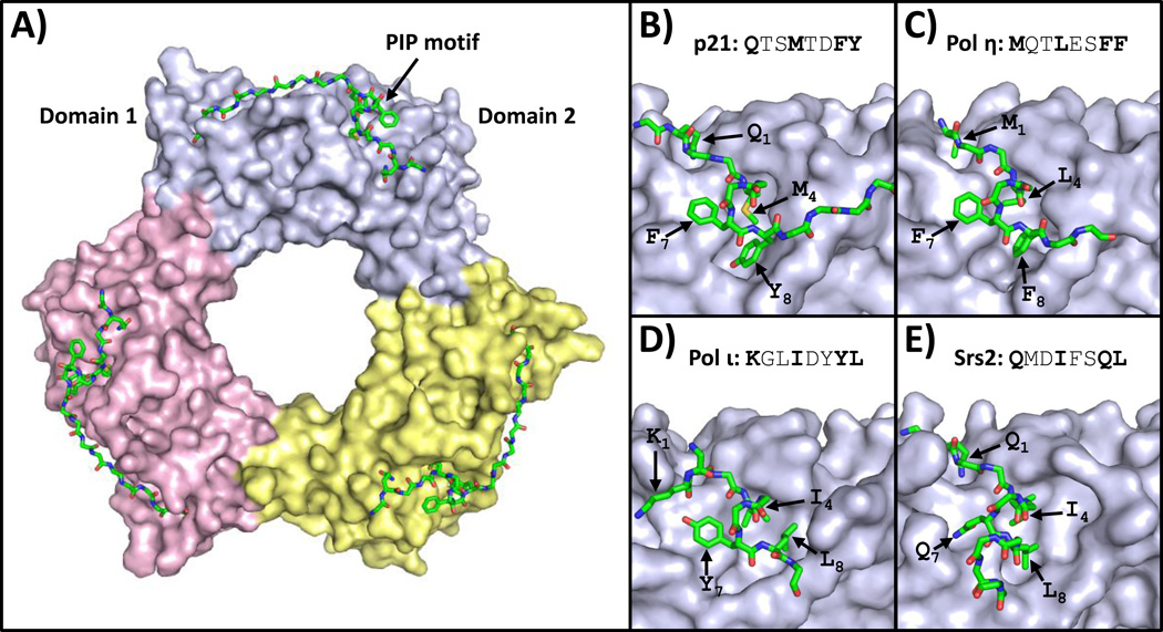
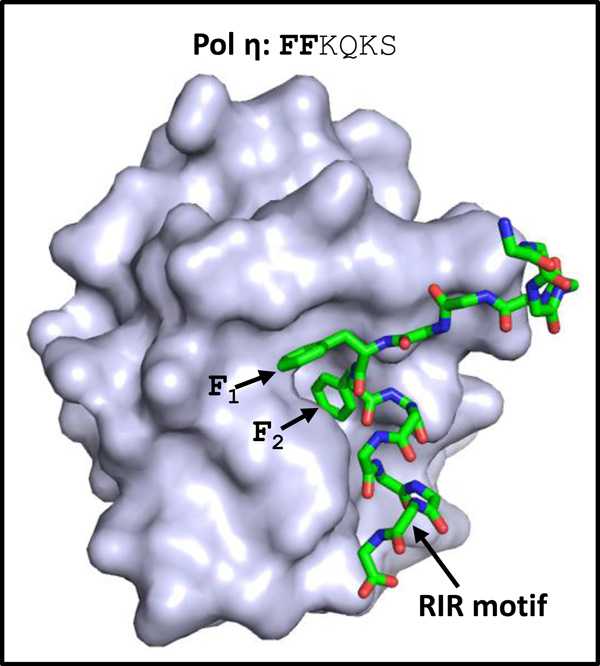
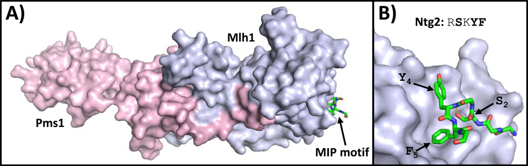
Many proteins responsible for genome maintenance interact with one another via short sequence motifs. The best known of these are PIP motifs, which mediate interactions with the replication protein PCNA. Others include RIR motifs, which bind the translesion synthesis protein Rev1, and MIP motifs, which bind the mismatch repair protein Mlh1. Although these motifs have similar consensus sequences, they have traditionally been viewed as separate motifs, each with their own target protein. In this article, we review several recent studies that challenge this view. Taken together, they imply that these different motifs are not distinct entities. Instead, there is a single, broader class of motifs, which we call "PIP-like" motifs, which have overlapping specificities and are capable of binding multiple target proteins. Given this, we must reassess the role of these motifs in forming the network of interacting proteins responsible for genome maintenance.

Keywords: DNA recombination; DNA repair; DNA replication; Rev1; translesion synthesis.


Figures

Figure 1
Figure 1
Structures of PCNA bound to PIP motifs. (A) An overview of the structure of human PCNA bound to peptides containing the PIP motif of p21 (PDB ID: 1AXC) is shown [28]. The three PCNA subunits are depicted in the surface representation (light blue, yellow, and pink). The PIP motif is depicted in the stick representation (with carbon, nitrogen, oxygen, and sulfur atoms colored green, blue, red, and yellow, respectively). (B) A close up view of human PCNA bound to the p21 PIP motif is shown [28]. The side chains of residues in positions 1, 4, 7, and 8 are indicated. (C) A close up view of human PCNA bound to the pol η PIP motif (PDB ID: 2ZVK) is shown [29]. (D) A close up view of human PCNA bound to the pol ι PIP motif (PDB ID: 2ZVM) is shown [29]. (E) A close up view of yeast PCNA bound to the Srs2 PIP motif (PDB ID: SV62) is shown [30].
Figure 2
Figure 2
Structure of Rev1 bound to a RIR motif. The structure of the CTD of human Rev1 bound to a peptide containing the RIR motif of pol η (PDB ID: 2LSK) is shown [20]. The human Rev1 CTD is depicted in the surface representation (light blue). The peptide containing the RIR motif of human pol η is depicted in the stick representation (with carbon, nitrogen, and oxygen atoms colored green, blue, and red, respectively). The side chains of residues in positions 1 and 2 are indicated.
Figure 3
Figure 3
Structure of Mlh1 bound to a MIP motif. (A) An overview of the structure of the CTD of yeast MutLα bound to a peptide containing the MIP motif of Ntg2 (PDB ID: 4FMN) is shown [23]. The Mlh1 and Pms1 subunits are depicted in the surface representation (light blue and pink, respectively). The peptide containing the MIP motif of Ntg2 is depicted in the stick representation (with carbon, nitrogen, and oxygen atoms colored green, blue, and red, respectively). The side chains of residues in positions 2, 4, and 5 and the hydrogen bond between the serine at position 2 and the backbone at position 4 are indicated.

References

    1. Tsurimoto T. PCNA binding proteins. Front Biosci. 1999;4:D849–D858. - PubMed
    1. Maga G, Hubscher U. Proliferating cell nuclear antigen (PCNA): a dancer with many partners. J Cell Sci. 2003;116:3051–3060. - PubMed
    1. Moldovan GL, Pfander B, Jentsch S. PCNA, the maestro of the replication fork. Cell. 2007;129:665–679. - PubMed
    1. Naryzhny SN. Proliferating cell nuclear antigen: a proteomics view. Cell Mol Life Sci. 2008;65:3789–3808. - PMC - PubMed
    1. Zhuang Z, Ai Y. Processivity factor of DNA polymerase and its expanding role in normal and translesion DNA synthesis. Biochim Biophys Acta. 2010;1804:1081–1093. - PMC - PubMed

MeSH terms