Review
. 2016 Aug 19;353(6301):766-71.
doi: 10.1126/science.aag2638.

Multifaceted interactions between adaptive immunity and the central nervous system

Affiliations
Review

Multifaceted interactions between adaptive immunity and the central nervous system

Jonathan Kipnis. Science. .

Abstract

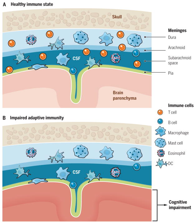
Neuroimmunologists seek to understand the interactions between the central nervous system (CNS) and the immune system, both under homeostatic conditions and in diseases. Unanswered questions include those relating to the diversity and specificity of the meningeal T cell repertoire; the routes taken by immune cells that patrol the meninges under healthy conditions and invade the parenchyma during pathology; the opposing effects (beneficial or detrimental) of these cells on CNS function; the role of immune cells after CNS injury; and the evolutionary link between the two systems, resulting in their tight interaction and interdependence. This Review summarizes the current standing of and challenging questions related to interactions between adaptive immunity and the CNS and considers the possible directions in which these aspects of neuroimmunology will be heading over the next decade.


Figures

Fig. 1
Fig. 1. Meningeal immunity in “surveillance” of brain function
(A) Representation of meninges (pia mater, lining the brain parenchyma; dura mater, attached to skull; arachnoid, attached to dura mater; subarachnoid space, space between arachnid and pia mater, where the CSF flows) and their coverage by the immune cells. Recent evidence suggests that meningeal immune cells, primarily T cells, affect brain function. (B) Elimination of meningeal T cells by using genetically modified mice, pharmacologically trapping T cells in the deep cervical lymph nodes, or preventing their migration to meningeal spaces results in impaired cognitive function. The precise mechanism of how meningeal T cells regulate cognitive function is still not fully understood. DC, dendritic cell.
Fig. 2
Fig. 2. Meningeal and parenchymal access of immune cells
(A) During the steady state, Tcells (and presumably other immune cells) circulate through the meningeal spaces. Their primary entry site is via the meningeal blood vessels, where the immune cells need to cross the blood-meningeal barrier (BMB) to enter the meningeal space. Blood-borne immune cells do not cross the blood-brain barrier (BBB) in a healthy situation. (B) Choroid plexus endothelial cells are fenestrated, which allows immune cells to easily cross them. For the immune cells to make their way into the CSF, however, they need to also cross a tight barrier of choroid plexus epithelial cell layer connected by tight junctions. (C and C′) Under pathological conditions such as inflammation, immune cells extravasate through the meningeal vessels and then cross the pial layer to infiltrate the brain parenchyma (C) or, more plausibly, the meningeal inflammatory environment results in the production of chemokines that, upon diffusion into the parenchyma (across pia), recruit peripheral immune cells across the BBB (C′).
Fig. 3
Fig. 3. Schematic representation of the glymphatic system
Periarterial space (formed between a blood vessel’s endothelial cells and the astrocytic endfeet processes) allows CSF to follow the arteries into the parenchyma. CSF, along with macromolecules within it, diffuses from the periarterial spaces as an interstitial fluid into the parenchyma, “washes” the parenchyma, and is reabsorbed into perivenular space, to be then carried back and mixed with the CSF.
Fig. 4
Fig. 4. CNS drainage: New concepts for old
(A) Before the discovery of meningeal lymphatic vessels, the old concept of CNS drainage was based on the fact that water from CSF is drained through arachnoid granulations, whereas macromolecules and immune cells from the CNS and the CSF are drained through the cribriform plate into nasal lymphatics and, from there, to CNS-draining deep cervical lymph nodes. (B) Discovery of the meningeal lymphatic vessels led to the hypothesis that they may drain meningeal immune cells and macromolecules from the parenchyma and the CSF, whereas the contribution of the cribriform plate as a drainage route for immune cells under homeostatic conditions needs to be reassessed. This route may be more active during neuroinflammatory conditions. Additional studies are needed to better characterize the contribution of each route of drainage for immune cells and macromolecules from the CNS and the CSF under homeostatic and pathological conditions.

References

    1. Medawar PB. Br J Exp Pathol. 1948;29:58–69. - PMC - PubMed
    1. Hašek M, Chutna J, Sládecek M, Lodin Z. Nature. 1977;268:68–69. - PubMed
    1. Wekerle H. Curr Top Microbiol Immunol. 2006;305:25–50. - PubMed
    1. Carson MJ, Doose JM, Melchior B, Schmid CD, Ploix CC. Immunol Rev. 2006;213:48–65. - PMC - PubMed
    1. del Río-Hortega P. In: Cytology and Cellular Pathology of the Nervous System. Penfield W, editor. Paul B. Hoebaer; New York: 1932. pp. 483–534.

Publication types