Review
. 2016 Aug 19;353(6301):783-5.
doi: 10.1126/science.aaf6260.

Inflammatory neuroprotection following traumatic brain injury

Affiliations
Review

Inflammatory neuroprotection following traumatic brain injury

Matthew V Russo et al. Science. .

Abstract

Traumatic brain injury (TBI) elicits an inflammatory response in the central nervous system (CNS) that involves both resident and peripheral immune cells. Neuroinflammation can persist for years following a single TBI and may contribute to neurodegeneration. However, administration of anti-inflammatory drugs shortly after injury was not effective in the treatment of TBI patients. Some components of the neuroinflammatory response seem to play a beneficial role in the acute phase of TBI. Indeed, following CNS injury, early inflammation can set the stage for proper tissue regeneration and recovery, which can, perhaps, explain why general immunosuppression in TBI patients is disadvantageous. Here, we discuss some positive attributes of neuroinflammation and propose that inflammation be therapeutically guided in TBI patients rather than globally suppressed.


Figures

Fig. 1
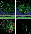
Fig. 1. Myeloid cell dynamics following brain injury
(A and B) Two-photon z-stacks captured through a thinned skull (blue) window of CX3CR1gfp/+ mice show the morphology of macrophages and microglia (green) in the meninges and brain parenchyma, respectively. (A) Naïve microglia are ramified, whereas meningeal macrophages have a wormlike appearance. (B) Within minutes to hours after a meningeal contusion injury, meningeal macrophages rapidly disappear (die) and activated microglia migrate to the glia limitans and assume a “jellyfish” morphology that allows them to fill spaces previously occupied by dead astrocytes. These cells participate in phagocytosis and reduce leakage from the meninges into the underlying brain parenchyma. See corresponding movie S1. (C and D) Lysozyme-Mgfp/+ myelomonocytic cells (neutrophils/monocytes in green) are recruited to sites of meningeal cell death (red) beginning ~1 hour after TBI. A two-photon z-stack captured at 24 hours post injury demonstrates that nearly all the myelomonocytic cells responding to cell death are in the (C) meninges (top 30 μm of z-stack) as opposed to the (D) parenchyma (bottom 30 μm of the same z-stack). See corresponding movie S3.
Fig. 2
Fig. 2. Scavenging macrophages after brain injury
(A and B) Meningeal macrophages repopulate the meninges beginning ~2 days post injury, where they assume the role of scavengers. Time-lapse two-photon movies of the meningeal space show (A) a single macrophage that had previously engulfed a dead cell extending a process to phagocytose another cell at 2 days post injury and (B) a macrophage probing and sampling a dead cell at 4 days post injury. See corresponding movie S2.

Similar articles

Cited by

References

    1. Corps KN, Roth TL, McGavern DB. JAMA Neurol. 2015;72:355–362. - PMC - PubMed
    1. Johnson VE, et al. Brain. 2013;136:28–42. - PMC - PubMed
    1. Homsi S, et al. J Neurotrauma. 2010;27:911–921. - PubMed
    1. Dohi K, et al. J Neuroinflammation. 2010;7:41. - PMC - PubMed
    1. Hsieh CL, et al. J Neurotrauma. 2014;31:1677–1688. - PMC - PubMed

Publication types