

Radiolabeled Phosphonium Salts as Mitochondrial Voltage Sensors for Positron Emission Tomography Myocardial Imaging Agents
- PMID: 27540422
- PMCID: PMC4977252
- DOI: 10.1007/s13139-016-0397-x
Radiolabeled Phosphonium Salts as Mitochondrial Voltage Sensors for Positron Emission Tomography Myocardial Imaging Agents
Abstract
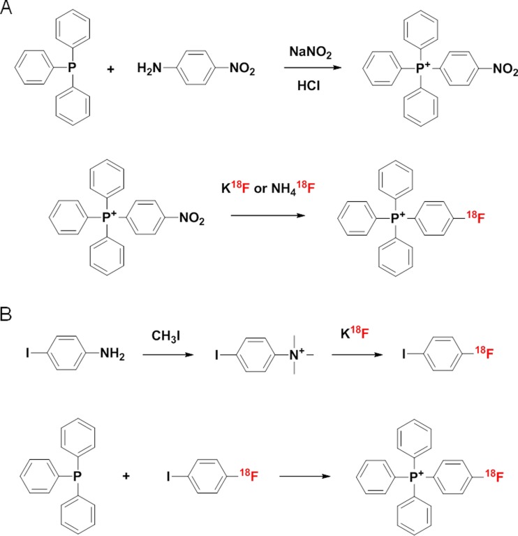
Despite substantial advances in the diagnosis of cardiovascular disease, (18)F-labeled positron emission tomography (PET) radiopharmaceuticals remain necessary to diagnose heart disease because clinical use of current PET tracers is limited by their short half-life. Lipophilic cations such as phosphonium salts penetrate the mitochondrial membranes and accumulate in mitochondria of cardiomyocytes in response to negative inner-transmembrane potentials. Radiolabeled tetraphenylphosphonium cation derivatives have been developed as myocardial imaging agents for PET. In this review, a general overview of these radiotracers, including their radiosynthesis, in vivo characterization, and evaluation is provided and clinical perspectives are discussed.
Keywords: 18F-Fluoroalkylphosphonium cations; Mitochondrial voltage sensors; Myocardial imaging agents; Positron emission tomography; Tetraphenylphosphonium cation derivatives.
Figures


Similar articles
-
Emerging Tracers for Nuclear Cardiac PET Imaging.Nucl Med Mol Imaging. 2018 Aug;52(4):266-278. doi: 10.1007/s13139-018-0521-1. Epub 2018 May 8. Nucl Med Mol Imaging. 2018. PMID: 30100939 Free PMC article. Review.
-
Synthesis of [18F]-labeled (6-fluorohexyl)triphenylphosphonium cation as a potential agent for myocardial imaging using positron emission tomography.Bioconjug Chem. 2012 Mar 21;23(3):431-7. doi: 10.1021/bc2004439. Epub 2012 Feb 9. Bioconjug Chem. 2012. PMID: 22264022
-
Radiosynthesis and evaluation of 18F-labeled aliphatic phosphonium cations as a myocardial imaging agent for positron emission tomography.Nucl Med Commun. 2015 Jul;36(7):747-54. doi: 10.1097/MNM.0000000000000315. Nucl Med Commun. 2015. PMID: 25850717
-
Highly efficient one-pot labeling of new phosphonium cations with fluorine-18 as potential PET agents for myocardial perfusion imaging.Mol Pharm. 2014 Nov 3;11(11):3823-31. doi: 10.1021/mp500216g. Epub 2014 Jun 5. Mol Pharm. 2014. PMID: 24852080
-
64Cu-labeled phosphonium cations as PET radiotracers for tumor imaging.Bioconjug Chem. 2011 Aug 17;22(8):1459-72. doi: 10.1021/bc200106p. Epub 2011 Jul 1. Bioconjug Chem. 2011. PMID: 21696200 Free PMC article. Review.
Cited by
-
Emerging Tracers for Nuclear Cardiac PET Imaging.Nucl Med Mol Imaging. 2018 Aug;52(4):266-278. doi: 10.1007/s13139-018-0521-1. Epub 2018 May 8. Nucl Med Mol Imaging. 2018. PMID: 30100939 Free PMC article. Review.
-
Tumor Targeting Effect of Triphenylphosphonium Cations and Folic Acid Coated with Zr-89-Labeled Silica Nanoparticles.Molecules. 2020 Jun 25;25(12):2922. doi: 10.3390/molecules25122922. Molecules. 2020. PMID: 32630467 Free PMC article.
-
KSNM60: The History of Radiopharmaceutical Sciences in Korea.Nucl Med Mol Imaging. 2022 Jun;56(3):114-126. doi: 10.1007/s13139-022-00744-8. Epub 2022 Apr 5. Nucl Med Mol Imaging. 2022. PMID: 35607629 Free PMC article. Review.
-
Research Progress on 18F-Labeled Agents for Imaging of Myocardial Perfusion with Positron Emission Tomography.Molecules. 2017 Mar 30;22(4):562. doi: 10.3390/molecules22040562. Molecules. 2017. PMID: 28358340 Free PMC article. Review.
-
Mitochondria-Targeted Triphenylphosphonium-Based Compounds: Syntheses, Mechanisms of Action, and Therapeutic and Diagnostic Applications.Chem Rev. 2017 Aug 9;117(15):10043-10120. doi: 10.1021/acs.chemrev.7b00042. Epub 2017 Jun 27. Chem Rev. 2017. PMID: 28654243 Free PMC article. Review.
References
-
- Fovino LN, Saladini G, Mormino GP, Saladini F, Razzolini R, Evangelista L. Risk stratification and prognostic assessment by myocardial perfusion-gated SPECT in patients with left bundle-branch block and low-intermediate cardiac risk. Ann Nucl Med. 2012;26:559–70. doi: 10.1007/s12149-012-0613-4. - DOI - PubMed
Publication types
LinkOut - more resources
Full Text Sources
Other Literature Sources