Review
. 2016 Jul 28:5:F1000 Faculty Rev-1882.
doi: 10.12688/f1000research.8829.1. eCollection 2016.

An overview of cutaneous T cell lymphomas

Affiliations
Review

An overview of cutaneous T cell lymphomas

Nooshin Bagherani et al. F1000Res. .

Abstract

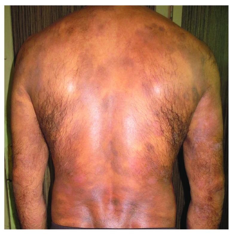
Cutaneous T cell lymphomas (CTCLs) are a heterogeneous group of extranodal non-Hodgkin's lymphomas that are characterized by a cutaneous infiltration of malignant monoclonal T lymphocytes. They typically afflict adults with a median age of 55 to 60 years, and the annual incidence is about 0.5 per 100,000. Mycosis fungoides, Sézary syndrome, and primary cutaneous peripheral T cell lymphomas not otherwise specified are the most important subtypes of CTCL. CTCL is a complicated concept in terms of etiopathogenesis, diagnosis, therapy, and prognosis. Herein, we summarize advances which have been achieved in these fields.

Keywords: Sézary syndrome; cutaneous T cell lymphoma; mycosis fungoides.


Conflict of interest statement

Competing interests: The authors declare that they have no competing interests.

No competing interests were disclosed.

Figures

Figure 1.
Figure 1.. Mycosis fungoides in a 40-year-old man manifested as generalized atrophic patches.
Figure 2.
Figure 2.. In pathological view, the cutaneous T cell lymphomas are characterized by haloed lymphocytes, exocytosis, epidermotropism, Pautrier’s microabscess, large hyperconvoluted, hyperchromatic lymphocytes in the epidermis, and lymphocytes aligned within the basal layer.
Figure 2A (left) A lymphocytic infiltrate is present in the dermis and extending into the overlying epidermis with minimal overlying spongiosis. Figure 2B (center) Lymphocytes with surrounding haloes are present in the epidermis as single cells and small clusters (Pautrier’s microabscesses). There is minimal accompanying spongiosis. Figure 2C (right) Psoriasiform epidermal hyperplasia with epidermotropism of haloed lymphocytes is seen in this case of patch-stage mycosis fungoides.
Figure 3.
Figure 3.. Etiopathogenesis of cutaneous T cell lymphoma.
CTCL, cutaneous T cell lymphoma.

References

    1. Rodd AL, Ververis K, Karagiannis TC: Current and Emerging Therapeutics for Cutaneous T-Cell Lymphoma: Histone Deacetylase Inhibitors. Lymphoma. 2012;2012:1–10, 290685. 10.1155/2012/290685 - DOI
    1. Litvinov IV, Netchiporouk E, Cordeiro B, et al. : Ectopic expression of embryonic stem cell and other developmental genes in cutaneous T-cell lymphoma. Oncoimmunology. 2014;3(11):e970025. 10.4161/21624011.2014.970025 - DOI - PMC - PubMed
    1. Lauenborg B, Christensen L, Ralfkiaer U, et al. : Malignant T cells express lymphotoxin α and drive endothelial activation in cutaneous T cell lymphoma. Oncotarget. 2015;6(17):15235–49. 10.18632/oncotarget.3837 - DOI - PMC - PubMed
    1. Nahidi Y, Meibodi NT, Ghazvini K, et al. : Evaluation of the Association Between Epstein-Barr Virus and Mycosis Fungoides. Indian J Dermatol. 2015;60(3):321. 10.4103/0019-5154.156423 - DOI - PMC - PubMed
    1. Gómez Venegas ÁA, Vargas Rubio RD: Unusual involvement in mycosis fungoides: Duodenal papilla. Rev Esp Enferm Dig. 2015. 10.17235/reed.2015.3831/2015 - DOI - PubMed

LinkOut - more resources