. 2017 Jul;54(5):3893-3905.
doi: 10.1007/s12035-016-0053-6. Epub 2016 Aug 19.

Metalloproteinases ADAM10 and ADAM17 Mediate Migration and Differentiation in Glioblastoma Sphere-Forming Cells

Affiliations

Metalloproteinases ADAM10 and ADAM17 Mediate Migration and Differentiation in Glioblastoma Sphere-Forming Cells

Elodie J Siney et al. Mol Neurobiol. 2017 Jul.

Abstract

Glioblastoma is the most common form of primary malignant brain tumour. These tumours are highly proliferative and infiltrative resulting in a median patient survival of only 14 months from diagnosis. The current treatment regimens are ineffective against the small population of cancer stem cells residing in the tumourigenic niche; however, a new therapeutic approach could involve the removal of these cells from the microenvironment that maintains the cancer stem cell phenotype. We have isolated multipotent sphere-forming cells from human high grade glioma (glioma sphere-forming cells (GSCs)) to investigate the adhesive and migratory properties of these cells in vitro. We have focused on the role of two closely related metalloproteinases ADAM10 and ADAM17 due to their high expression in glioblastoma and GSCs and their ability to activate cytokines and growth factors. Here, we report that ADAM10 and ADAM17 inhibition selectively increases GSC, but not neural stem cell, migration and that the migrated GSCs exhibit a differentiated phenotype. We also observed a correlation between nestin, a stem/progenitor marker, and fibronectin, an extracellular matrix protein, expression in high grade glioma tissues. GSCs adherence on fibronectin is mediated by α5β1 integrin, where fibronectin further promotes GSC migration and is an effective candidate for in vivo cancer stem cell migration out of the tumourigenic niche. Our results suggest that therapies against ADAM10 and ADAM17 may promote cancer stem cell migration away from the tumourigenic niche resulting in a differentiated phenotype that is more susceptible to treatment.

Keywords: Cancer stem cell; Cell differentiation; Cell migration; Disintegrin; Extracellular matrix; Glioma.


Conflict of interest statement

Funding

Medical Research Council capacity building studentship to E.J.S.; Wessex Medical Trust (N06), The Royal Society (RG090173) and the Wessex Brain Tumour Fund to S.W.-M.

Conflict of Interest

The authors declare that they have no competing interests.

Figures

Fig. 1
Fig. 1
ADAM10 and ADAM17 are expressed in patient-derived GSCs and anti-ADAM17 inhibits protease function. a Representative confocal images of metalloproteinases ADAM10 (A10, green) and ADAM17 (A17, green) and stem cell markers SOX2 (red), CD133 (red) and nestin (Nes; red) staining of GSCs cultured as monolayers from four cell lines (G002, G049, G100 and G112). Nuclei stained with DAPI (blue), scale bar = 20 μm. N.B. G112 ADAM17 and nestin staining are from different wells, hence the absence of a merge picture. b The protease activity of purified rhADAM17 (left) or G002 cell lysates (right) pre-treated with either IgG isotype control (10 μg/ml), anti-ADAM17 (10 μg/ml) or TAPI-2 (10 μg/ml). Two separate G002 cell lysates were collected at passages 3 and 5, respectively, and tested in triplicate; the purified rhADAM17 was tested once in triplicate. **P < 0.01, ***P < 0.001 compared to IgG control using one-way ANOVA followed by Tukey’s posthoc test. c Tumourspheres from G112 after 11 days in vitro, scale bar = 100 μm
Fig. 2
Fig. 2
ADAM10 and ADAM17 inhibition increases GSC, but not NSC; directional migration and the migrated GSCs are more differentiated. a Migration of neural stem cells from foetal sample H1450 (NSC) and isolated GSCs from patient sample G100 (GSC) towards selected chemoattractants. Lower transwell chambers contained bovine serum albumin (BSA, 0.1 g/ml), laminin (LN, 10 μg/ml), growth factors (GF: EGF 20 ng/ml, FGF 10 ng/ml and heparin 2 μg/ml) or foetal bovine serum (FBS, 0.1 %). N = 3 wells per condition and per cell line; ***P < 0.001 and ****P < 0.0001 compared to BSA using two-way ANOVA followed by Tukey’s posthoc test. b Chemoattraction of two NSC (H1445 and H1450) and four GSC (G002, G036, G037 & G049) lines with either anti-ADAM10 (0.2 μg/ml), anti-ADAM17 (2 μg/ml) or no treatment (control) in lower chamber. Cell numbers in lower chambers were normalised to the NSC control to give percent cell migration. N = 4 wells per condition and per cell line; *P < 0.05, **P < 0.01 compared to the control using two-way ANOVA followed by Bonferroni’s posthoc test. c 24-h toxicity test of ADAM10 and ADAM17 blocking antibodies on GSCs from G002. 2.5 × 105 cells were added into wells containing IgG (2 μg/ml), anti-ADAM10 (0.2 μg/ml) anti-ADAM17 (2 μg/ml) or anti-ADAM10 and anti-ADAM17 combined. Cell viability was calculated using trypan blue to visualise and discount dead cells. N = 3 wells per condition; P = 0.8292 using one-way ANOVA. d Percentage of migrated and non-migrated GSCs expressing stem cell/progenitor marker nestin, putative brain tumour stem cell marker CD133 or neuronal marker βIII-tubulin from two GSC lines (G037 and G065). N = 2 wells per condition and per cell line; **P < 0.01, ***P < 0.001 using the Mann-Whitney test. e Sphere formation ability of migrated vs. non-migrated GSCs, from patient sample G002, following transwell migration assay. 5 × 103 cells from the upper (non-migrated) and lower (migrated) chambers were plated in triplicate. At day 11, spheres >100 μm were counted and averaged. ****P < 0.0001 using unpaired t test
Fig. 3
Fig. 3
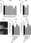
Extracellular matrix proteins alter the expression of differentiation markers in GSCs. a Immunostaining of five tissue samples (G065, G071, G083, G097, G099) for laminin (LN), fibronectin (FN) and vitronectin (VN) in green, nuclei stained with DAPI (blue); the star indicates FN expression in distinct regions; the arrow indicates diffuse FN in tissue. Scale bar = 50 μm. bd Percentage of GSCs positive for b nestin, median values LN 95.9 %, FN 82.4 % and VN 94.0 % , c S100β, median values LN 0.0 %, FN 12.8 % and VN 11.5 % and d βIII-tubulin, median values LN 3.3 %, FN 14.7 % and VN 60.3 % from G100 GSC line cultured on LN, FN or VN for 14 days and immunostained for the three markers. N = 3 wells per condition; *P < 0.05 using Kruskal-Wallis followed by Tukey’s posthoc test
Fig. 4
Fig. 4
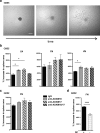
ADAM10 or ADAM17 inhibition increases cell migration out of tumourspheres on three different ECMs. Tumoursphere spreading assay: Individual tumourspheres (100 μm in diameter) were incubated with 1 μM Ara-C and either IgG (2 μg/ml), anti-ADAM10 (0.2 μg/ml), anti-ADAM17 (2 μg/ml) or anti-ADAM10 and ADAM17 combined. Spheres were imaged five times at regular intervals between t = 0 h and t = 45 h; sphere area was measured using Image J. Cell migration out of the sphere was calculated as ((t = 45 h − t = 0 h)/t = 0 h) × 100 to give percent increase in sphere area. a Phase contrast images of sphere migration from a single G065 sphere on LN at 0 h, 20 h and 45 h, scale bar = 100 μm. b Cell migration out of G065 spheres was tested on wells pre-coated with three ECMs; laminin (LN), fibronectin (FN) or vitronectin (VN) at 25 μg/ml. N = 4–6 spheres per condition; *P < 0.05 using one-way ANOVA followed by Bonferroni’s posthoc test. c Cell migration out of G002 spheres was tested on wells pre-coated with FN (25 μg/ml), N = 4–6 spheres per condition; P > 0.05 using one-way ANOVA. d Cell migration out of G002 spheres on fibronectin in the absence or presence of growth factors (GF) when treated with IgG only. N = 5–6 spheres per condition; **P < 0.0001 using unpaired t test
Fig. 5
Fig. 5
ADAM10 and ADAM17 inhibition increases GSC migration towards fibronectin. a Immunostaining of G065, G071, G083, G097 and G099 tissue samples for stem/progenitor cell marker nestin (green), stem cell marker Sox2 (red), astrocyte marker S100β (green), neuronal marker βIII-tubulin (red), nuclei stained with DAPI (blue), scale bar = 50 μm. b Correlation between nestin and fibronectin (FN) intensity expression in five tissues, G065, G071, G083, G097 and G099 (Pearson product moment correlation test, r = 0.943, P = 0.016, Sigma plot). c Haptotactic migration of G100 GSCs through ECM protein-coated transwell over 24 h. N = 4 wells per condition; **P < 0.01, ***P < 0.001 using one-way ANOVA followed by Tukey’s posthoc test. d Haptotactic migration of four GSC lines (G002, G100, G109, G112) over 24 h through fibronectin in absence of growth factors. Cells were treated with IgG isotype control (2 μg/ml), anti-ADAM10 (0.2 μg/ml), anti-ADAM17 (2 μg/ml) or anti-ADAM10 and ADAM17 combined. N = 4 wells per condition and per cell line; **P < 0.01, ***P < 0.001 using one-way ANOVA followed by Tukey’s posthoc test
Fig. 6
Fig. 6
GSC adhesion to fibronectin is mediated by integrin α5β1 and interaction with ADAM10 and ADAM17. Adhesion of GSCs to fibronectin over 1.5 h. Dissociated cells from G100 GSC line were incubated with antibodies blocking α (a) or β (b) integrins and IgG isotype control. N = 6–8 wells per condition; **P < 0.01, ***P < 0.001 compared to IgG control, using one-way ANOVA followed by Tukey’s posthoc test. Expression of β1 integrin (c) and DAPI (d) in G100 tumoursphere adhered to fibronectin; scale bar =30 μm. (ef) Adhesion of G100 GSCs to fibronectin over 1.5 h. Cells were treated with β1-blocking antibody with either ADAM10 and ADAM17 blocking antibodies (e) or recombinant human (rh) ADAM10 and ADAM17 (f). N = 4–5 wells per condition (e), N = 8 wells per condition (f); *P < 0.05, **P < 0.01, ***P < 0.001 using one-way ANOVA followed by Tukey’s posthoc test

Similar articles

Cited by

References

    1. Singh SK, et al. Identification of a cancer stem cell in human brain tumors. Cancer Res. 2003;63:5821–5828. - PubMed
    1. Chen J, et al. A restricted cell population propagates glioblastoma growth after chemotherapy. Nature. 2012;488(7412):522–526. doi: 10.1038/nature11287. - DOI - PMC - PubMed
    1. Piccirillo SG, et al. Bone morphogenetic proteins inhibit the tumorigenic potential of human brain tumour-initiating cells. Nature. 2006;444(7120):761–765. doi: 10.1038/nature05349. - DOI - PubMed
    1. Burger JA, Burger M, Kipps TJ. Chronic lymphocytic leukemia B cells express functional CXCR4 chemokine receptors that mediate spontaneous migration beneath bone marrow stromal cells. Blood. 1999;94(11):3658–3667. - PubMed
    1. Bulstrode H, et al. A-disintegrin and metalloprotease (ADAM) 10 and 17 promote self-renewal of brain tumor sphere forming cells. Cancer Lett. 2012;326(1):79–87. doi: 10.1016/j.canlet.2012.07.022. - DOI - PubMed

Publication types

MeSH terms

LinkOut - more resources