. 2016 Nov 18;11(11):2981-2990.
doi: 10.1021/acschembio.6b00507. Epub 2016 Sep 28.

Molecular Determinants of Tubulin's C-Terminal Tail Conformational Ensemble

Affiliations

Molecular Determinants of Tubulin's C-Terminal Tail Conformational Ensemble

Kathryn P Wall et al. ACS Chem Biol. .

Abstract

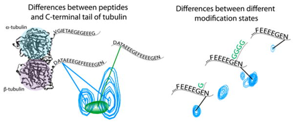
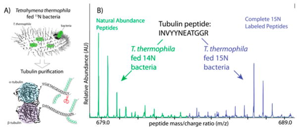
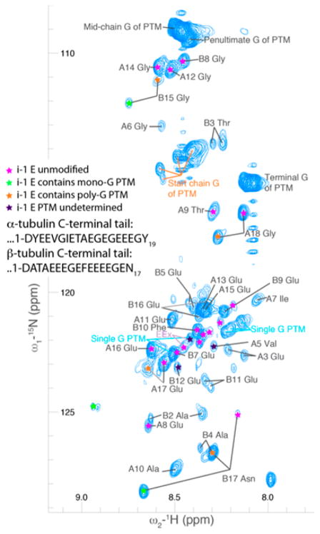
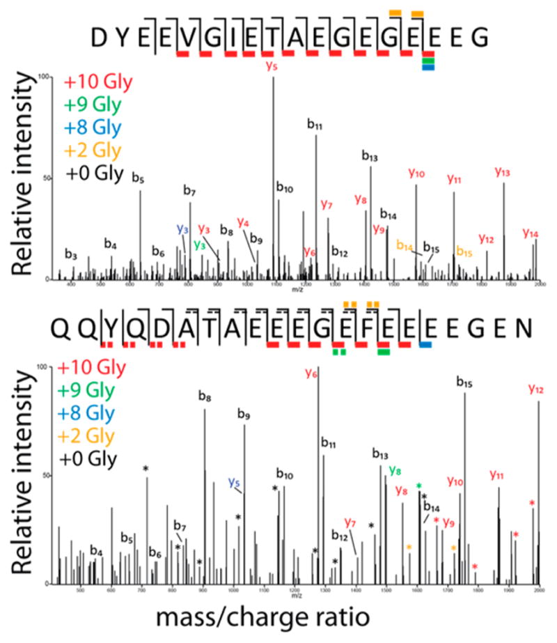
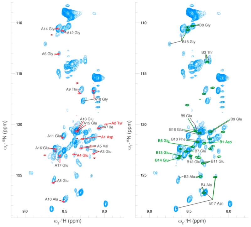
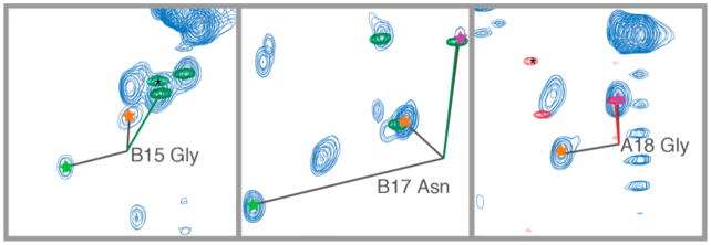
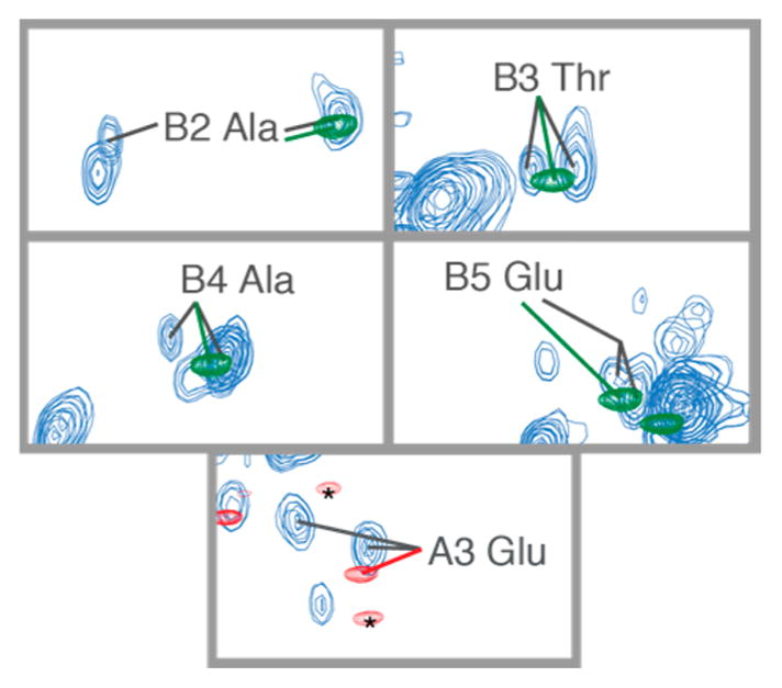
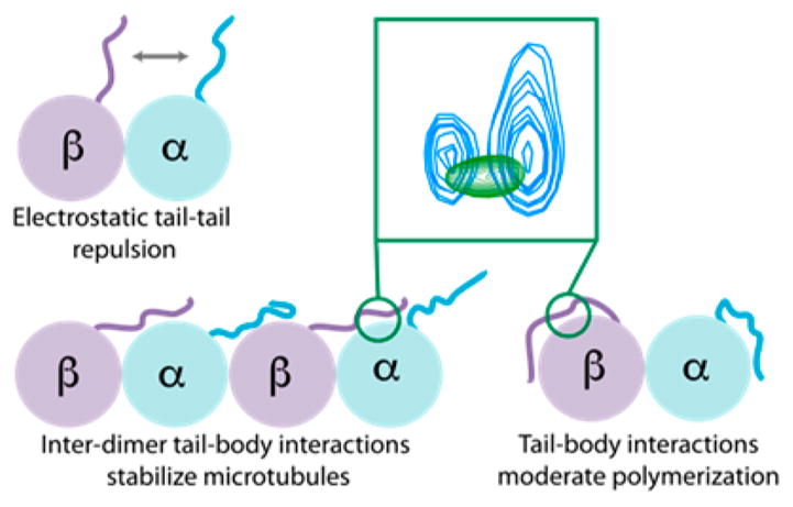
Tubulin is important for a wide variety of cellular processes including cell division, ciliogenesis, and intracellular trafficking. To perform these diverse functions, tubulin is regulated by post-translational modifications (PTM), primarily at the C-terminal tails of both the α- and β-tubulin heterodimer subunits. The tubulin C-terminal tails are disordered segments that are predicted to extend from the ordered tubulin body and may regulate both intrinsic properties of microtubules and the binding of microtubule associated proteins (MAP). It is not understood how either interactions with the ordered tubulin body or PTM affect tubulin's C-terminal tails. To probe these questions, we developed a method to isotopically label tubulin for C-terminal tail structural studies by NMR. The conformational changes of the tubulin tails as a result of both proximity to the ordered tubulin body and modification by mono- and polyglycine PTM were determined. The C-terminal tails of the tubulin dimer are fully disordered and, in contrast with prior simulation predictions, exhibit a propensity for β-sheet conformations. The C-terminal tails display significant chemical shift differences as compared to isolated peptides of the same sequence, indicating that the tubulin C-terminal tails interact with the ordered tubulin body. Although mono- and polyglycylation affect the chemical shift of adjacent residues, the conformation of the C-terminal tail appears insensitive to the length of polyglycine chains. Our studies provide important insights into how the essential disordered domains of tubulin function.


Conflict of interest statement

Notes

The authors declare no competing financial interest.

Figures

Figure 1
Figure 1
NMR chemical shifts (peak position) of C-terminal tail residues differing from those same residues within peptides of the same sequences. Mono- and polyglycylation (green Gs) affect the chemical shift of adjacent residues but do not appear to have a significant influence on the conformational ensemble of the domain.
Figure 2
Figure 2
Labeling method and confirmation of heavy isotope incorporation. (A) We fed heavy bacteria grown on 15N to T. thermophila. We then purified endogenous T. thermophila tubulin; our protocol resulted in full incorporation of 15N into tubulin. (B) Mass spectrometry showing two degrees of incorporation of 15N into the tubulin peptide (INVYYNEATGGR). The highest green peak appears at the m/z position expected for the fully 14N, 12C peptide. The cluster of three peaks result from the incorporation of single heavy atoms, primarily 13C carbon, which has a natural abundance of 1.1%. In blue are the fully 15N labeled peptides, with a similar peptide distribution to the natural abundance peptides, but at a mass increased by the number of nitrogen atoms in the peptide.
Figure 3
Figure 3
A 2D projection of a 3D HCNO spectrum of the C-terminal tails of tubulin in the context of the full TOG-purified tubulin dimer. Several modification states are represented (denoted by colored stars), resulting in more than one peak for several residues. Distinct clusters of peaks appeared for glycine residues in polyglycine modifications (the beginning, middle, penultimate, and final residues of the chain) as well as for monoglycine modifications. The residues at the beginning of the chain (those directly conjugated to a glutamate) were separated into two clusters, corresponding to monoglycine (light blue text) and polyglycine (orange text) modifications. Peaks are numbered relative to the start of the C-terminal tail, as indicated in the inset; the corresponding residues are β-tubulin D427 (B1) and α-tubulin D431 (A1) in the T. thermophila sequences for BTU1 and ATU1, respectively.
Figure 4
Figure 4
Tandem mass spectrometry (MS-MS) spectra using CID fragmentation of α- (top) and β-tubulin(bottom) C-terminal tail peptides, each modified with 10 glycine residues. Fragments containing the C-terminus are “y” ions, while those containing the N-terminus are labeled as “b” ions. The fragments are color coded according to how many glycine residues were present on that fragment. For the α-tubulin peptide, most modifications occurred on the final two glutamic acid residues, while the modifications were more dispersed between the final six residues for β-tubulin.
Figure 5
Figure 5
A comparison of the 2D projection of a 3D HCNO spectrum of the C-terminal tails of tubulin in the context of the full TOG-purified dimer as compared to bacterially produced GST-fusion peptides of the C-terminal tail sequences. The spectrum of the GST-α fusion peptide is shown in red, overlaid with the tubulin spectrum; similarly, the spectrum of the GST-β fusion peptide is shown in green, overlaid with the tubulin spectrum. Significant differences between the tubulin and peptide spectrum could be due to the presence of post-translational modification on our endogenous tubulin samples or due to interactions of the C-terminal tails with the rest of the tubulin dimer. As the majority of our endogenous sample was unmodified, we expect interactions with the rest of the dimer played the dominant role. Peaks with a small black asterisk have been assigned to the linker region between the peptides and GST and thus do not overlay with the C-terminal tails.
Figure 6
Figure 6
Insets of the spectra of Figure 5 with modification states of previous residue annotated, comparing the chemical shifts of C-terminal tails of tubulin with GST-fusion peptides. The peptides overlaid closely with the peaks corresponding to the unmodified form of the C-terminal tail residue. We observed significant chemical shift changes due to the change in the local chemical environment of the residue due to the presence of glycine modifications. Modifications are indicated by stars as in Figure 4.
Figure 7
Figure 7
Residues closest to the ordered tubulin body that appeared to be present in two conformational ensembles, presumably due to interaction with the tubulin body surface. These residues appeared in the spectrum as two peaks, neither of which overlayed exactly with the peptide peak.
Figure 8
Figure 8
Tubulin C-terminal tails, which can modulate intrinsic properties of microtubules through their intertail electrostatic repulsion and inter- and intradimer tail–body interactions. Our data suggest a significant role for tail–body interactions.

References

    1. Tompa P, Fuxreiter M. Fuzzy complexes: polymorphism and structural disorder in protein–protein interactions. Trends Biochem Sci. 2008;33:2–8. - PubMed
    1. Borg M, Mittag T, Pawson T, Tyers M, Forman-Kay JD, Chan HS. Polyelectrostatic interactions of disordered ligands suggest a physical basis for ultrasensitivity. Proc Natl Acad Sci U S A. 2007;104:9650–9655. - PMC - PubMed
    1. Uversky VN. Multitude of binding modes attainable by intrinsically disordered proteins: a portrait gallery of disorder-based complexes. Chem Soc Rev. 2011;40:1623. - PubMed
    1. Sugase K, Dyson HJ, Wright PE. Mechanism of coupled folding and binding of an intrinsically disordered protein. Nature. 2007;447:1021–1025. - PubMed
    1. Hough LE, Dutta K, Sparks S, Temel DB, Kamal A, Tetenbaum-Novatt J, Rout MP, Cowburn D. The molecular mechanism of nuclear transport revealed by atomic scale measurements. eLife. 2015;4:e10027. - PMC - PubMed

Publication types

LinkOut - more resources