. 2016 Sep 13;7(37):58832-58847.
doi: 10.18632/oncotarget.11315.

Boar seminal plasma exosomes maintain sperm function by infiltrating into the sperm membrane

Affiliations

Boar seminal plasma exosomes maintain sperm function by infiltrating into the sperm membrane

Jian Du et al. Oncotarget. .

Abstract

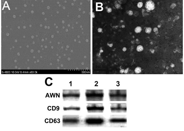
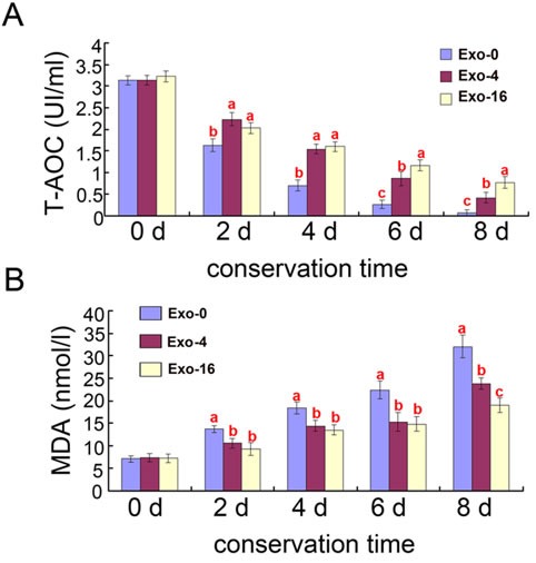
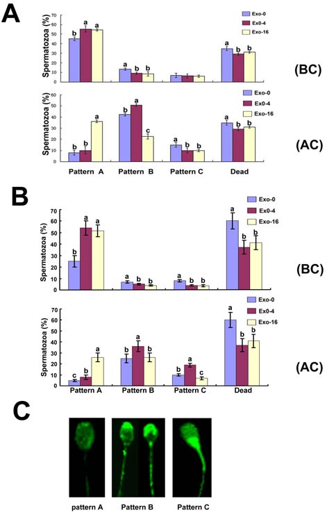
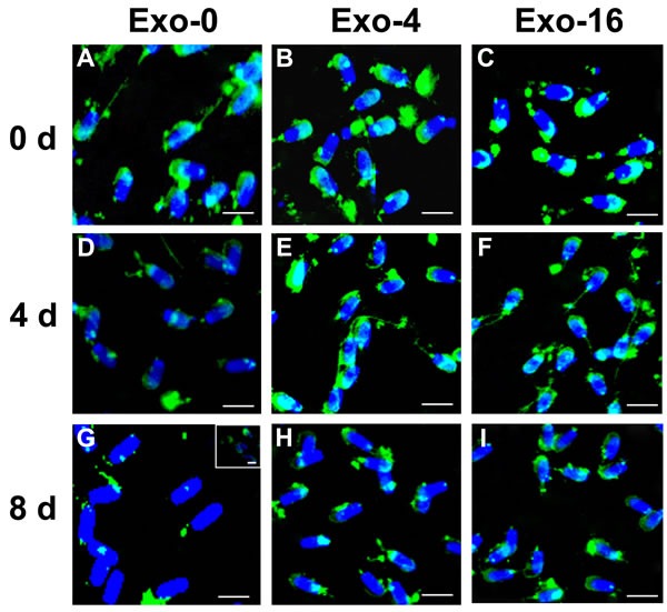
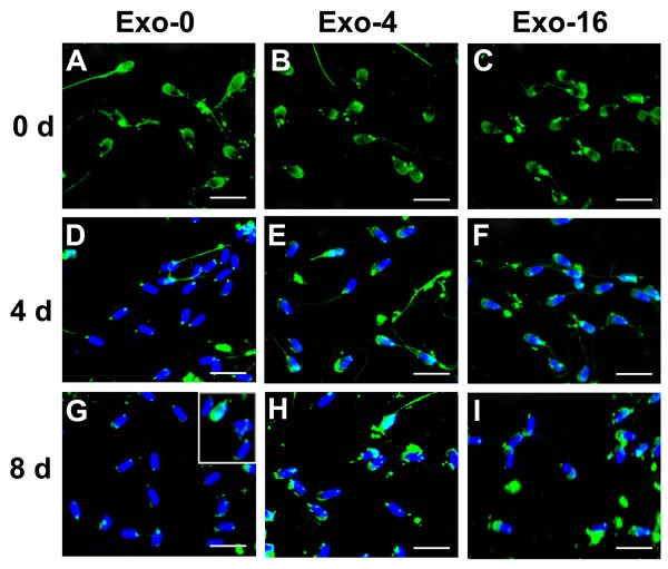
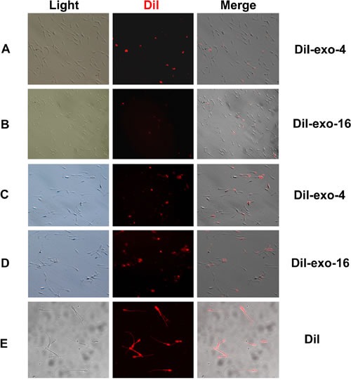
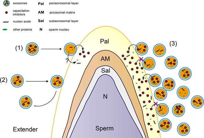
Seminal plasma ingredients are important for maintenance of sperm viability. This study focuses on the effect of boar seminal plasma exosomes on sperm function during long-term liquid storage. Boar seminal plasma exosomes had typical nano-structure morphology as measured by scanning electron microscopy (SEM) and molecular markers such as AWN, CD9 and CD63 by western blot analysis. The effect on sperm parameters of adding different ratio of boar seminal plasma exosomes to boar sperm preparations was analyzed. Compared to the diluent without exosomes, the diluent with four times or sixteen times exosomes compared to original semen had higher sperm motility, prolonged effective survival time, improved sperm plasma membrane integrity (p < 0.05), increased total antioxidant capacity (T-AOC) activity and decreased malondialdehyde (MDA) content. The diluent containing four times concentration of exosomes compared to original semen was determined to inhibit premature capacitation, but not to influence capacitation induced in vitro. Inhibition of premature capacitation is likely related to the concentration of exosomes which had been demonstrated to transfer proteins including AWN and PSP-1 into sperm. In addition, using fluorescence microscopy and scanning electron microscopy analysis, it was demonstrated that exosomes in diluent were directly binding to the membrane of sperm head which could improve sperm plasma membrane integrity.

Keywords: Pathology Section; boar sperm quality; capacitation; liquid storage; seminal plasma exosomes.


Conflict of interest statement

CONFLICTS OF INTEREST

All authors read and approved the final manuscript. The authors declare no competing financial interests.

Figures

Figure 1
Figure 1. Identification of boar seminal plasma exosomes
A., B. Morphology of exosomes by scanning electron microscope. Morphology of negative stained exosomes (B); C. Molecular markers of exosomes (AWN, CD9 and CD63) were detected by Western blot. 1, 2, 3 lanes represented three repetitions.
Figure 2
Figure 2. Effects of different concentrations of exosomes on T-AOC and MDA content in boar sperm during liquid storage at 17°C
A. Effects of different concentrations of exosomes on T-AOC activity. B. Effects of different concentrations of exosomes on MDA content. Different superscripts within the same time demonstrate significant differences (p < 0.05), and the same superscripts demonstrate insignificant differences (p > 0.05). Exo-0, Exo-4, Exo-16 represented the diluent with 0, 4 and 16 times concentration of exosomes of original semen, respectively.
Figure 3
Figure 3. CTC staining patterns of sperm in the diluent with Exo-0, Exo-4 and Exo-16 exosomes
A, CTC staining patterns of sperm on the 4th day in the diluent with Exo-0, Exo-4 and Exo-16 exosomes. (BC): before capacitation; (AC): at 3 hours after sperm capacitation was induced. B, CTC staining patterns of sperm on the 8th day in the diluent with Exo-0, Exo-4 and Exo-16 exosomes. Capacitation fluid was Tyrode's medium with 3 mg/mL BSA (see Materials and Methods). (BC): before capacitation; (AC): at 3 hours after sperm capacitation was induced. C, illustration of spermatozoa different fluorescence patterns of spermatozoa samples. Pattern A. faint fluorescence uniformly distributed over the sperm head denoting non-capacitated spermatozoa. Pattern B. the acrosomal region of the sperm heads fluoresce brightly denoting capacitated spermatozoa. Pattern C. the acrosomal region of the sperm head is non-fluorescent while the post-acrosomal region of the sperm head appeared strong fluorescence and this indicated a capacitated, acrosome-reacted spermatozoa. Dead represented the percentage of spermatozoa with no movement of the flagellum. Exo-0, Exo-4, Exo-16 represented the diluent with 0, 4 and 16 times concentration of exosomes of original semen, respectively. Sperm head length: 7.0 μm.
Figure 4
Figure 4. Immunofluorescence detection of AWN on sperm incubated in the diluent without exosomes (Exo-0) and with different concentrations of exosomes (Exo-4 and Exo-16) on day 0, and after 4 days and 8 days at 17°C
In the diluent without exosomes (Exo-0), AWN was present on the acrosome membrane of all sperm on day 0 A. Partial AWN protein existed on acrosome membrane of sperm D. All sperm had lost AWN on acrosome membrane by day 8 G. In the diluent with different concentrations of exosomes (Exo-4 and Exo-16), respectively, AWN located on the acrosome membrane of all sperm on day 0 B. and C. and day 4 E. and F. AWN dispersed in front area of sperm heads on day 8 H. and I. Arrowheads represented AWN location on acrosome membrane. Exo-0, Exo-4 and Exo-16 represented the diluent without exosomes, the diluent with four times exosomes concentration, and sixteen times exosomes concentration of original semen, respectively. Sperm head length: 7.0 μm.
Figure 5
Figure 5. Immunofluorescence detection of PSP-I on sperm incubated in the diluent without exosomes (Exo-0) and with different concentrations of exosomes (Exo-4 and Exo-16) on day 0, and after 4 days and 8 days at 17°C
In the diluent without exosomes, PSP-1 located on acrosome membrane and apical area of sperm tail on day 0 A. Partial PSP-1 protein existed on the acrosome membrane and principal area of sperm tail on day 4 D. All sperm had lost PSP-1 on acrosome membrane and only remain PSP-1 remained in the apical area of the middle piece of sperm tail on day 8 G.. In the diluent with different concentrations of exosomes (Exo-4 or Exo-16), respectively, PSP-1 existed on the acrosome membrane and principal areas of the sperm tail on day 0 B., C. and day 4 E., F. and sperm lost part of the PSP-1 on the acrosome membrane on day 8 H., I. Arrowheads and asterisk represent PSP-1 locations on acrosome membranes and on the apical area of the middle piece of sperm tails, respectively. Exo-0, Exo-4 and Exo-16 represented the diluent without exosomes, four times exosomes concentration and sixteen times exosomes concentration of original semen, respectively. Sperm head length: 7.0 μm.
Figure 6
Figure 6. Red fluorescence sperm was detected during incubation with different concentration of DiI-exo (DiI-exo-4 and DiI-exo-16) at 17°C
The sperm samples were treated by 0.125% trypsin and then washed 3 times using the diluent. Finally, the 2 μL of sperm samples were dropped on cover slides and observed using fluorescence microscopy. A. Sperm was incubated one day in the diluent with DiI-exo-4; B. Sperm was incubated one day in the diluent with DiI-exo-16; C. Sperm was incubated 4 days in the diluent with DiI-exo-4; D. Sperm was incubated 4 days in the diluent with DiI-exo-16. E. Sperm incubated 3 hours in the diluent with 0.005 mM DiI dye displayed red fluorescence. DiI-exo-4, DiI-exo-16 represented the diluent with four and sixteen times concentration of exosomes of original semen, respectively. And the exosomes were labeled DiI dye. Scale: sperm head length 7.0 μm.
Figure 7
Figure 7. The ultrastructure of sperm at 4 d in different conditions by scanning electron microscopy (SEM)
A. A-i, A-ii: Ultrastructure of membrane surface of sperm head in the diluent with Exo-1. B. B-i, B-ii: Ultrastructure of membrane surface of sperm head in the diluent with exosomes (Exo-4 or Exo-16). Arrowheads indicated the nano-granules (exosomes).
Figure 8
Figure 8. A speculative model of exosomes for protection of sperm function
(1) It was a dynamic process that exosomes was released from and band with sperm membrane. (2) The elements in exosomes could be accumulated in sperm through exosomes transferring into membrane of sperm. (3) Excessive exosomes in diluent led to transfer more exosomes ingredients such as AWN and PSP-1 proteins (as capacitation inhibitors) into sperm membrane in vitro.

References

    1. Koch S, Acebron SP, Herbst J, Hatiboglu G, Niehrs C. Post-transcriptional Wnt Signaling Governs Epididymal Sperm Maturation. Cell. 2015;163:1225–1236. - PubMed
    1. Poliakov A, Spilman M, Dokland T, Amling CL, Mobley JA. Structural heterogeneity and protein composition of exosome-like vesicles (prostasomes) in human semen. The Prostate. 2009;69:159–167. - PubMed
    1. Suzuki K, Asano A, Eriksson B, Niwa K, Nagai T, Rodriguez-Martinez H. Capacitation status and in vitro fertility of boar spermatozoa: effects of seminal plasma, cumulus-oocyte-complexes-conditioned medium and hyaluronan. International journal of andrology. 2002;25:84–93. - PubMed
    1. Abney TO, Williams WL. Inhibition of sperm capacitation by intrauterine deposition of seminal plasma decapacitation factor. Biology of reproduction. 1970;2:14–17. - PubMed
    1. Mendoza N, Casao A, Perez-Pe R, Cebrian-Perez JA, Muino-Blanco T. New insights into the mechanisms of ram sperm protection by seminal plasma proteins. Biology of reproduction. 2013;88:149. - PubMed

LinkOut - more resources