

The effect of epigenetic silencing and TP53 mutation on the expression of DLL4 in human cancer stem disorder
- PMID: 27542210
- PMCID: PMC5325341
- DOI: 10.18632/oncotarget.11316
The effect of epigenetic silencing and TP53 mutation on the expression of DLL4 in human cancer stem disorder
Abstract
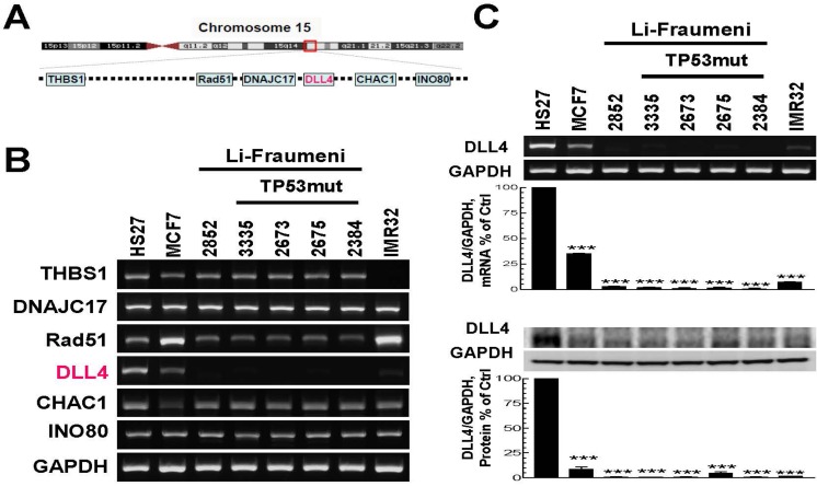
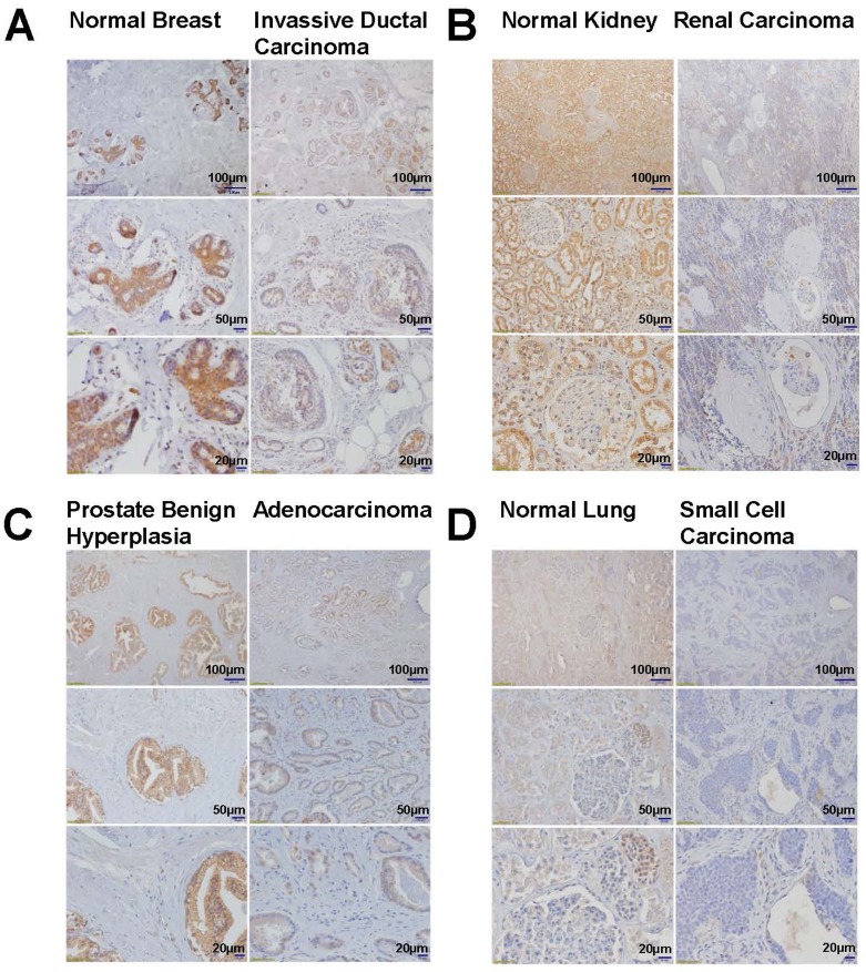
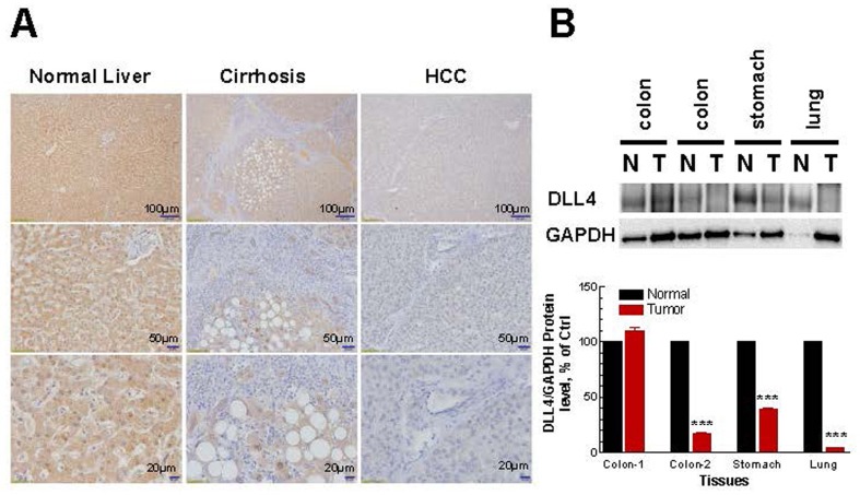
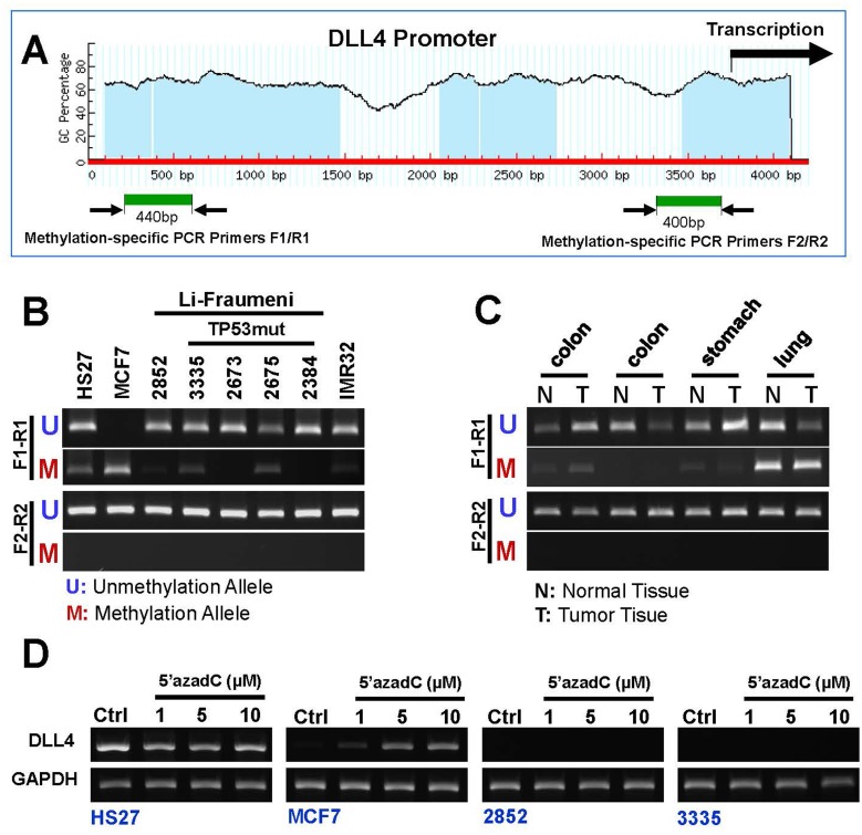
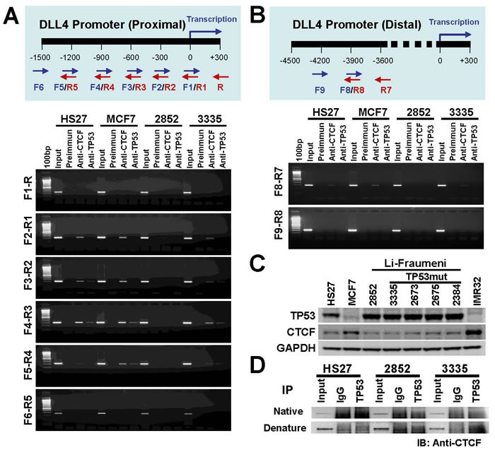
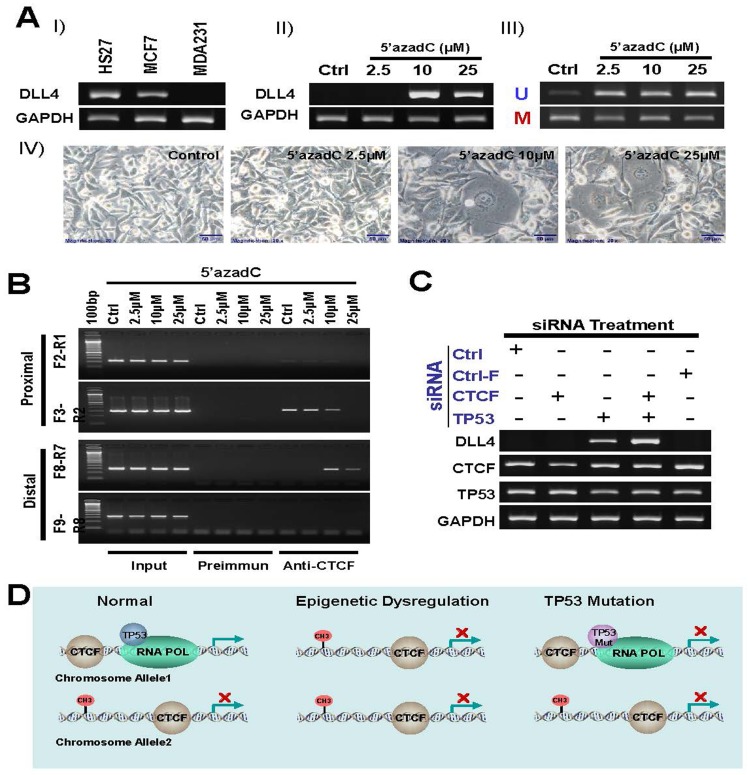
The Li-Fraumeni Syndrome (LFS), a genetically rare heterogeneous cancer syndrome, is characterized primarily by a germline p53 (TP53) gene mutation. We recently discovered a balanced reciprocal chromosomal translocation t(11;15)(q23;q15) in the non-cancerous skin fibroblasts of a bilateral breast cancer patient in LFS family. This prompted us to investigate the breakpoint region of the translocation, which uncovered a gene that encodes a Notch ligand, DLL4, (locus at 15q15.1), a key target in tumor vasculature. We analyzed DLL4 gene expression and protein level in LFS non-cancerous skin fibroblast cell lines and non-LFS cancer cell lines. DLL4 is abrogated in all the LFS cells and drastically down-regulated in breast (MCF7) and brain (IMR32) cancer cells and tumor tissue samples. However, DNA methylation studies revealed that DLL4 promoter is silenced only in MCF7 but not in LFS cells. We further investigated the regulation of DLL4 gene expression by ChIP assays, which demonstrated that p53 binds to DLL4 promoter through its association with CTCF, a chromosomal networking protein CCCTC binding factor. This implies a possible karyotype-phenotype correlation with respect to DLL4 in LFS and breast cancer initiation and progression. The drastic reduction or absence in the expression of DLL4 in LFS as well as breast and brain cancer cells is significant and supports the concept that this ligand may also play a role in cancer immune-surveillance; and its resuscitation in the tumor microenvironment may stimulate T-cell immunity and suppress tumor growth. Therefore, DLL4 may provide a strong platform as an immuno-therapeutic target in LFS and cancer patients.
Keywords: DLL4; Notch; angiogenesis; epigenetics; p53.
Conflict of interest statement
We declare no conflict of interest.
Figures
Similar articles
-
Balanced t(11;15)(q23;q15) in a TP53+/+ breast cancer patient from a Li-Fraumeni syndrome family.Cancer Genet Cytogenet. 2006 Jul 1;168(1):50-8. doi: 10.1016/j.cancergencyto.2005.12.009. Cancer Genet Cytogenet. 2006. PMID: 16772121
-
The TP53 gene promoter is not methylated in families suggestive of Li-Fraumeni syndrome with no germline TP53 mutations.Cancer Genet Cytogenet. 2009 Aug;193(1):63-6. doi: 10.1016/j.cancergencyto.2009.04.014. Cancer Genet Cytogenet. 2009. PMID: 19602465
-
TP53 and CDKN1A mutation analysis in families with Li-Fraumeni and Li-Fraumeni like syndromes.Fam Cancer. 2017 Apr;16(2):243-248. doi: 10.1007/s10689-016-9935-z. Fam Cancer. 2017. PMID: 27714481
-
Germline TP53 mutations and Li-Fraumeni syndrome.Hum Mutat. 2003 Mar;21(3):313-20. doi: 10.1002/humu.10185. Hum Mutat. 2003. PMID: 12619118 Review.
-
Li-Fraumeni Syndrome Disease Model: A Platform to Develop Precision Cancer Therapy Targeting Oncogenic p53.Trends Pharmacol Sci. 2017 Oct;38(10):908-927. doi: 10.1016/j.tips.2017.07.004. Epub 2017 Aug 14. Trends Pharmacol Sci. 2017. PMID: 28818333 Free PMC article. Review.
Cited by
-
CHAC1 in urological tumors: contextual dualism and therapeutic implications.Front Oncol. 2025 Aug 8;15:1610915. doi: 10.3389/fonc.2025.1610915. eCollection 2025. Front Oncol. 2025. PMID: 40860820 Free PMC article. Review.
-
Functional roles of CTCF in breast cancer.BMB Rep. 2017 Sep;50(9):445-453. doi: 10.5483/bmbrep.2017.50.9.108. BMB Rep. 2017. PMID: 28648147 Free PMC article.
-
The TP53-Related Signature Predicts Immune Cell Infiltration, Therapeutic Response, and Prognosis in Patients With Esophageal Carcinoma.Front Genet. 2021 Jun 16;12:607238. doi: 10.3389/fgene.2021.607238. eCollection 2021. Front Genet. 2021. PMID: 34234806 Free PMC article.
-
Breast cancer in patients with Li-Fraumeni syndrome - a case-series study and review of literature.Breast Cancer (Dove Med Press). 2017 Mar 23;9:207-215. doi: 10.2147/BCTT.S134241. eCollection 2017. Breast Cancer (Dove Med Press). 2017. PMID: 28356770 Free PMC article.
-
Role of Jagged1/STAT3 signalling in platinum-resistant ovarian cancer.J Cell Mol Med. 2019 Jun;23(6):4005-4018. doi: 10.1111/jcmm.14286. Epub 2019 Apr 16. J Cell Mol Med. 2019. PMID: 30993885 Free PMC article.
References
-
- Muller PA, Vousden KH. P53 Mutations in Cancer. Nature cell biology. 2013;15:2–8. - PubMed
-
- Masciari S, Dillon DA, Rath M, Robson M, Weitzel JN, Balmana J, Gruber SB, Ford JM, Euhus D, Lebensohn A, Telli M, Pochebit SM, Lypas G, et al. Breast cancer phenotype in women with TP53 germline mutations: a Li-Fraumeni syndrome consortium effort. Breast cancer research and treatment. 2012;133:1125–1130. - PMC - PubMed
-
- Maillard I, Fang T, Pear WS. Regulation of lymphoid development, differentiation, and function by the Notch pathway. Annual Review of Immunology. 2005;23:945–974. - PubMed
MeSH terms
Substances
Grants and funding
LinkOut - more resources
Full Text Sources
Other Literature Sources
Research Materials
Miscellaneous
