. 2016 Sep 20;7(38):61366-61377.
doi: 10.18632/oncotarget.11351.

Role of high expression levels of STK39 in the growth, migration and invasion of non-small cell type lung cancer cells

Affiliations

Role of high expression levels of STK39 in the growth, migration and invasion of non-small cell type lung cancer cells

Zhao Li et al. Oncotarget. .

Abstract

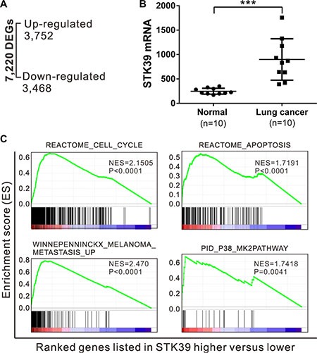
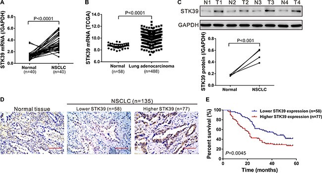
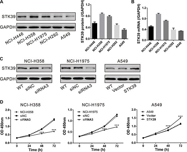
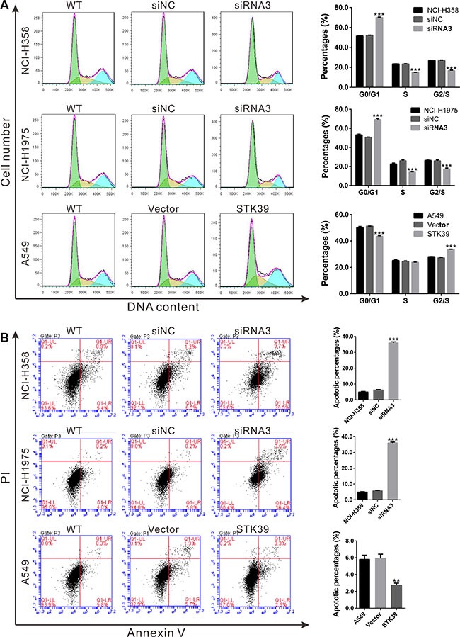
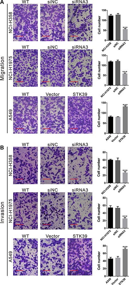
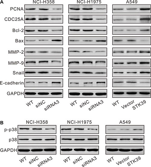
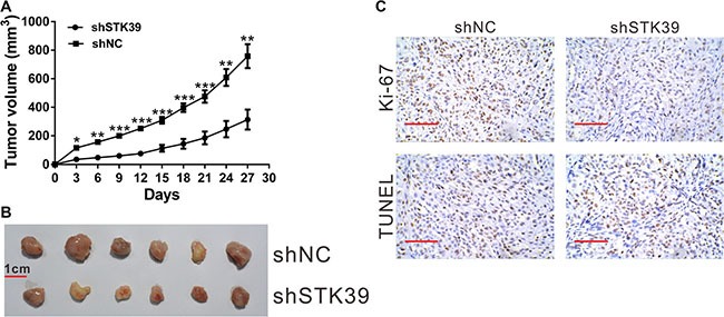
Non-small cell type lung cancer (NSCLC) is the most common malignancy and the leading cause of cancer related mortality. In this study, serine/threonine kinase 39 (STK39) was identified as an up-regulated gene in NSCLC tissues by next-generation RNA sequencing. Although STK39 gene polymorphisms may be prognostic of overall survival in patients with early stage NSCLC, the roles of STK39 in NSCLC cancer are poorly understood. In the current study, Genome Set Enrichment Analysis (GSEA) on the RNA-seq data of NSCLC specimens indicated that cancer-related process and pathways, including metastasis, cell cycle, apoptosis and p38 pathway, were significantly correlated with STK39 expression. STK39 expression was significantly increased in NSCLC cases and its protein expression was positively correlated with the poor tumor stage, large tumor size, advanced lymphnode metastasis and poor prognosis. Down-regulation of STK39 in NSCLC cells significantly decreased cell proliferation by blocking of cell cycle and inducing apoptosis. We also found that STK39 knockdown in NSCLC cells remarkably repressed cell migration and invasion. On the contrary, overexpression of STK39 in NSCLC cells had inverse effects on cell behaviors. Taken together, STK39 acts as a tumor oncogene in NSCLC and can be a potential biomarker of carcinogenesis.

Keywords: NSCLC; STK39; metastasis; proliferation.


Conflict of interest statement

Conflicts of Interest: None declared.

Figures

Figure 1
Figure 1. RNA sequencing data analysis
(A) DEGs were identified by RNA sequencing. (B) RNA-sequencing data showed that STK39 mRNA expression was significantly higher in NSCLC tissues than in paired non-cancerous tissues (n = 10). (C) GSEA analysis in NSCLC patients with higher STK39 expression versus lower STK39 expression. NES, normalized enrichment score.
Figure 2
Figure 2. STK39 overexpression correlates with poor survival in patients with NSCLC
(A) STK39 mRNA levels were determined in 40 pairs of NSCLC and non-cancerous tissues using real-time PCR. (B) STK39 expression in lung adenocarcinoma and normal tissues based on TCGA dataset (P < 0.0001). (C) Representative STK39 protein expression in unaffected tissues (N1, N2, N3 and N4) and NSCLC (T1, T2, T3 and T4). (D) STK39 protein expression was assessed by immunohistochemistry staining in NSCLC tissues. Scale bar: 100 μm. (E) Kaplan-Meier survival analysis showed that patients with lower STK39 expression level have a better prognosis than that of patients with higher STK39 expression (P < 0.01).
Figure 3
Figure 3. STK39 promotes cell proliferation in NSCLC cells
(A, B) STK39 protein and mRNA expression in 5 NSCLC cell lines was analyzed by Western blot (A) and real-time PCR (B), respectively. Data were based on at least 3 independent experiments. (C) Expression of STK39 in NCI-H358, NCI-H1975 and A549 cells was analyzed by Western blot. NCI-H358 and NCI-H1975 cells were transfected with STK39 siRNA (siRNA3) or control siRNA (siNC); A549 cells was infected with STK39 expression lentivirus or control vector lentivirus (Vector). (D) Cell proliferation was detected at 0, 24, 48 and 72 h after siRNA transfection or lentiviral infection in NCI-H358, NCI-H1975 and A549 cells. Data were based on at least 3 independent experiments, and shown as mean ± SD. ***P < 0.001.
Figure 4
Figure 4. STK39 accelerates G1/S phase transition and inhibits cell apoptosis in NSCLC cells
NCI-H358 and NCI-H1975 cells were transfected with STK39 siRNA (siRNA3) or control siRNA (siNC); A549 cells was infected with STK39 expression lentivirus or control vector lentivirus (Vector). At 24 h after siRNA transfection or lentiviral infection, cells were collected. (A) Cell cycle distribution was analyzed using PI staining and flow cytometry. (B) Cell apoptosis was measured by Annexin V-PI double staining followed by flow cytometry analysis. **P < 0.01, ***P < 0.001.
Figure 5
Figure 5. STK39 promotes the cellular migration and invasion of NSCLC cells
Migration (A) and invasion (B) assay were performed in Transwell chambers. For invasion assay, the upper chamber was pre-coated with Matrigel. Scale bar: 100 μm ***P < 0.001.
Figure 6
Figure 6. Mechanisms how STK39 exerts its function in NSCLC
NCI-H358 and NCI-H1975 cells were transfected with STK39 siRNA (siRNA3) or control siRNA (siNC); A549 cells was infected with STK39 expression lentivirus or control vector lentivirus (Vector). (A) At 48 h after siRNA transfection or lentiviral infection, cells were collected. Protein levels of key proteins in cell cycle, cell apoptosis and metastasis were determined by Western blot. Representative blots of 3 independent experiments were shown. (B) Phosphorylation of p38 was detected by Western blot.
Figure 7
Figure 7. STK39 knockdown suppressed NSCLC cell proliferation in vivo
Knockdown of STK39 in NCI-H358 cells significantly inhibited tumor growth in nude mice xenograft model. (A) The growth curves of xenograft tumors in different groups. (B) At day 27, mice were sacrificed and tumors were resected (n = 6). Comparison of excised tumors. Scale bar: 1 cm. (C) Xenograft tumors with Ki67 immunostaining and TUNEL staining. Scale bar: 100 μm. *P < 0.05, **P < 0.01, ***P < 0.001.

References

    1. Jemal A, Siegel R, Ward E, Hao Y, Xu J, Thun MJ. Cancer statistics, 2009. CA Cancer J Clin. 2009;59:225–249. - PubMed
    1. Jemal A, Bray F, Center MM, Ferlay J, Ward E, Forman D. Global cancer statistics. CA Cancer J Clin. 2011;61:69–90. - PubMed
    1. Koudelakova V, Kneblova M, Trojanec R, Drabek J, Hajduch M. Non-small cell lung cancer—genetic predictors. Biomedical papers of the Medical Faculty of the University Palacky, Olomouc, Czechoslovakia. 2013;157:125–136. - PubMed
    1. Wang Z, Gerstein M, Snyder M. RNA-Seq: a revolutionary tool for transcriptomics. Nature reviews Genetics. 2009;10:57–63. - PMC - PubMed
    1. Meyerson M, Gabriel S, Getz G. Advances in understanding cancer genomes through second-generation sequencing. Nature Reviews Genetics. 2010;11:685–696. - PubMed

MeSH terms