. 2016 Sep 6;7(36):58516-58530.
doi: 10.18632/oncotarget.11305.

Targeting ALDH1A1 by disulfiram/copper complex inhibits non-small cell lung cancer recurrence driven by ALDH-positive cancer stem cells

Affiliations

Targeting ALDH1A1 by disulfiram/copper complex inhibits non-small cell lung cancer recurrence driven by ALDH-positive cancer stem cells

Xinwei Liu et al. Oncotarget. .

Abstract

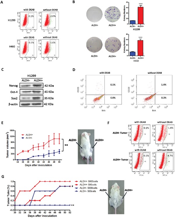
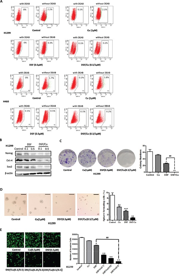
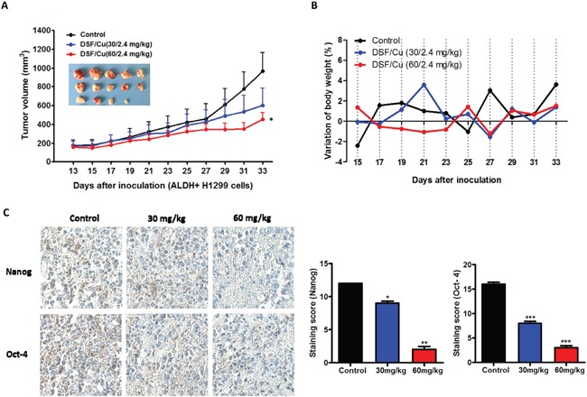
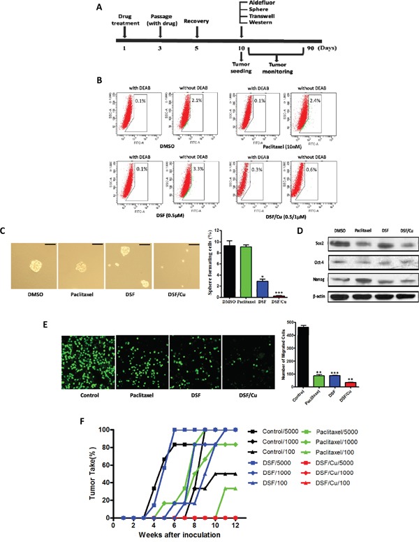
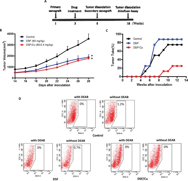
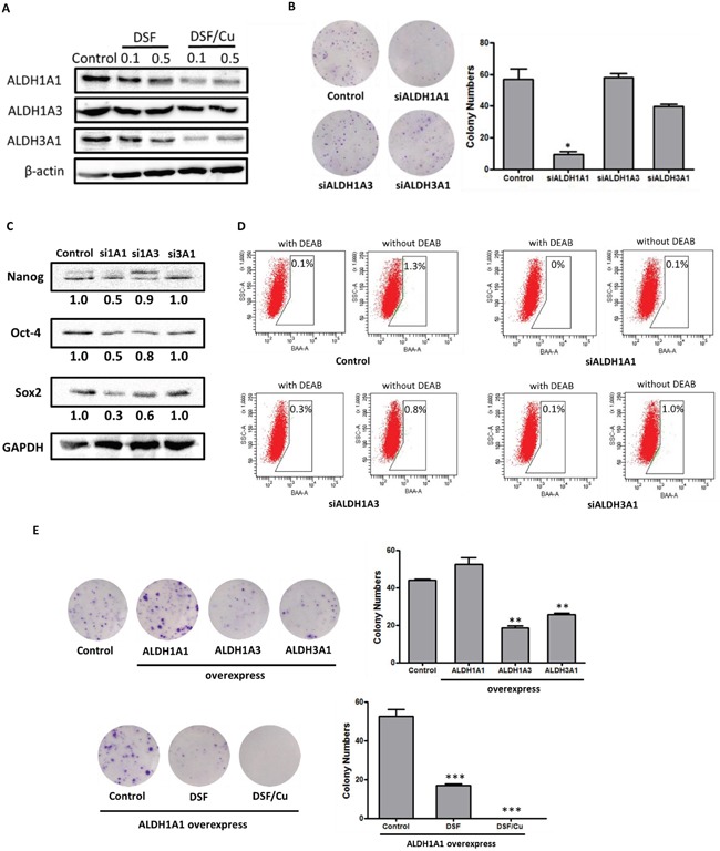
The existence of cancer stem cells (CSCs) in non-small cell lung cancer (NSCLC) has profound implications for cancer therapy. In this study, a disulfiram/copper (DSF/Cu) complex was evaluated in vitro and in vivo for its efficacy to inhibit CSCs, which drive recurrence of NSCLC. First, we investigated whether DSF/Cu could inhibit ALDH-positive NSCLC stem cells in vitro and tumors derived from sorted ALDH-positive CSCs in vivo. DSF/Cu (0.5/1 μmol/l) significantly inhibited the expression of stem cell transcription factors (Sox2, Oct-4 and Nanog) and reduced the capacities of NSCLC stem cells for self-renewal, proliferation and invasion in vitro. Regular injections with DSF/Cu (60/2.4 mg/kg) reduced the size of tumors derived from sorted ALDH-positive stem cells. Two other NOD/SCID xenograft models were used to determine whether DSF/Cu could target NSCLC stem cells and inhibit tumor recurrence in vivo. DSF/Cu treatment eliminated ALDH-positive cells and inhibited tumor recurrence, which was reflected by reduced tumor growth in recipient mice that were inoculated with tumor cells derived from DSF/Cu-treated cells or primary xenografts. RNA interference and overexpression of ALDH isozymes suggested that ALDH1A1, which plays a key role in ALDH-positive NSCLC stem cells, might be the target of the DSF/Cu complex. Collectively, our data demonstrate that DSF/Cu targets ALDH1A1 to inhibit NSCLC recurrence driven by ALDH-positive CSCs. Thus, the DSF/Cu complex may represent a potential therapeutic strategy for NSCLC patients.

Keywords: ALDH1A1; NSCLC; cancer stem cell; disulfiram/copper; recurrence.


Conflict of interest statement

No potential conflicts of interest were disclosed.

Figures

Figure 1
Figure 1. ALDH-positive cells represent cancer stem cells in some NSCLC cell lines
A. ALDH-positive and ALDH-negative cells were isolated from the indicated NSCLC cell lines by FACS. Brightly fluorescent ALDH-expressing cells (ALDH-positive cells) were detected in the green fluorescence channel (BAA) using flow cytometry. DEAB, a specific inhibitor of ALDH, was used to establish the baseline fluorescence of these cells and to define the ALDH-positive region. B. Analysis of cell colony numbers in colony forming assays of ALDH-positive and ALDH-negative cells (***P < 0.001, χ2 test). C. Analysis of stem cell transcription factors by western blotting. D. Double staining of Aldefluor and CD133 (PE) in NCI-H1299 cells. E. Comparison of primary xenograft formation by sorted ALDH-positive and ALDH-negative NCI-H1299 cells in NOD/SCID mice (**P < 0.01, 2-tailed t test). F. The percentage of ALDH-positive cells in xenograft tumors derived from ALDH-positive and ALDH-negative cells. G. Comparison of tumor take (%) in NOD/SCID mice with secondary xenografts of ALDH-positive and ALDH-negative cells taken from primary xenograft tumors (**P < 0.01, χ2 test, compared with the ALDH+ 500 cells group; #P < 0.05, χ2 test, compared with the ALDH+ 5000 cells group).
Figure 2
Figure 2. DSF/Cu inhibits the stemness of NSCLCs in vitro
A. Inhibitory effect of DSF (0.5 μM), Cu (1 μM) and DSF/Cu (0.5/1 μM) on the ALDH-positive cell population from the NSCLC cells lines H1299 and H460. B. DSF (0.1 μM, and 0.5 μM) and DSF/Cu (0.1/1 μM, and 0.5/1 μM) inhibit the expression of the stem cell transcription factors Nanog, Sox2 and Oct-4 expression in H1299 cells. C. Inhibitory effect of DSF (0.5 μM), Cu (1 μM) and DSF/Cu (0.5/1 μM) on the colony forming ability of H1299 cells (*P < 0.05, **P < 0.01, ##P < 0.01, one-way ANOVA). D. Inhibitory effect of DSF and DSF/Cu on tumorsphere formation. Tumorspheres were incubated with DSF (0.5 μM), Cu (1 μM) and DSF/Cu (0.5/1 μM) for 7 days. Scale bar, 100 μm. (**P < 0.01, ***P < 0.001, one-way ANOVA). E. Inhibitory effect of DSF/Cu on transwell invasion assay. Cells were incubated with DSF (0.5 μM), Cu (0.5 μM) and DSF/Cu (0.1/0.5 μM, 0.25/0.5 μM and 0.5/0.5 μM) in transwells for 24 hours (*P < 0.05, **P < 0.01, one-way ANOVA).
Figure 3
Figure 3. DSF/Cu inhibits ALDH-positive NSCLC stem cells in vivo
A. DSF/Cu decreases the size of tumors derived from ALDH-positive NSCLC xenografts. Images of the tumors that developed in NOD/SCID mice from each treatment group are also shown. (*P < 0.05, one-way ANOVA). B. DSF/Cu has no apparent toxicity as determined by body weight. C. Representative images of immunohistochemical staining of tumor tissue for the stem cell transcription factors Nanog and Oct-4. The staining intensity was scored as 0 (negative), 1 (weak), 2 (medium) and 3 (strong). Extent of staining was scored as 0 (0%), 1 (1-25%), 2 (26-50%), 3 (51-75%) and 4 (76-100%), according to the percentage of the positively stained areas in relation to the whole carcinoma area (*P < 0.05, **P < 0.01, ***P < 0.001, one-way ANOVA).
Figure 4
Figure 4. Effects of DSF/Cu pretreatment on NSCLC stem cell numbers in vitro and tumor seeding in vivo
A. Schematic representation of treatment scheme. Cells were pretreated with drugs, then allowed to recover before experimental testing. B. Inhibitory effect of drug pretreatment on ALDH-positive cell populations before inoculation. C. Inhibitory effect of drug pretreatment on tumorsphere formation. Scale bar, 50 μm. (*P < 0.05, ***P < 0.001, χ2 test). D. Inhibitory effect of drug pretreatment on the expression of stem cell transcription factors. E. Inhibitory effect of drug treatment on transwell migration assays (**P < 0.01, ***P < 0.001, one-way ANOVA). F. Latency periods and tumor take percentage in xenograft mice receiving cells pretreated with the indicated drugs.
Figure 5
Figure 5. Effects of DSF and DSF/Cu treatment in vivo and tumor growth on secondary xenografts
A. Schematic representation of the treatment scheme. B. Drug treatment reduces the size of primary xenograft tumors (*P < 0.05, 2-tailed t test). C. Percentage of tumor take and latency period in mice receiving secondary xenografts from each treatment group. D. Percentage of ALDH-positive cells in secondary xenograft tumors from each treatment group.
Figure 6
Figure 6. ALDH1A1 plays a key role in ALDH-positive NSCLC stem cells
A. DSF (0.1 μM, and 0.5 μM) and DSF/Cu (0.1/1 μM, and 0.5/1 μM) inhibits the expression of the ALDH isozymes ALDH1A1, ALDH1A3 and ALDH3A1. B. The effects of siRNA knockdown of ALDH isozymes on colony-forming efficiency (*P < 0.05, one-way ANOVA). C. The levels of stem cell transcription factors in siRNA-treated cells. D. ALDH activity in siRNA-treated cells. E. Top row: the effects of overexpression of ALDH isozymes on the colony-forming efficiency of NCI-H1299 cells; bottom row: the inhibitory effects of DSF (0.5 μM) and DSF/Cu (0.5/1 μM) on ALDH1A1-overexpressing NCI-H1299 cells (**P < 0.01, ***P < 0.001, one-way ANOVA).

References

    1. Reya T, Morrison SJ, Clarke MF, Weissman IL. Stem cells, cancer, and cancer stem cells. Nature. 2001;414:105–11. - PubMed
    1. Clarke MF, Dick JE, Dirks PB, Eaves CJ, Jamieson CH, Jones DL, Visvader J, Weissman IL, Wahl GM. Cancer stem cells—perspectives on current status and future directions: AACR Workshop on cancer stem cells. Cancer Res. 2006;66:9339–44. - PubMed
    1. Al-Hajj M, Wicha MS, ito-Hernandez A, Morrison SJ, Clarke MF. Prospective identification of tumorigenic breast cancer cells. Proc Natl Acad Sci U S A. 2003;100:3983–8. - PMC - PubMed
    1. Singh SK, Clarke ID, Terasaki M, Bonn VE, Hawkins C, Squire J, Dirks PB. Identification of a cancer stem cell in human brain tumors. Cancer Res. 2003;63:5821–8. - PubMed
    1. Lee CJ, Dosch J, Simeone DM. Pancreatic cancer stem cells. J Clin Oncol. 2008;26:2806–12. - PubMed

MeSH terms