Targeting a novel cancer-driving protein (LAPTM4B-35) by a small molecule (ETS) to inhibit cancer growth and metastasis
- PMID: 27542271
- PMCID: PMC5295449
- DOI: 10.18632/oncotarget.11325
Targeting a novel cancer-driving protein (LAPTM4B-35) by a small molecule (ETS) to inhibit cancer growth and metastasis
Abstract
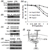
Our previous studies demonstrated that LAPTM4B-35 is overexpressed in a variety of solid cancers including hepatocellular carcinoma (HCC), and is an independent factor for prognosis. LAPTM4B-35 overexpression causes carcinogenesis and enhances cancer growth, metastasis and multidrug resistance, and thus may be a candidate for therapeutic targeting. The present study shows ethylglyoxal bisthiosemicarbazon (ETS) has effective anticancer activity through LAPTM4B-35 targeting. Bel-7402 and HepG2 cell lines from human HCC were used as cell models in which LAPTM4B-35 is highly expressed, and a human fetal liver cell line was used as a control. The results showed ETS has a specific and pronounced lethal effect on HCC cells, but not on fetal liver cells in culture. ETS also attenuated growth and metastasis of human HCC xenograft in nude mice, and extended the life span of mice with HCC. ETS induced HCC cell apoptosis, and upregulated a large number of proapoptotic genes and downregulated antiapoptotic genes. When endogenous overexpression of LAPTM4B-35 was knocked down with RNAi, the killing effect of ETS on HepG2 cells was significantly attenuated. ETS also inhibited phosphorylation of LAPTM4B-35 Tyr285, which involves in activation of the PI3K/Akt signaling pathway induced by LAPTM4B-35 overexpression. In addition, the induction of alterations in quantity of c-Myc, Bcl-2, Bax, cyclinD1 and Akt-p molecules in HepG2 cells by LAPTM4B-35 overexpression could be reversed by ETS.
Conclusion: ETS is a promising candidate for treatment of HCC through LAPTM4B-35 protein targeting.
Keywords: LAPTM4B-35; apoptosis; cancer targeted therapy; ethylglyoxal bisthiosemicarbazon (ETS); hepatocellular-carcinoma.
Conflict of interest statement
The authors have declared no conflicts of interest.
Figures


Similar articles
-
Overexpression of LAPTM4B-35 promotes growth and metastasis of hepatocellular carcinoma in vitro and in vivo.Cancer Lett. 2010 Aug 28;294(2):236-44. doi: 10.1016/j.canlet.2010.02.006. Epub 2010 Mar 3. Cancer Lett. 2010. PMID: 20202745
-
Arenobufagin, a natural bufadienolide from toad venom, induces apoptosis and autophagy in human hepatocellular carcinoma cells through inhibition of PI3K/Akt/mTOR pathway.Carcinogenesis. 2013 Jun;34(6):1331-42. doi: 10.1093/carcin/bgt060. Epub 2013 Feb 7. Carcinogenesis. 2013. PMID: 23393227
-
Total alkaloids of Rubus aleaefolius Poir inhibit hepatocellular carcinoma growth in vivo and in vitro via activation of mitochondrial-dependent apoptosis.Int J Oncol. 2013 Mar;42(3):971-8. doi: 10.3892/ijo.2013.1779. Epub 2013 Jan 18. Int J Oncol. 2013. PMID: 23338043
-
The Potential Use of Anticancer Peptides (ACPs) in the Treatment of Hepatocellular Carcinoma.Curr Cancer Drug Targets. 2020;20(3):187-196. doi: 10.2174/1568009619666191111141032. Curr Cancer Drug Targets. 2020. PMID: 31713495 Review.
-
LAPTM4B: an oncogene in various solid tumors and its functions.Oncogene. 2016 Dec 15;35(50):6359-6365. doi: 10.1038/onc.2016.189. Epub 2016 May 23. Oncogene. 2016. PMID: 27212036 Free PMC article. Review.
Cited by
-
LAPTM4B promotes the progression of bladder cancer by stimulating cell proliferation and invasion.Oncol Lett. 2021 Nov;22(5):765. doi: 10.3892/ol.2021.13026. Epub 2021 Sep 8. Oncol Lett. 2021. PMID: 34589144 Free PMC article.
-
The role and regulatory mechanism of lysosome associated protein transmembrane 4β in tumors.Front Oncol. 2025 Mar 31;15:1552007. doi: 10.3389/fonc.2025.1552007. eCollection 2025. Front Oncol. 2025. PMID: 40231269 Free PMC article. Review.
-
Increased levels of LAPTM4B, VEGF and survivin are correlated with tumor progression and poor prognosis in breast cancer patients.Oncotarget. 2017 Jun 20;8(25):41282-41293. doi: 10.18632/oncotarget.17176. Oncotarget. 2017. PMID: 28476037 Free PMC article.
-
Total Flavonoids of Chuju Decrease Oxidative Stress and Cell Apoptosis in Ischemic Stroke Rats: Network and Experimental Analyses.Front Neurosci. 2021 Dec 9;15:772401. doi: 10.3389/fnins.2021.772401. eCollection 2021. Front Neurosci. 2021. PMID: 34955724 Free PMC article.
References
-
- Shao GZ, Zhou RL, Zhang QY, Zhang Y, Liu JJ, Rui JA, Wei X, Ye DX. Molecular cloning and characterization of LAPTM4B, a novel gene upregulated in hepatocellular carcinoma. Oncogene. 2003;22:5060–5069. - PubMed
-
- Rou Li Zhou. LAPTM4B: A Novel Diagnostic Biomarker and Therapeutic Target for Hepatocellular Carcinoma. In: Wan-Yee Lau., editor. <Hepatocellular Carcinoma Basic Research>. InTech Press; 2012. pp. 1–34. ISBN 978-953-51-0023-2.
-
- Yang H, Xiong FX, Qi RZ, Liu ZW, Lin M, Rui JA, Zhou RL. LAPTM4B-35 Is a Novel Prognostic Factor of Hepatocellular Carcinoma. Journal of Surgical Oncology. 2010;101:363–369. - PubMed
-
- Kasper G, Vogel A, Klaman I, Gröne J, Petersen I, Weber B, Castaños-Vélez E, Staub E, Mennerich D. The human LAPTM4b transcript is upregulated in various types of solid tumours and seems to play a dual functional role during tumour progression. Cancer Lett. 2005;16:93–103. - PubMed
MeSH terms
Substances
Grants and funding
LinkOut - more resources
Full Text Sources
Other Literature Sources
Medical
Molecular Biology Databases
Research Materials

