. 2016 Sep 30;291(40):20900-20910.
doi: 10.1074/jbc.M116.720268. Epub 2016 Aug 19.

Mechanistic Basis of Glutaminase Activation: A KEY ENZYME THAT PROMOTES GLUTAMINE METABOLISM IN CANCER CELLS

Affiliations

Mechanistic Basis of Glutaminase Activation: A KEY ENZYME THAT PROMOTES GLUTAMINE METABOLISM IN CANCER CELLS

Yunxing Li et al. J Biol Chem. .

Abstract

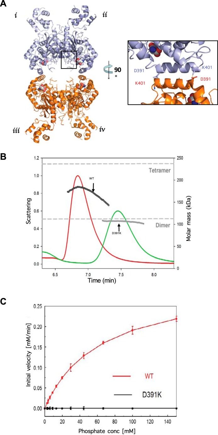
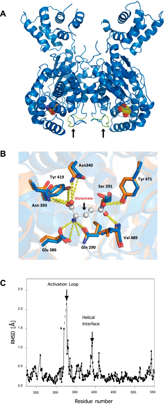
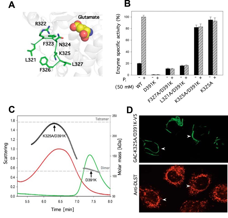
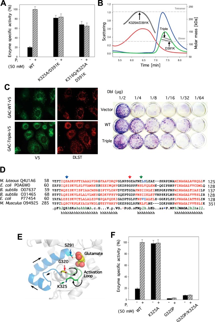
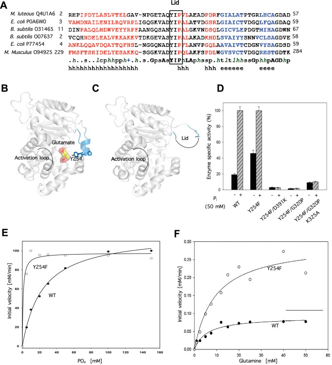
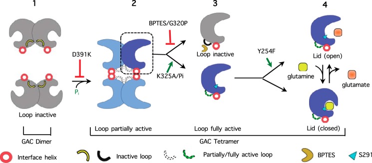
Glutamine-derived carbon becomes available for anabolic biosynthesis in cancer cells via the hydrolysis of glutamine to glutamate, as catalyzed by GAC, a splice variant of kidney-type glutaminase (GLS). Thus, there is significant interest in understanding the regulation of GAC activity, with the suggestion being that higher order oligomerization is required for its activation. We used x-ray crystallography, together with site-directed mutagenesis, to determine the minimal enzymatic unit capable of robust catalytic activity. Mutagenesis of the helical interface between the two pairs of dimers comprising a GAC tetramer yielded a non-active, GAC dimer whose x-ray structure displays a stationary loop ("activation loop") essential for coupling the binding of allosteric activators like inorganic phosphate to catalytic activity. Further mutagenesis that removed constraints on the activation loop yielded a constitutively active dimer, providing clues regarding how the activation loop communicates with the active site, as well as with a peptide segment that serves as a "lid" to close off the active site following substrate binding. Our studies show that the formation of large GAC oligomers is not a pre-requisite for full enzymatic activity. They also offer a mechanism by which the binding of activators like inorganic phosphate enables the activation loop to communicate with the active site to ensure maximal rates of catalysis, and promotes the opening of the lid to achieve optimal product release. Moreover, these findings provide new insights into how other regulatory events might induce GAC activation within cancer cells.

Keywords: enzyme mechanism; glutaminase; metabolism; mutagenesis; protein structure.


Figures

FIGURE 1.
FIGURE 1.
A single mutation in the helical interface of glutaminase results in an inactive, dimeric form of the enzyme. A, x-ray crystal structure of wild-type human GAC (PDB 5D3O) showing the anti-parallel salt bridge at the helical interface of two dimers (inset). B, SEC-MALS analysis of wild-type GAC and GAC(D391K). The signal from the 90° scattering detector is shown as red (wild-type GAC) and green (GAC(D391K)) lines (left, y axis). Arrows indicate the average molecular weight as calculated (each second) across the protein elution peak (right, y axis). Theoretical molecular weights based on the primary sequences for the GAC dimer and the tetramer are indicated as horizontal dashed lines. Protein samples (100 μm) were injected into the SEC-MALS system for analysis. C, comparison of specific activities of 50 nm wild-type GAC or GAC(D391K) with increasing phosphate concentration. The GAC catalyzed conversion of glutamine to glutamate was assayed as described under ”Experimental Procedures.“ Results are representative of three independent titrations.
FIGURE 2.
FIGURE 2.
Structure determination of GAC(D391K) and comparison with wild-type GAC coordinates. A, ribbon depiction of GAC(D391K) showing the location of the active site based on the structure for the product of the GAC-catalyzed reaction, glutamate (yellow), bound to wild-type GAC (PDB 3SS5), and the resolved activation loop (colored in green indicated with black arrows) near the helical interface. B, details of the glutamine/glutamate binding site (bound glutamate is shown) where wild-type GAC-tetramer (orange) and GAC(D391K)-dimer (blue) contact residues are superimposed. C, r.m.s. deviation plot of carbon atom coordinates in the two crystalline forms of GAC illustrating the apparent flexibility of the activation loop in wild-type but not GAC(D391K) dimeric GAC. The dashed line indicates the region lacking resolvable electron density in the tetrameric structure. Differences in the backbone structures also occur at the helical interface and the substrate lid (see text).
FIGURE 3.
FIGURE 3.
Alanine scanning of the activation loop reveals lysine 325 as a critical residue for GAC enzyme activity. A, numbered residues constituting the activation loop and their proximity to the active site. B, alanine substitution in the inactive dimeric GAC(D391K) background and the resulting effects on glutaminase activity in the presence or absence of 50 mm phosphate. The specific activity of all recombinant proteins (50 nm) with or without 50 mm phosphate was tested and the relative activity normalized with respect to wild-type GAC. Results are the average of three independent determinations with error bars representing standard error. C, SEC-MALS determination of GAC(K325A/D391K) oligomer size distribution demonstrates that the activation loop mutant induces the formation of a heterogeneous population of tetramers. The signals from the 90° scattering detector are shown as red (GAC(K325A/D391K)) and green (GAC(D391K)) lines (left, y axis). Arrows indicate the average molecular weight as calculated (each second) across the protein elution peak (right, y axis). Theoretical molecular weights based on the primary sequence for the dimer and the tetramer are indicated as horizontal dashed lines. D, subcellular localization of V5-tagged GAC(K325A/D391K) (green, upper panel) compared with the mitochondrial marker, DLST (red, lower panel) in SKBR3 cells. Arrows highlight macroscopic oligomeric forms of GAC(K325A/D391K) or the distribution of the mitochondrial marker DLST.
FIGURE 4.
FIGURE 4.
Uncoupling the necessity of GAC oligomerization for enzyme activation and the connection between the activation loop and the glutaminase active site. A, engineering a constitutively active dimeric form of GAC. The specific activity of each recombinant GAC (50 nm) was assayed with or without 50 mm phosphate. B, SEC-MALS analysis of GAC(D391K), GAC(K325A/D391K), and the triple mutant GAC(K316Q/K325A/D391K). The signals from the 90° scattering detector are shown as red (GAC(K325A/D391K)), green (GAC(D391K)), and blue (GAC(K316Q/K325A/D391K)) lines (left, y axis). Arrows are pointing to the average molecular weight, which is calculated (each second) across the protein elution peak (right, y axis). Theoretical molecular weight based on primary sequence for the dimer and tetramer are indicated as horizontal dashed lines. C, subcellular localization of V5-tagged wild-type GAC or the triple mutant GAC(K316Q/K325A/D391K) (green, left panels) compared with the mitochondrial marker, DLST (red, right panels) in SKBR3 cells. The K316Q substitution restores wild-type-like mitochondrial localization to GAC. Right panel, Dbl-induced focus formation is enhanced by co-expression of either GACWT or GAC(K316Q/K325A/D391K). NIH-3T3 cells were co-transfected with the indicated amounts of Dbl plasmid and 1 μg of either GACWT or mutant plasmid. Cells were grown for 12 days, fixed with 3.7% formaldehyde in PBS, and stained with 1% crystal violet in methanol to visualize foci. D, primary sequence alignment of several bacterial glutaminases whose structures are known illustrating the conservation of the transducing peptide containing the active site serine 286 (black arrow), glycine 320 (red arrow), and lysine 325 in mammalian glutaminase (green arrow). E, relative positions of the catalytic site serine 291 to the activation loop residue lysine 325, and glycine 320 within the connecting peptide between serine 291 and lysine 325. Colored arrowheads point to residues and correspond to those indicated in the sequence alignment shown in D. F, glycine 320 is critical for activation loop communication to serine 291 in the GAC active site. The specific activity of all the proteins at 50 nm was assayed with and without added phosphate (50 mm) and standard errors are based on three independent experiments.
FIGURE 5.
FIGURE 5.
Phosphate activation of GAC influences substrate (glutamine) accessibility and product (glutamate) release. A, primary sequence alignment guided by the available glutaminase structures as described in the legend to Fig. 4A highlighting the conservation of the YIP motif constituting the substrate pocket lid. B, proximal relationship of the activation loop and the substrate accessibility lid when either glutamate or glutamine is bound (PDB 3SS5). C, ligand free depiction of GAC where the dashed line segments designate unresolved regions in the crystal structure (PDB 3SS3). D, effect of YIP motif disruption on the activity of recombinant GAC. The specific activity of all purified proteins at 50 nm was assayed with and without added inorganic phosphate (50 mm). E, YIP motif lid disruption results in a higher apparent affinity of inorganic phosphate for GAC(Y254F). Dose dependence of phosphate activation of 50 nm Y254F versus GACWT. The titrations and fits are representative of three independent experiments. F, initial velocity analysis showing the Vmax difference between Y254F versus GACWT. GAC was assayed at 300 nm with increasing substrate (glutamine) concentrations. Michaelis-Menten parameters for the best fits shown by the solid curves are: GAC(WT) Km = 5.6 mm, Vmax = 0.09 mm/min, and for GAC(Y254F) Km = 9.5 mm, Vmax = 0.30 mm/min. Results are representative of three independent trials.
FIGURE 6.
FIGURE 6.
Proposed mechanism of GAC activation illustrating critical points of up-regulation (green) and inhibition (red). 1) The dimeric GAC species has an activation loop orientation that does not support catalysis. 2) Tetrameric GAC has an activation loop that supports catalysis and an open active site lid that facilitates more rapid substrate binding and product release. 3) Close-up of a monomeric species within the tetramer: BPTES or the G320P substitution stabilizes the activation loop in an orientation that does not support catalysis, whereas the K325A mutant or the presence of inorganic phosphate results in the positioning of the activation loop required for catalysis. 4) Close-up of a monomeric species within the tetramer. Upon the binding of glutamine, the active site lid closes over the substrate. The Y254F substitution promotes catalysis by facilitating the opening of the active site lid allowing for accelerated substrate binding and release of product.

References

    1. DeBerardinis R. J., Lum J. J., Hatzivassiliou G., and Thompson C. B. (2008) The biology of cancer: metabolic reprogramming fuels cell growth and proliferation. Cell Metab. 7, 11–20 - PubMed
    1. Cantor J. R., and Sabatini D. M. (2012) Cancer cell metabolism: one hallmark, many faces. Cancer Discov. 2, 881–898 - PMC - PubMed
    1. Hensley C. T., Wasti A. T., and DeBerardinis R. J. (2013) Glutamine and cancer: cell biology, physiology, and clinical opportunities. J. Clin. Invest. 123, 3678–3684 - PMC - PubMed
    1. Warburg O. (1956) On the origin of cancer cells. Science 123, 309–314 - PubMed
    1. Vander Heiden M. G., Cantley L. C., and Thompson C. B. (2009) Understanding the Warburg effect: the metabolic requirements of cell proliferation. Science 324, 1029–1033 - PMC - PubMed

MeSH terms

LinkOut - more resources