. 2016 Sep 30;291(40):21222-21233.
doi: 10.1074/jbc.M116.749515. Epub 2016 Aug 19.

Binding Sites for Acylated Trehalose Analogs of Glycolipid Ligands on an Extended Carbohydrate Recognition Domain of the Macrophage Receptor Mincle

Affiliations

Binding Sites for Acylated Trehalose Analogs of Glycolipid Ligands on an Extended Carbohydrate Recognition Domain of the Macrophage Receptor Mincle

Hadar Feinberg et al. J Biol Chem. .

Abstract

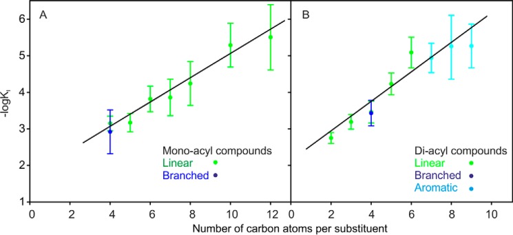
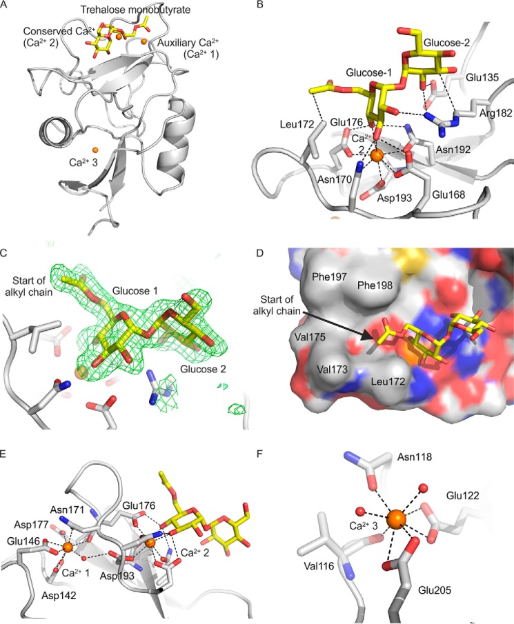
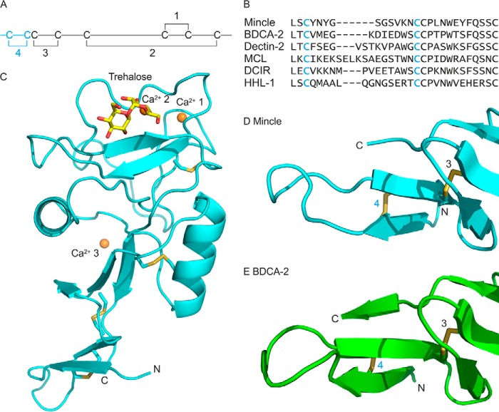
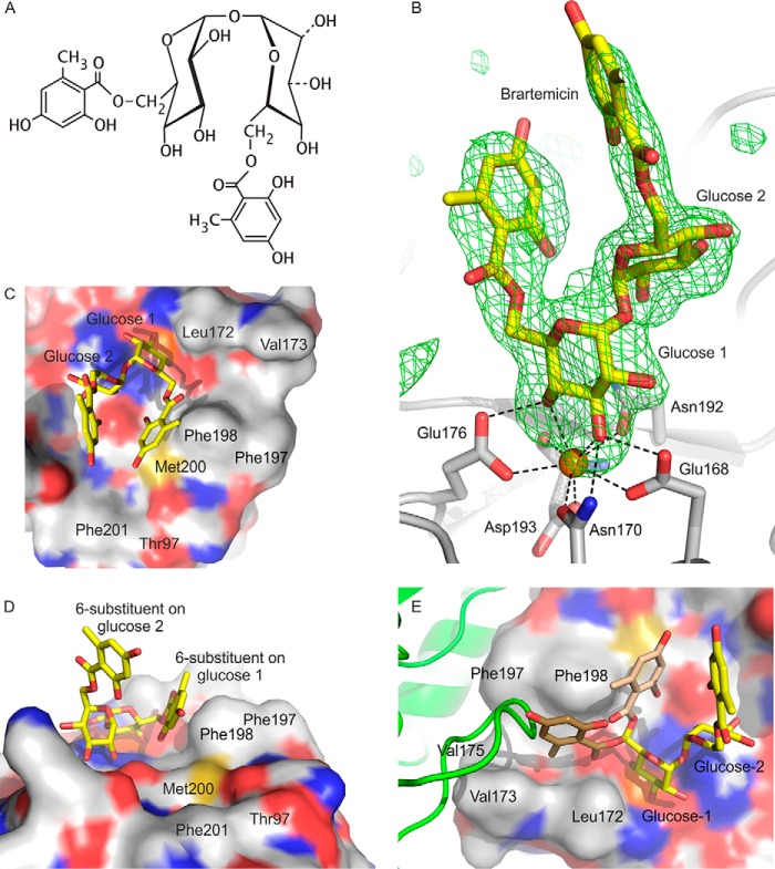
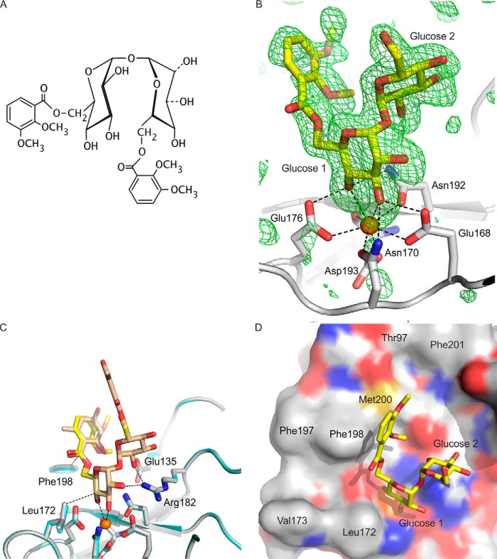
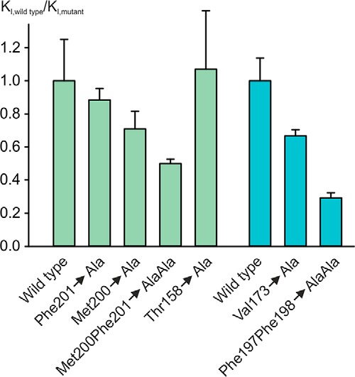
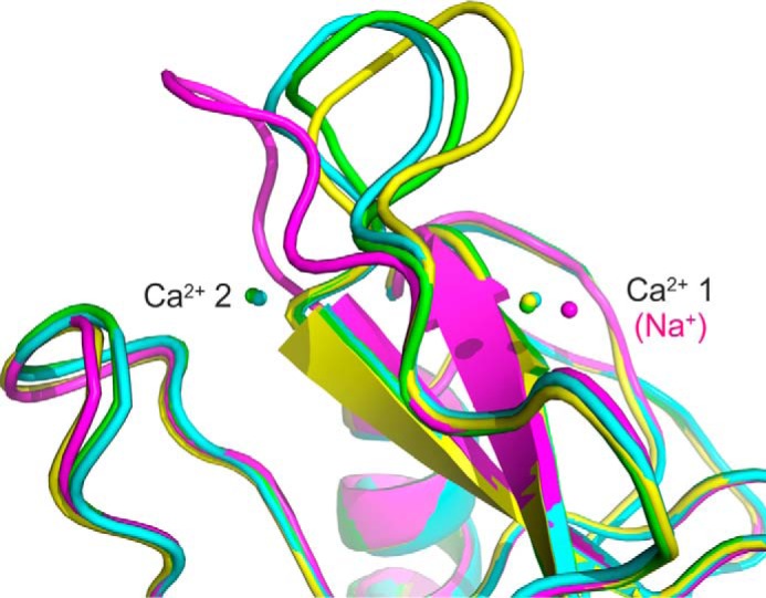
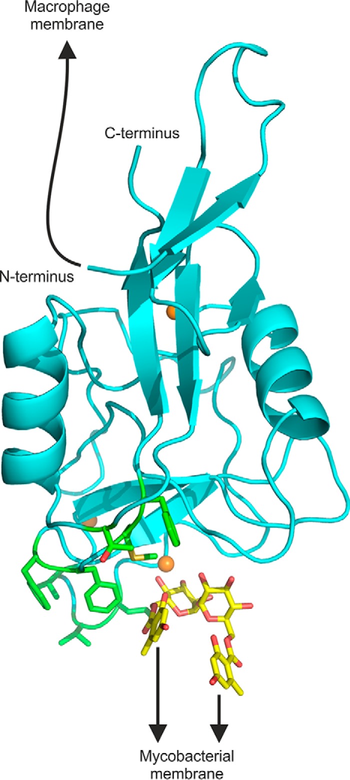
The macrophage receptor mincle binds to trehalose dimycolate on the surface of Mycobacterium tuberculosis Signaling initiated by this interaction leads to cytokine production, which underlies the ability of mycobacteria to evade the immune system and also to function as adjuvants. In previous work the mechanism for binding of the sugar headgroup of trehalose dimycolate to mincle has been elucidated, but the basis for enhanced binding to glycolipid ligands, in which hydrophobic substituents are attached to the 6-hydroxyl groups, has been the subject of speculation. In the work reported here, the interaction of trehalose derivatives with bovine mincle has been probed with a series of synthetic mimics of trehalose dimycolate in binding assays, in structural studies by x-ray crystallography, and by site-directed mutagenesis. Binding studies reveal that, rather than reflecting specific structural preference, the apparent affinity of mincle for ligands with hydrophobic substituents correlates with their overall size. Structural and mutagenesis analysis provides evidence for interaction of the hydrophobic substituents with multiple different portions of the surface of mincle and confirms the presence of three Ca2+-binding sites. The structure of an extended portion of the extracellular domain of mincle, beyond the minimal C-type carbohydrate recognition domain, also constrains the way the binding domains may interact on the surface of macrophages.

Keywords: carbohydrate-binding protein; glycolipid; lectin; mycobacteria; tuberculosis.


Figures

FIGURE 1.
FIGURE 1.
Relationship between apparent affinity for mincle and total size of acyl groups attached to 6-OH groups of trehalose. Affinities for monoacylated (A) and diacylated (B) trehalose derivatives were determined in binding competition assays. Binding of radiolabeled mannose-conjugated serum albumin to biotin-tagged CRD from mincle immobilized in streptavidin-coated wells was detected in the presence of soluble competing ligands. KI values measured in the competition experiments closely approximate the KD values (11). Straight lines were fitted to the data by least-squares fitting. Results are reported as the means ± S.D. for n = 3–4 separate experiments, each performed in duplicate.
FIGURE 2.
FIGURE 2.
Structure of mincle CRD bound to trehalose monobutyrate. A, overall structure of complex. B, close-up view of ligand-binding site, with glucose residue 1 in the primary sugar-binding site, ligated to the conserved Ca2+ (Ca2+ 2), and glucose residue 2 in the secondary sugar-binding site. C, Fo-Fc electron density omit map, calculated by omitting the trehalose monobutyrate ligand from the model, contoured at 3.0 σ, showing partial monobutyrate structure attached to the 6-OH group of glucose residue 1. D, surface of CRD showing the position of the carboxyl end of the monobutyrate substituent at the end of the hydrophobic groove formed by residues Phe-197 and Phe-198 on one side and Leu-172 and Val-173 on the other. E, close-up view showing amino acid residues ligated to the auxiliary Ca2+ (Ca2+ 1), including residues that bridge to the conserved Ca2+ (Ca2+ 2). F, close-up view of Ca2+ 3, showing amino acid side chain and backbone ligands. Copy C of the protein is shown. Protein in schematic representations as well as carbon atoms of stick and surface representations are presented in gray, carbon atoms of the sugars are yellow, oxygen atoms are red, nitrogen atoms are blue, and Ca2+ ions are orange. The electron density map is represented as green mesh.
FIGURE 3.
FIGURE 3.
Structure of the extended CRD from mincle. A, arrangement of disulfide bonds in C-type CRDs. The additional fourth disulfide bond in the extended CRD is highlighted in cyan. B, sequence comparisons of N-terminal extensions in CRDs. Sequences shown are from bovine mincle and human blood dendritic cell antigen 2, human dectin-2, macrophage C-type lectin (MCL, also designated dectin-3), human dendritic cell immunoreceptor (DCIR), and human hepatic lectin-1 (HHL-1, major subunit of the asialoglycoprotein receptor). C, overall structure of the extended CRD, showing bound trehalose and three Ca2+. Structures of the N-terminal extensions of mincle (D) and BDCA-2, PDB entry 4ZES (E) are shown. Proteins in schematic representations are shown in cyan for the extended CRD from mincle and green for BDCA-2. Disulfide bonds are highlighted in yellow, and other atoms are colored as in Fig. 2.
FIGURE 4.
FIGURE 4.
Structure of brartemicin bound to the extended CRD of mincle. A, chemical structure of brartemicin. B, omit map showing the position of brartemicin bound to the extended CRD from mincle. Electron density in the Fo-Fc map is contoured at 3.0 σ. C and D, surface representation of brartemicin-binding site from above and side. E, view of the binding site showing a loop from a symmetry-related molecule (green) occupying part of the hydrophobic groove adjacent to glucose residue 1. The arrangement of the 6-substituent attached to glucose residue 1 observed in the crystal structure is shown in light brown, and a model for an alternative conformation that occupies the same space as the loop from the adjacent molecule in the crystal structure is shown in dark brown. Atoms are represented as in Fig. 2.
FIGURE 5.
FIGURE 5.
Structure of brartemicin analog bound to the extended CRD of mincle. A, chemical structure of brartemicin analog. B, Fo-Fc omit map showing the brartemicin analog with electron density contoured at 3 σ. C, overlay of brartemicin and brartemicin analog structures. In this panel, carbon atoms in brartemicin are colored light brown, and carbon atoms of the analog are shown in yellow, with protein carbon atoms of the CRD shown in gray for the brartemicin complex and cyan for the analog complex. D, surface representation of brartemicin analog in the binding site of mincle. Atoms are represented as in Fig. 2.
FIGURE 6.
FIGURE 6.
Mutagenesis of hydrophobic binding surface. Affinity of mutants for mono-octanoyltrehalose was measured in binding competition assays. KI values for each mutant are normalized to the KI value for trehalose with that mutant to eliminate the impact of changes on the affinity for trehalose. The only instance in which there is more than a 20% change in the affinity for trehalose is in the case of changing Met-200 to alanine, which increases the KI for trehalose by 3.0 ± 0.1-fold. This effect probably reflects the role of Met-200 in positioning Glu-135, which interacts with glucose residue 2 in the extended sugar-binding site. Results in blue, recalculated from (7), are for the hydrophobic groove, whereas results in green are for the additional hydrophobic surface that interacts with brartemicin. The graph shows the ratio of the normalized KI values for the wild type CRD compared with the mutants, so smaller numbers reflect reduced affinity. Results are reported as the means ± S.D. for n = 3–4 separate experiments, each performed in duplicate.
FIGURE 7.
FIGURE 7.
Flexibility in the side of the hydrophobic groove. Superposition of mincle CRD structures shows complexes of the minimal CRD with citrate (magenta), with trehalose (yellow), and with trehalose monobutyrate (green) as well as the extended CRD complexed with trehalose (cyan).
FIGURE 8.
FIGURE 8.
Model showing orientation of extended N terminus and ligand-binding sites. A composite model was generated by positioning brartemicin, from the crystal structure of brartemicin with the CRD, in the binding site of the extended CRD with bound trehalose. The model highlights portions of the extended CRD that interact with the macrophage membrane and with glycolipid targets at the surface of mycobacteria. The N terminus is linked to the macrophage membrane by a 19-amino acid sequence not seen in the crystal structure. The binding site for mycobacterial ligands is indicated based on the orientation of brartemicin in the binding site and highlighting of hydrophobic residues implicated in ligand binding in green. Other atoms are colored as in Fig. 2.

References

    1. Matsumoto M., Tanaka T., Kaisho T., Sanjo H., Copeland N. G., Gilbert D. J., Jenkins N. A., and Akira S. (1999) A novel LPS-inducible C-type lectin is a transcriptional target of NF-IL6 in macrophages. J. Immunol. 163, 5039–5048 - PubMed
    1. Ishikawa E., Ishikawa T., Morita Y. S., Toyonaga K., Yamada H., Takeuchi O., Kinoshita T., Akira S., Yoshikai Y., and Yamasaki S. (2009) Direct recognition of the mycobacterial glycolipid, trehalose dimycolate, by C-type lectin Mincle. J. Exp. Med. 206, 2879–2888 - PMC - PubMed
    1. Matsunaga I., and Moody D. B. (2009) Mincle is a long sought receptor for mycobacterial cord factor. J. Exp. Med. 206, 2865–2868 - PMC - PubMed
    1. Lang R. (2013) Recognition of the mycobacterial cord factor by mincle: relevance for granuloma formation and resistance to tuberculosis. Front. Immunol. 4, 5. - PMC - PubMed
    1. Wells C. A., Salvage-Jones J. A., Li X., Hitchens K., Butcher S., Murray R. Z., Beckhouse A. G., Lo Y.-L., Manzanero S., Cobbold C., Schroder K., Ma B., Orr S., Stewart L., Lebus D., et al. (2008) The macrophage-inducible C-type lectin, mincle, is an essential component of the innate immune response to Candida albicans. J. Immunol. 180, 7404–7413 - PubMed