. 2016 Aug 20;17(1):31.
doi: 10.1186/s12860-016-0109-5.

Prolactin-induced PAK1 tyrosyl phosphorylation promotes FAK dephosphorylation, breast cancer cell motility, invasion and metastasis

Affiliations

Prolactin-induced PAK1 tyrosyl phosphorylation promotes FAK dephosphorylation, breast cancer cell motility, invasion and metastasis

Alan Hammer et al. BMC Cell Biol. .

Abstract

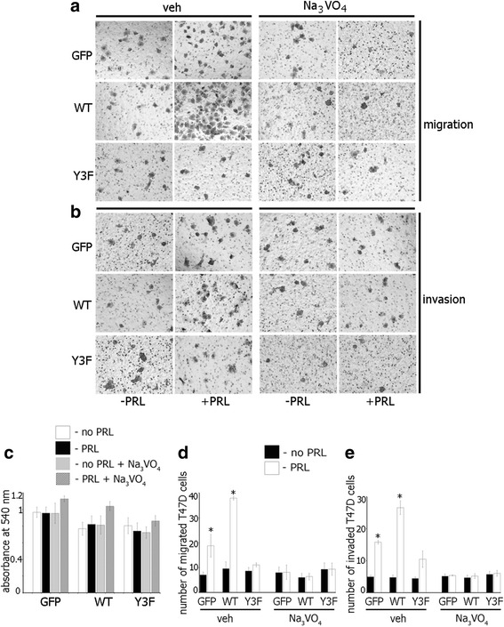
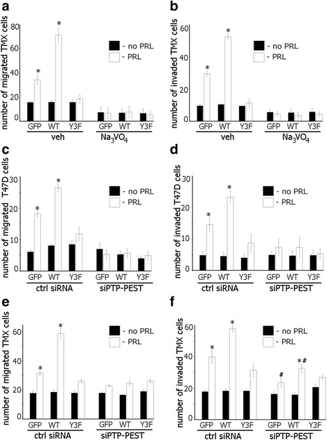
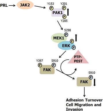
Background: The serine/threonine kinase PAK1 is an important regulator of cell motility. Both PAK1 and the hormone/cytokine prolactin (PRL) have been implicated in breast cancer cell motility, however, the exact mechanisms guiding PRL/PAK1 signaling in breast cancer cells have not been fully elucidated. Our lab has previously demonstrated that PRL-activated tyrosine kinase JAK2 phosphorylates PAK1 on tyrosines 153, 201, and 285, and that tyrosyl phosphorylated PAK1 (pTyr-PAK1) augments migration and invasion of breast cancer cells.

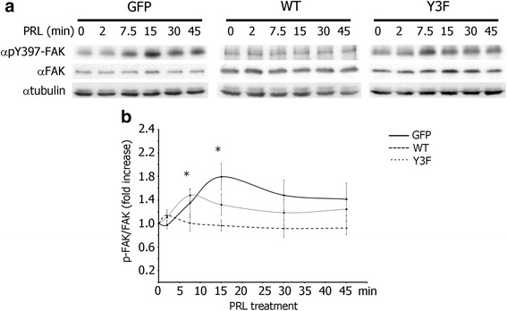
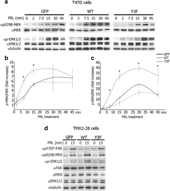
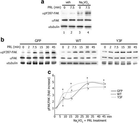
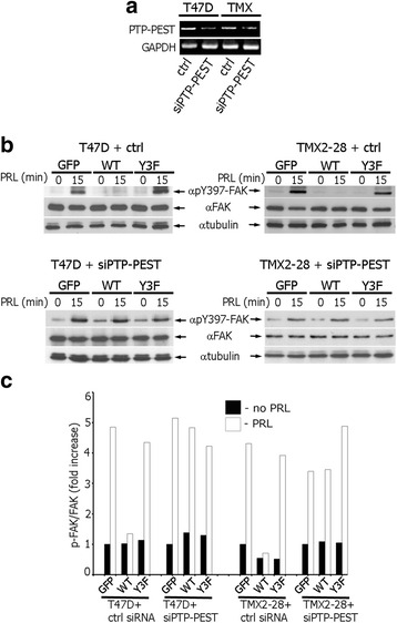
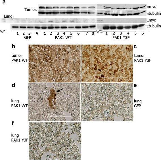
Results: Here we further investigate the mechanisms by which pTyr-PAK1 enhances breast cancer cell motility in response to PRL. We demonstrate a distinct reduction in PRL-induced FAK auto-phosphorylation in T47D and TMX2-28 breast cancer cells overexpressing wild-type PAK1 (PAK1 WT) when compared to cells overexpressing either GFP or phospho-tyrosine-deficient mutant PAK1 (PAK1 Y3F). Furthermore, pTyr-PAK1 phosphorylates MEK1 on Ser298 resulting in subsequent ERK1/2 activation. PRL-induced FAK auto-phosphorylation is rescued in PAK1 WT cells by inhibiting tyrosine phosphatases and tyrosine phosphatase inhibition abrogates cell motility and invasion in response to PRL. siRNA-mediated knockdown of the tyrosine phosphatase PTP-PEST rescues FAK auto-phosphorylation in PAK1 WT cells and reduces both cell motility and invasion. Finally, we provide evidence that PRL-induced pTyr-PAK1 stimulates tumor cell metastasis in vivo.

Conclusion: These data provide insight into the mechanisms guiding PRL-mediated breast cancer cell motility and invasion and highlight a significant role for pTyr-PAK1 in breast cancer metastasis.

Keywords: Breast cancer cells; FAK; PAK1; Prolactin; Tyrosyl phosphorylation.


Figures

Fig. 1
Fig. 1
Tyrosyl phosphorylation of PAK1 negatively regulates PRL-induced FAK auto-phosphorylation. a Whole cell lysates (WCL) of T47D cells stably overexpressing GFP, PAK1 WT, or PAK1 Y3F treated with PRL (200 ng/ml) for the indicated times were probed for FAK auto-phosphorylation using αpY397-FAK antibody. The expression levels of γtubulin were used as an internal loading control. b Graph represents the densitometric analysis of the bands obtained for pY397-FAK normalized to total FAK for at least 3 independent experiments. The solid line represents T47D GFP cells, the dashed line represents T47D PAK1 WT cells, and the dotted line represents T47D PAK1 Y3F cells. Bars represent mean ± SE. *P < 0.05 compared with the same cells not treated with PRL
Fig. 2
Fig. 2
Tyrosyl phosphorylation of PAK1 promotes S298-MEK1 phosphorylation and ERK activation in response to PRL. a WCL of T47D cells stably overexpressing GFP, PAK1 WT, or PAK1 Y3F treated with PRL (200 ng/ml) for the indicated times were probed for MEK phosphorylation using αpS298-MEK and ERK1/2 activation using αphospho-ERK1/2 (pT202/Y204) antibodies. b, c Graphs represent the densitometric analysis of the bands obtained for phospho-MEK (b) or phospho-ERK1/2 (c) normalized to total MEK or ERK1/2, respectively, for at least 3 independent experiments. Bars represent mean ± SE . *P < 0.05 compared with cells expressing GFP with the same treatment. d WCL of TMX2-28 cells stably overexpressing GFP, PAK1 WT, or PAK1 Y3F treated with PRL (200 ng/ml) for the indicated times were probed with the indicated antibodies. The expression levels of γtubulin were used as an internal loading control
Fig. 3
Fig. 3
Protein tyrosine phosphatase inhibition rescues PRL-mediated FAK auto-phosphorylation in T47D WT cells. a Tyrosine phosphatase inhibition by Na3VO4 permits PRL-induced FAK auto-phosphorylation in PAK1 WT cells. WCL of T47D PAK1 WT cells treated with either vehicle (veh) or Na3VO4 (100 ng/ml) for 1 h before PRL (200 ng/ml) treatment were probed for FAK auto-phosphorylation by αpY397-FAK antibody. b FAK is auto-phosphorylation in T47D GFP, PAK1 WT, and PAK1 Y3F cells in response to PRL in the presence of Na3VO4. The cells were treated with Na3VO4 as in A and with PRL (200 ng/ml) for the indicated times. FAK auto-phosphorylation was assessed as in A. The expression levels of γtubulin were used as an internal loading control. c Graph represents the densitometric analysis of the bands obtained for pY397-FAK normalized to total FAK for at least 3 independent experiments. Bars represent mean ± SE. *P < 0.05 compared with the same cells not treated with PRL
Fig. 4
Fig. 4
Silencing of tyrosine phosphatase PTP-PEST rescues FAK auto-phosphorylation in T47D and TMX2-28 cells. a PTP-PEST siRNA reduces PTP-PEST mRNA in T47D and TMX2-28 cells. T47D and TMX2-28 cells were transfected with either PTP-PEST siRNA or control non-coding siRNA (ctrl) and mRNA levels were assessed by RT-PCR using PTP-PEST-specific primers. GAPDH primers were used an internal control. b WCL of T47D and TMX2-28 clones transfected with control or PTP-PEST siRNAs and treated with PRL (200 ng/ml) for 0 or 15 min were probed for FAK auto-phosphorylation using αpY397-FAK antibody. The expression levels of γtubulin were used as an internal loading control. c Graph represents the densitometric analysis of the bands obtained for pY397-FAK normalized to total FAK
Fig. 5
Fig. 5
Protein tyrosine phosphatase inhibition impedes PRL-mediated T47D cell migration and invasion. a, b Equal amounts of T47D GFP, PAK WT, or PAK1 Y3F cells were loaded into the upper part of the Boyden chamber uncovered (a) or covered with Matrigel (b) with or without Na3VO4 (100 ng/ml). PRL (200 ng/ml) was added to the lower part. Representative brightfield images of the cells migrated/invaded to the lower chamber were taken in 48 h. A LUCPlan FLN 40X objective lens and wide field WHN 10X eyepiece on an inverted Olympus IX81 microscope were used. (c) Na3VO4 (100 ng/ml) treatment on T47D cells for 48 h has no cytotoxic effect. d, e The number of cells that migrated to the lower surface of the chamber toward PRL (white bar) or vehicle (black bar) after 48 h was counted and plotted. Bars represent mean ± SE. *P < 0.05 compared with the same cells not treated with PRL
Fig. 6
Fig. 6
Silencing of the tyrosine phosphatase PTP-PEST reduces PRL-mediated cell migration and invasion. a Tyrosine phosphatase inhibition abolishes PRL-induced TMX2-28 cell migration a and invasion b. TMX2-28 GFP, PAK WT, or PAK1 Y3F cells were assessed as in Fig. 5. cf Equal amount of T47D (c, d) or TMX2-28 (e, f) clones were transfected with either control or PTP-PEST siRNA and loaded into the upper part of the Boyden chamber covered (d, f) or not (c, e) with Matrigel. The number of cells that migrated/invaded to the lower chamber toward PRL (white bar) or vehicle (black bar) after 48 h was counted and plotted. Bars represent mean ± SE. *P < 0.05 compared with the same cells not treated with PRL. #P < 0.05 compared with the same cells treated with PRL but transfected with control siRNA (f)
Fig. 7
Fig. 7
PAK1 tyrosyl phosphorylation stimulates PRL-induced tumor metastasis in vivo. a myc-PAK1 was detected in all tumor lysates isolated from myc-PAK1 WT and myc-PAK1 Y3F inoculated mice. PRL-induced tyrosyl phosphorylation of PAK1increased tumor metastasis, as 3 out of 8 lungs from WT mice contained myc-PAK1 (lanes 2, 4 and 5). No myc-PAK1 was detected in any of the lungs from the Y3F or GFP mice. Anti-tubulin antibody was used as a loading control. Whole cell lysate (WCL) of TMX2-28 PAK1 WT cells was loaded as a control for PAK1-myc position in the gels. bf Representative images of myc-PAK1 detected with anti-myc in breast tumor (b, c) and lung tissue (df). myc-PAK1 was detected in breast tumors of myc-PAK1 WT- (b) and Y3F- (c) and lung of myc-PAK1 WT- inoculated mice (d) but not in lungs of GFP (e) or PAK1 Y3F-inoculated mice (f). The arrow highlights metastatic nodule. Counterstaining with hematoxylin was omitted. Scale bar is 200 μm in (b) and 100 μm in (ce)
Fig. 8
Fig. 8
Proposed mechanism for the role of PRL-activated PAK1 in breast cancer cell migration. PRL binding to the PRLR results in activation of the non-receptor tyrosine kinase JAK2. JAK2 tyrosyl phosphorylates PAK1 on Y153, 201, and 285, enhancing PAK1 kinase and scaffolding activities. PRL treatment also leads to FAK auto-phosphorylation at Y397. Activated PAK1 phosphorylates MEK1 at S298, resulting in increased MEK1/ERK binding and enhanced ERK activity. Active ERK phosphorylates FAK at S910, leading to dephosphorylation of FAK at Y397 by the tyrosine phosphatase PTP-PEST as shown by Zheng et al. (2009). FAK dephosphorylation decreases FAK kinase activity and promotes adhesion turnover and breast cancer cell migration

Similar articles

Cited by

References

    1. Marano RJ, Ben-Jonathan N. Minireview: extrapituitary prolactin: an update on the distribution, regulation, and functions. Mol Endocrinol. 2014;28(5):622–633. doi: 10.1210/me.2013-1349. - DOI - PMC - PubMed
    1. Clevenger CV, Furth PA, Hankinson SE, Schuler LA. The role of prolactin in mammary carcinoma. Endocr Rev. 2003;24(1):1–27. doi: 10.1210/er.2001-0036. - DOI - PMC - PubMed
    1. Bernichtein S, Touraine P, Goffin V. New concepts in prolactin biology. J Endocrinol. 2010;206(1):1–11. doi: 10.1677/JOE-10-0069. - DOI - PubMed
    1. Holtkamp W, Nagel GA, Wander HE, Rauschecker HF, von Heyden D. Hyperprolactinemia is an indicator of progressive disease and poor prognosis in advanced breast cancer. Int J Cancer. 1984;34(3):323–328. doi: 10.1002/ijc.2910340307. - DOI - PubMed
    1. Bhatavdekar JM, Shah NG, Balar DB, Patel DD, Bhaduri A, Trivedi SN, et al. Plasma prolactin as an indicator of disease progression in advanced breast cancer. Cancer. 1990;65(9):2028–2032. doi: 10.1002/1097-0142(19900501)65:9<2028::AID-CNCR2820650924>3.0.CO;2-9. - DOI - PubMed

Publication types

MeSH terms

Substances