. 2016 Dec;30(12):3997-4006.
doi: 10.1096/fj.201600728R. Epub 2016 Aug 19.

Complement-induced activation of the cardiac NLRP3 inflammasome in sepsis

Affiliations

Complement-induced activation of the cardiac NLRP3 inflammasome in sepsis

Miriam Kalbitz et al. FASEB J. 2016 Dec.

Abstract

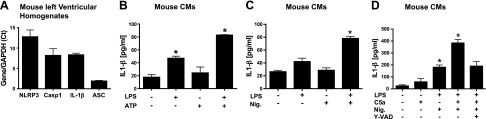
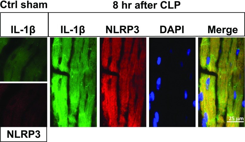
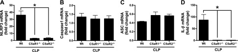
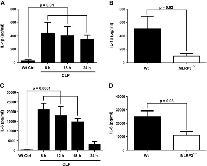
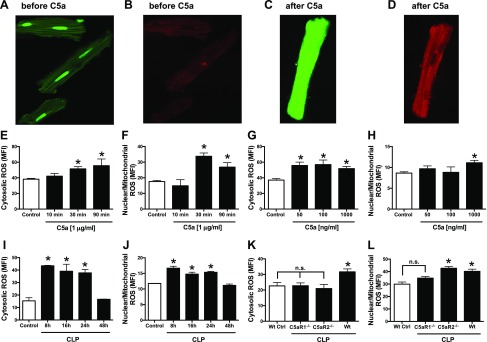
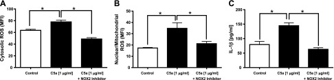
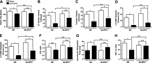
Cardiac dysfunction develops during sepsis in humans and rodents. In the model of polymicrobial sepsis induced by cecal ligation and puncture (CLP), we investigated the role of the NLRP3 inflammasome in the heart. Mouse heart homogenates from sham-procedure mice contained high mRNA levels of NLRP3 and IL-1β. Using the inflammasome protocol, exposure of cardiomyocytes (CMs) to LPS followed by ATP or nigericin caused release of mature IL-1β. Immunostaining of left ventricular frozen sections before and 8 h after CLP revealed the presence of NLRP3 and IL-1β proteins in CMs. CLP caused substantial increases in mRNAs for IL-1β and NLRP3 in CMs which are reduced in the absence of either C5aR1 or C5aR2. After CLP, NLRP3-/- mice showed reduced plasma levels of IL-1β and IL-6. In vitro exposure of wild-type CMs to recombinant C5a (rC5a) caused elevations in both cytosolic and nuclear/mitochondrial reactive oxygen species (ROS), which were C5a-receptor dependent. Use of a selective NOX2 inhibitor prevented increased cytosolic and nuclear/mitochondrial ROS levels and release of IL-1β. Finally, NLRP3-/- mice had reduced defects in echo/Doppler parameters in heart after CLP. These studies establish that the NLRP3 inflammasome contributes to the cardiomyopathy of polymicrobial sepsis.-Kalbitz, M., Fattahi, F., Grailer, J. J., Jajou, L., Malan, E. A., Zetoune, F. S., Huber-Lang, M., Russell, M. W., Ward, P. A. Complement-induced activation of the cardiac NLRP3 inflammasome in sepsis.

Keywords: C5a; C5a receptors; CLP; IL-1β.


Figures

Figure 1.
Figure 1.
mRNA levels of inflammasome components in mouse heart homogenates and activation of NLRP3 inflammasome in CMs. A) Using LV homogenates from mouse hearts, mRNAs were measured for NLRP3, caspase-1, ASC, and IL-1β by qRT-PCR and developed as a ratio to mRNA for Gapdh (set as 1.0). B) Mouse CMs were incubated with buffer or with LPS (1 µg/ml for 4 h at 37°C) or the combination, with the endpoint being IL-1β release. C, D) CM release of IL-1β was measured after cell exposure to buffer or LPS (primer), followed by addition of activator (ATP or nigericin, 1 mM or 10 µm, respectively, for 45 min at 37°C). Where indicated, 10 µm Y-VAD, a global caspase inhibitor, was also used. For each bar, n ≥ 5 samples. *P < 0.05
Figure 2.
Figure 2.
Activation of NLRP3 inflammasome in LV mouse CMs after CLP. Immunofluorescence analysis of LV frozen sections from sham-procedure cointrol (ctrl sham) mouse WT heart (left boxes) or hearts 8 h after CLP. Magnification, ×63. Analysis involved immunostaining for IL-1β (green), NLRP3 (red), nuclear staining by DAPI, and the merge image for all 3 stains. Staining for IL-1β and NLRP3 was very faint in sham-procedure control hearts, in striking contrast to hearts 8 h after CLP.
Figure 3.
Figure 3.
In sepsis, NLRP3 inflammasome activation in mouse CMs is reduced in absence of either C5aR1 or C5aR2. Activation of the NLRP3 inflammasome components NLRP3 (A), caspase-1 (B), and ASC (C), as well as IL-β (D), in mouse CMs before (not shown) and 16 h after CLP. In these studies, qRT-PCR was used in LV heart homogenates from WT, and C5aR1−/− or C5aR2−/−, 16 h after CLP. Results are ratios of NLRP3 inflammasome component mRNA to Gapdh mRNA (the latter being set at 1.0). For each bar, n ≥ 4 samples. *P < 0.05.
Figure 4.
Figure 4.
Reduced plasma cytokines in NLRP3−/− mice after CLP. Plasma levels of IL-1β (A, B) after CLP were reduced in NLRP3 8–24 h after CLP. A) Time course of plasma levels of IL-1β after CLP. B) Plasma levels of IL-1β were obtained 18 h after CLP. C, D) A similar protocol was used with a focus on plasma IL-6 8 h after CLP. All assays were performed by ELISA. For each bar, n ≥ 5 plasma samples.
Figure 5.
Figure 5.
Buildup of ROS in nuclear/mitochondrial and cytosolic areas of CMs. Fluorescent dyes that detect ROS in mitochondria/nuclei (B, D, F, H, J, L) or in cytosol (A, C, E, G, I, K) of rat CMs were used, with endpoints being fluorescence in CMs, as determined by confocal microscopy. A, B) Sham-procedure CMs exposed to buffer. C, D) CMs exposed to rC5a (1 µg/ml for 30 min). E, F) CMs exposed to C5a 10, 30, and 90 min. G, H) CMs exposed to increasing amounts of C5a for 90 min. I, J) CMs from WT mice obtained at various time points after CLP. K, L) CMs obtained from WT, C5aR1−/−, and C5aR2−/− mice 16 h after CLP. For each bar, n > 5 samples. *P < 0.05.
Figure 6.
Figure 6.
NOX2 inhibitor blocks buildup of ROS in CMs and release of IL-1β. A, B) Cytosolic ROS (A) and nuclear/mitochondrial ROS (B) in absence or presence of NOX2 inhibitor in rat CMs exposed to C5a for 1 h at 37°C. C) Companion studies focused on IL-1β release from C5a-activated CMs in the presence or absence of NOX2 inhibitor. CMs exposed to C5a for 8 h. For each bar, n ≥ 4 samples. *P < 0.05.
Figure 7.
Figure 7.
ECHO/Doppler parameters in WT and NLRP3−/− mice 8 h after CLP. HR (A), LV SV (B), CO (C), LV VolS (D), LV VolD (E), E/A ratio (F), isovolumic relaxation (G), and E/E′sa (H) represent selected measures of systolic and diastolic heart function in mice before and 8 h after CLP. For each bar, n = 5 mice. ns, nonsignificant. *P < 0.05.

References

    1. Franchi L., Eigenbrod T., Muñoz-Planillo R., Nuñez G. (2009) The inflammasome: a caspase-1-activation platform that regulates immune responses and disease pathogenesis. Nat. Immunol. 10, 241–247 - PMC - PubMed
    1. Stutz A., Golenbock D. T., Latz E. (2009) Inflammasomes: too big to miss. J. Clin. Invest. 119, 3502–3511 - PMC - PubMed
    1. Kalbitz M., Grailer J. J., Fattahi F., Jajou L., Herron T. J., Campbell K. F., Zetoune F. S., Bosmann M., Sarma J. V., Huber-Lang M., Gebhard F., Loaiza R., Valdivia H. H., Jalife J., Russell M. W., Ward P. A. (2015) Role of extracellular histones in the cardiomyopathy of sepsis. FASEB J. 29, 2185–2193 - PMC - PubMed
    1. Martinon F., Pétrilli V., Mayor A., Tardivel A., Tschopp J. (2006) Gout-associated uric acid crystals activate the NALP3 inflammasome. Nature 440, 237–241 - PubMed
    1. Duewell P., Kono H., Rayner K. J., Sirois C. M., Vladimer G., Bauernfeind F. G., Abela G. S., Franchi L., Nuñez G., Schnurr M., Espevik T., Lien E., Fitzgerald K. A., Rock K. L., Moore K. J., Wright S. D., Hornung V., Latz E. (2010) NLRP3 inflammasomes are required for atherogenesis and activated by cholesterol crystals. Nature 464, 1357–1361 - PMC - PubMed

Publication types