Review
. 2017 Jan;26(1):32-39.
doi: 10.1002/pro.3022. Epub 2016 Sep 7.

How cryo-electron microscopy and X-ray crystallography complement each other

Affiliations
Review

How cryo-electron microscopy and X-ray crystallography complement each other

Hong-Wei Wang et al. Protein Sci. 2017 Jan.

Abstract

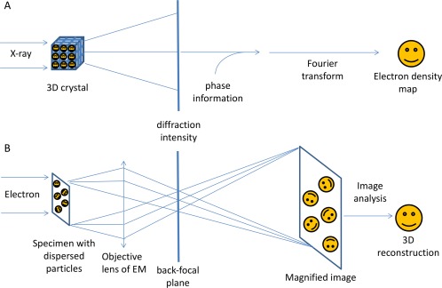
With the ability to resolve structures of macromolecules at atomic resolution, X-ray crystallography has been the most powerful tool in modern structural biology. At the same time, recent technical improvements have triggered a resolution revolution in the single particle cryo-EM method. While the two methods are different in many respects, from sample preparation to structure determination, they both have the power to solve macromolecular structures at atomic resolution. It is important to understand the unique advantages and caveats of the two methods in solving structures and to appreciate the complementary nature of the two methods in structural biology. In this review we provide some examples, and discuss how X-ray crystallography and cryo-EM can be combined in deciphering structures of macromolecules for our full understanding of their biological mechanisms.

Keywords: X-ray and electron scattering; X-ray crystallography; cryo-EM; protein structure determination; structural biology; structure determination methods.


Figures

Figure 1
Figure 1
Technical difference between X‐ray crystallography and single particle cryo‐EM. A: illustrates the physics and mathematical principles of X‐ray crystallography to solve a structure. Periodic arrays of molecules in a regular three‐dimensional lattice resulted in a “diffraction pattern” when illuminated by X‐rays. Because of the lack of focus lens for X‐rays, only intensities can be recorded, which resulted in the phase problem in X‐ray crystallography. Part (B) illustrates the physics and mathematical principles of EM in solving a structure, because of an electron's strong interaction with each atom's Coulomb potential, individual molecules within a specimen can be imaged directly. The virtue density of a biological molecule can be reconstructed from a set of 2D projections of the molecule with common structure, imaged at various orientations by the electron microscope. In the back‐focal plane of the objective lens, the diffraction pattern of a biological specimen can also be obtained.
Figure 2
Figure 2
Docking of atomic models into the 3D cryo‐EM map of Ryanodine receptor 1 complex at 10 Angstrom resolution. The 3D EM map of the Ryanodine receptor 1 (EMD‐5041) is shown in semi‐transparent rendering in two orthogonal views. The atomic model of a ryanodine receptor monomer (grey) is docked in the map. The atomic model of SPRY2 domain docked in the map with rigid docking algorithm is shown in red color. Its equivalent domain in the atomic model of the full‐length protein is shown in blue color. The atomic models of the two SPRY2 domain are in very similar orientation and location.
Figure 3
Figure 3
Cryo‐EM 3D reconstruction helps phasing in X‐ray crystallography. A: is the single particle cryo‐EM 3D reconstruction of the TLR13‐ssRNA at 4.8 Å resolution. Part (B) is the electron density map (blue mesh) of the TLR13‐ssRNA after phase extension from the initial cryo‐EM map to X‐ray crystallographic data at 2.3 Angstrom resolution. The main chain of the atomic model of TLR13 protein in the density is shown in green sticks. A few glycosylated sites with the sugar molecules are shown in stick model. Both (A) and (B) are shown in stereo pairs.
Figure 4
Figure 4
Domain structure locked in rigid state by crystal packing. Part (A) is a partial map from the single particle cryo‐EM 3D reconstruction of the influenza RdRP tetramer at 4.3 Å resolution in semi‐transparent rendering, with the crystal structure of an RdRP monomer docked in the map. The N‐terminal domain of PA subunit is not resolved in the 3D map so the atomic model protruding out from the map. Part (B) shows two adjacent RdRP monomers packed in the crystal lattice for X‐ray crystallography. The N‐terminal domain of PA subunit is in close contact with the PB subunit of the adjacent monomer. In both (A) and (B), PA subunit is in blue color, PB subunit is in cyan color, and the RNA is in yellow color.

References

    1. Shi Y (2014) A glimpse of structural biology through X‐ray crystallography. Cell 159:995–1014. - PubMed
    1. Kuhlbrandt W (2014) Biochemistry. The resolution revolution. Science 343:1443–1444. - PubMed
    1. Taylor GL (2010) Introduction to phasing. Acta Cryst D66:325–338. - PMC - PubMed
    1. Newman J (2006) A review of techniques for maximizing diffraction from a protein crystal in stilla. Acta Cryst D62:27–31. - PubMed
    1. Rossmann MG (2000) Fitting atomic models into electron‐microscopy maps. Acta Cryst D56:1341–1349. - PubMed

Publication types

MeSH terms