. 2016 Nov;11(11):1993-2005.
doi: 10.1016/j.jtho.2016.07.033. Epub 2016 Aug 17.

Malignant Mesothelioma Effusions Are Infiltrated by CD3+ T Cells Highly Expressing PD-L1 and the PD-L1+ Tumor Cells within These Effusions Are Susceptible to ADCC by the Anti-PD-L1 Antibody Avelumab

Affiliations

Malignant Mesothelioma Effusions Are Infiltrated by CD3+ T Cells Highly Expressing PD-L1 and the PD-L1+ Tumor Cells within These Effusions Are Susceptible to ADCC by the Anti-PD-L1 Antibody Avelumab

Swati Khanna et al. J Thorac Oncol. 2016 Nov.

Abstract

Introduction: The functional aspects of programmed death 1 (PD-1) and PD ligand 1 (PD-L1) immune checkpoints in malignant mesothelioma have not been studied.

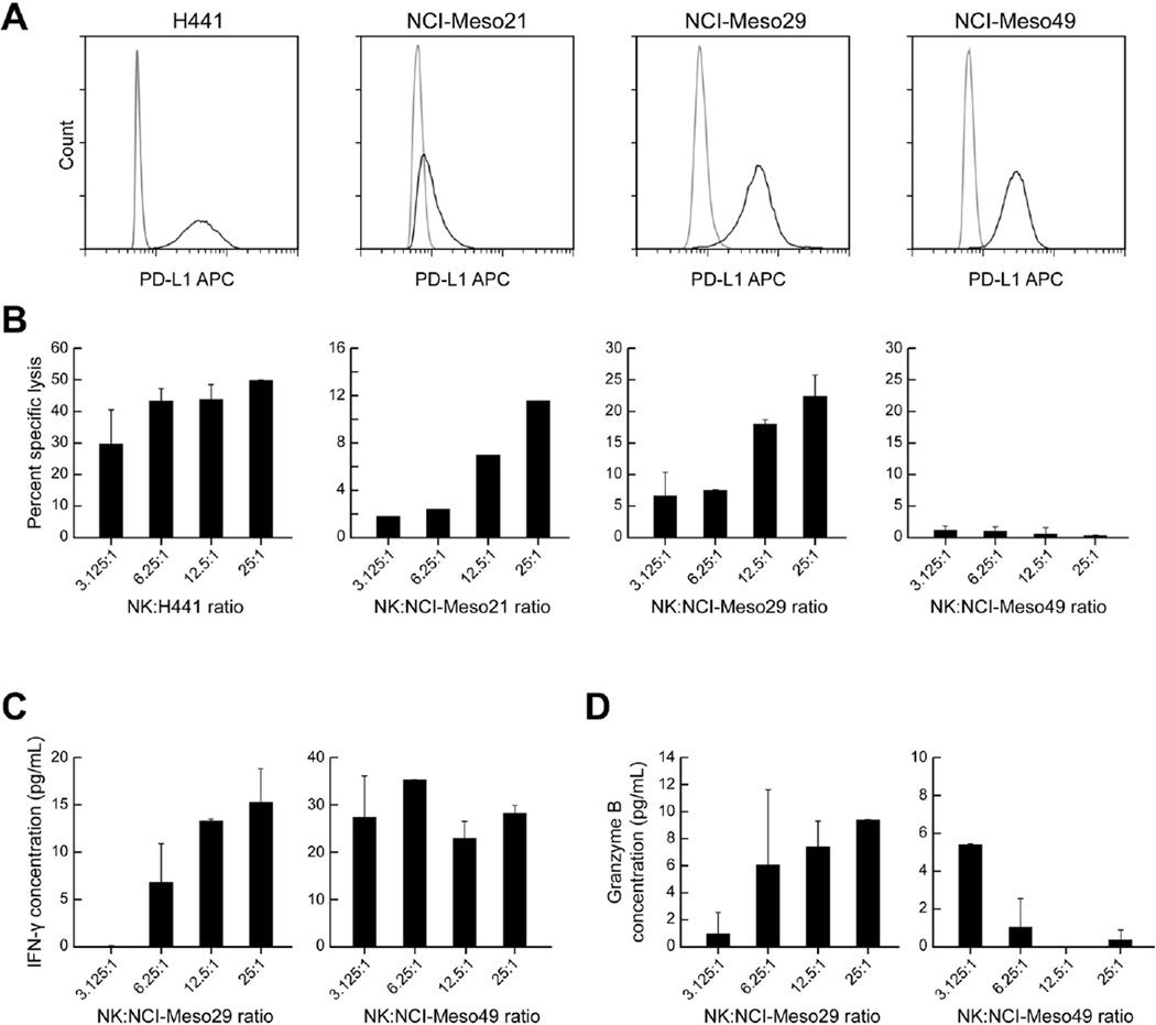
Methods: Tumor samples from 65 patients with mesothelioma were evaluated for PD-L1 expression by immunohistochemistry, and its prognostic significance was examined. Malignant effusions from patients with pleural and peritoneal mesothelioma were evaluated for PD-1-positive and PD-L1-positive infiltrating lymphocytes and their role in inducing PD-L1 expression in tumor cells. Antibody-dependent cellular cytotoxicity (ADCC) of avelumab, a fully humanized immunoglobulin G1 anti PD-L1 antibody against primary mesothelioma cell lines, was evaluated in presence of autologous and allogeneic natural killer cells.

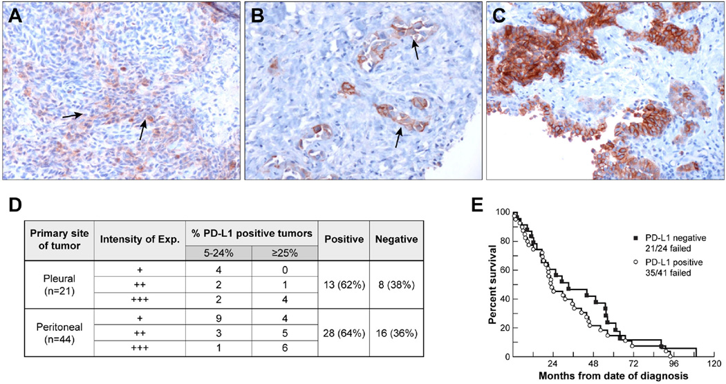
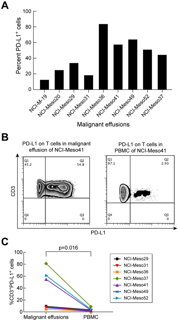
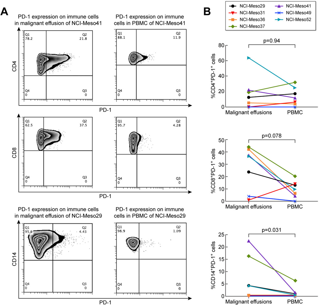
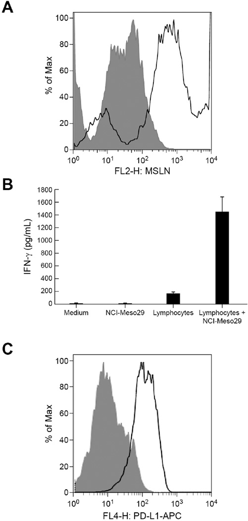
Results: Of 65 pleural and peritoneal mesothelioma tumors examined, 41 (63%) were PD-L1-positive, which was associated with slightly inferior overall survival compared to patients with PD-L1-negative tumors (median 23.0 versus 33.3 months, p = 0.35). The frequency of PD-L1 expression was similar in patients with pleural and peritoneal mesothelioma, with 62% and 64% of samples testing positive, respectively. In nine mesothelioma effusion samples evaluated, the fraction of cells expressing PD-L1 ranged from 12% to 83%. In seven patients with paired malignant effusion and peripheral blood mononuclear cell (PBMC) samples, PD-L1 expression was significantly higher on CD3-positive T cells present in malignant effusions as compared with PBMCs (p = 0.016). In addition, the numbers of CD14-positive PD-1-positive cells were increased in malignant effusions compared with PBMCs (p = 0.031). The lymphocytes present in malignant effusions recognized autologous tumor cells and induced interferon-γ-mediated PD-L1 expression on the tumor cell surface. Of the three primary mesothelioma cell lines tested, two were susceptible to avelumab-mediated ADCC in the presence of autologous natural killer cells.

Conclusions: Most pleural as well as peritoneal mesotheliomas express PD-L1. Malignant effusions in this disease are characterized by the presence of tumor cells and CD3-positive T cells that highly express PD-L1. In addition, mesothelioma tumor cells are susceptible to ADCC by the anti-PD-L1 antibody avelumab.

Keywords: ADCC; Avelumab; Mesothelioma; PD-1-PD-L1.


Conflict of interest statement

The authors disclose no potential conflicts of interest

Figures

Figure 1
Figure 1. PD-L1 expression on tumor samples from patients with pleural and peritoneal mesothelioma
Immunohistochemical analysis showing different degrees of PD-L1 expression in mesothelioma. (A) Sarcomatoid mesothelioma with 10% cell membrane positivity and weak (+) intensity (arrow). (B) Epithelioid mesothelioma with 10% cell membrane positivity and strong (+++) intensity (arrow). (C) Epithelioid mesothelioma with 60% cell membrane positivity and strong (+++) intensity. All images were acquired at a magnification of 200×. (D) Table summarizing tumor PD-L1 expression by immunohistochemistry. (E) Kaplan-Meier plot showing overall survival of patients with PD-L1-expressing and PD-L1 negative tumors. Patients with PD-L1 positive tumors had a slightly inferior overall survival compared to that of patients with PD-L1 negative tumors (median survival 23.0 months for PD-L1 positive tumors vs. 33.3 months for PD-L1 negative tumors; p=0.35).
Figure 2
Figure 2. PD-L1 expression on cells present in malignant mesothelioma effusions
(A)Percentage of basal PD-L1 expressing cells present in malignant effusions from nine different patients. (B) Representative example of PD-L1 expression on CD3+ T cells in malignant effusion and PBMC of patient NCI-Meso41. (C) Percentage of CD3+PD-L1+ cells in the malignant effusions and PBMCs of seven patients with paired effusion and PBMC samples.
Figure 3
Figure 3. PD-1 expression on immune cells in malignant effusions and PBMC of mesothelioma patients
(A) Representative example of PD-1 expression on CD4+ T cells and CD8+ T cells in malignant effusions and PBMC for patient NCI-Meso41 and on CD14+ monocytes for patient NCI-Meso29. (B) Percentage of CD4+PD-1+ cells, CD8+PD-1+ and CD14+PD-1+ cells in malignant effusions and PBMCs of seven patients with paired effusion and PBMC samples.
Figure 4
Figure 4. Induction of tumor PD-L1 expression by co-culture of autologous lymphocytes and tumor cells present in ascites of mesothelioma patient NCI-Meso29
(A) Tumor cells present in ascites were evaluated by their positivity for the tumor differentiation antigen mesothelin by flow cytometry using mouse anti-mesothelin primary antibody followed by goat anti-mouse IgG PE labeled secondary antibody. Shaded histogram represents the binding of isotype control antibody and solid histograms represent the binding of anti-mesothelin antibody. (B) Reactivity of lymphocytes with autologous tumor cells was evaluated by measuring IFN-γ released in the supernatants of co-cultures of lymphocytes and tumor cells. (C) Flow cytometry analysis of PD-L1 expression on autologous tumor cells obtained from ascites of patient NCI-Meso29 grown in the presence (solid) or absence (shaded) of autologous lymphocytes cultured from ascites and 106 IU/mL IL-2.
Figure 5
Figure 5. Antibody dependent cellular cytotoxicity of avelumab against primary mesothelioma cell lines
(A) Strong basal PD-L1 expression on the lung adenocarcinoma cell line H441 and weak PD-L1 expression on the NCI-Meso21 cell line. Strong expression of PD-L1 on NCI-Meso29 and NCI-Meso49 cell lines after treatment with IFN-γ for 24 hours. (B) Percent specific lysis of H441 and NCI-Meso21 cells by allogeneic NK cells and for NCI-Meso29 and NCI-Meso49 cells by autologous NK cells at different E:T ratios in the presence of 20 µg/mL avelumab. (C) IFN-γ released in the supernatants after 4 h co-cultures of autologous NCI-Meso29 tumor and NK cells, and autologous NCI-Meso49 tumor cells and NK cells. (D) Granzyme B levels in supernatants of autologous co-culture of tumor cells and NK cells for NCI-Meso29 and NCI-Meso49.

References

    1. Robinson BW, Lake RA. Advances in malignant mesothelioma. N Engl J Med. 2005;353:1591–1603. - PubMed
    1. Boutin C, Schlesser M, Frenay C, et al. Malignant pleural mesothelioma. Eur Respir J. 1998;12:972–981. - PubMed
    1. Vogelzang NJ, Rusthoven JJ, Symanowski J, et al. Phase III study of pemetrexed in combination with cisplatin versus cisplatin alone in patients with malignant pleural mesothelioma. J Clin Oncol. 2003;21:2636–2644. - PubMed
    1. Thomas A, Hassan R. Immunotherapies for non-small-cell lung cancer and mesothelioma. Lancet Oncol. 2012;13:e301–e310. - PMC - PubMed
    1. Ho M, Hassan R, Zhang J, et al. Humoral immune response to mesothelin in mesothelioma and ovarian cancer patients. Clin Cancer Res. 2005;11:3814–3820. - PubMed

MeSH terms