. 2016 Dec:96:12-21.
doi: 10.1016/j.nbd.2016.08.005. Epub 2016 Aug 17.

Isoflurane-induced inactivation of CREB through histone deacetylase 4 is responsible for cognitive impairment in developing brain

Affiliations

Isoflurane-induced inactivation of CREB through histone deacetylase 4 is responsible for cognitive impairment in developing brain

Tanusree Sen et al. Neurobiol Dis. 2016 Dec.

Abstract

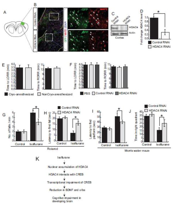
Anesthetics including isoflurane are known to induce neuronal dysfunction in the developing brain, however, the underlying mechanism is mostly unknown. The transcriptional activation of CREB (cyclic AMP response element binding protein) and the alterations in acetylation of histones modulated by several histone deacetylases such as HDAC4 (histone deacetylase 4) are known to contribute to synaptic plasticity in the brain. Here we have shown that administration of isoflurane (1.4%) for 2h leads to transcriptional inactivation of CREB which results in loss of dendritic outgrowth and decreased expression level of proteins essential for memory and cognitive functions, such as BDNF, and c-fos in the developing brain of mice at postnatal day 7 (PND7). To elucidate the molecular mechanism, we found that exposure to isoflurane leads to an increase in nuclear translocation of HDAC4, which interacts with CREB in the nucleus. This event, in turn, results in a decrease in interaction between an acetyltransferase, CBP, and CREB that ultimately leads to transcriptional inactivation of CREB. As a result, the expression level of BDNF, and c-fos were significantly down-regulated after administration of isoflurane in PND7 brain. Depletion of HDAC4 in PND7 brain rescues the transcriptional activation of CREB along with augmentation in the level of the expression level of BDNF and c-fos. Moreover, administration of lentiviral particles of HDAC4 RNAi in primary neurons rescues neurite outgrowth following isoflurane treatment. Taken together, our study suggests that HDAC4-induced transcriptional inactivation of CREB is responsible for isoflurane-induced cognitive dysfunction in the brain.

Keywords: CREB; HDAC; Isoflurane; Synapse.


Figures

Figure 1
Figure 1. Treatment with isoflurane decreases neurite outgrowth
(A) Primary neurons were overexpressed with GFP and treated with isoflurane. Neurons were imaged by confocal microscopy to monitor dendritic morphology. (B–C) Total (B) and average (C) dendritic length were measured after isoflurane treatment. (D) Branch point/cell analysis in neurons after isoflurane treatment. *p<0.05, n=9, one-way ANOVA, mean ± Standard Error Mean.
Figure 2
Figure 2. Treatment with isoflurane affects transcriptional activation of CREB
(A) Mice were exposed to isoflurane, and protein levels of bdnf and c-fos were measured by Western blot analysis. The protein levels were quantitated using image J software. (B–C) Real-time RT-PCR analysis was performed to measure the mRNA level of bdnf (B) and cfos (C) in primary neurons treated with isoflurane. *p<0.05, n=5, one-way ANOVA, mean ± Standard Error Mean. (D) ChIP assay was performed using lysates isolated from isoflurane-treated brain (cortex) lysates. The images are shown as inverted of the original images. Quantitative measurement of the intensity of PCR product after ChIP analysis. *p<0.05, n=5, one-way ANOVA, mean ± Standard Error Mean.
Figure 3
Figure 3. Decrease in phosphorylation of HDAC4 is responsible for its nuclear localization after isoflurane treatment
(A) Primary neurons were treated with isoflurane with or without treatment of TSA (100 nM) or sodium butyrate (20 mM). CREB binding to the bdnf promoter was monitored by ChIP assay. (B) Quantitative analysis of CREB binding to the bdnf promoter. *p<0.05, n=5, one-way ANOVA, mean ± Standard Error Mean. (C) Nuclear and cytosolic fractions were isolated after treatment with isoflurane in mice. The protein level of HDAC4, 5 and 9 was monitored in each fraction by western blot hybridization. Quantitative analysis of protein level of HDAC4, 5 and 9 in both nuclear and cytosolic fraction after isoflurane treatment. *p<0.05, n=5, one-way ANOVA, mean ± Standard Error Mean. (D) Confocal microscopic analysis to monitor HDAC4 in primary neurons after treatment with isoflurane. (E) The phosphorylation level of HDAC4 at its Ser632 and Ser246 residues were monitored by Western blot analysis in lysates isolated from primary neuron culture treated with or without isoflurane. Quantitation of phosphorylation of HDAC4 level after isoflurane treatment. *p<0.05, n=5, one-way ANOVA, mean ± Standard Error Mean.
Figure 4
Figure 4. HDAC4 interacts with CREB
(A) After isoflurane treatment, protein lysates were used for immunoprecipitation (IP) using anti-acetyl lysine antibody and protein level of H3 and H4 were measured by Western blot analysis. (B) Mice were treated with isoflurane and cortical lysates were precipitated with the anti-HDAC4 antibody. The protein level of CREB was monitored by Western blot hybridization. Quantitative analysis of protein intensity after coIP to analyze HDAC4 interaction with CREB. *p<0.05, n=5, one-way ANOVA, mean ± Standard Error Mean. (C) Confocal microscopic analysis of HDAC4 and CREB after isoflurane treatment in primary neurons. Arrowhead indicates co-localization of HDAC4 and CREB. (D) Mice were treated with isoflurane in a dose-dependent manner and interaction between HDAC4 and CREB was measured by co-IP studies. (E) Mice were treated with isoflurane and cortical lysates were precipitated with the anti-CBP antibody. The protein level of CREB was monitored by Western blot hybridization. Quantitative analysis of protein intensity after coIP to analyze HDAC4 interaction with CREB. *p<0.05, n=5, one-way ANOVA, mean ± Standard Error Mean. (F) Phosphorylation of CREB at residue S133 was monitored by Western blot analysis. *p<0.05, n=6, one-way ANOVA, mean ± Standard Error Mean.
Figure 5
Figure 5. Depletion of HDAC4 rescues transcriptional activity of CREB following exposure of isoflurane
(A) Depletion of HDAC4 after administration of HDAC4 RNAi in the cortex was confirmed by Western blot hybridization in both treated and untreated brain lysates. Quantitative analysis of HDAC4 level after administration of HDAC4 RNAi in the cortex. *p<0.05, n=5, one-way ANOVA, mean ± Standard Error Mean. (B) Co-IP analysis to measure binding between CREB and CBP in cortical lysates isolated from either HDAC4 RNAi or control RNA-treated brain lysates. Quantitative analysis of the intensity of CREB protein in cortex depletion of HDAC4 following isoflurane treatment. *p<0.05, n=5, one-way ANOVA, mean ± Standard Error Mean. (C) ChIP assay to monitor CREB binding to bdnf promoter in cortical lysates isolated from either HDAC4 RNAi or control RNA treated brain lysates. Quantitative analysis of CREB binding to bdnf promoter after depletion of HDAC4 following isoflurane treatment. *p<0.05, n=5, one-way ANOVA, mean ± Standard Error Mean. (D) Western blot analysis to measure protein level of BDNF, cfos, and Arc in cortical lysates isolated from either HDAC4 RNAi or control RNA treated brain lysates. (E) Time to measure LORR and RORR after exposure of isoflurane with or without TSA treatment. (F) Cells were treated with isoflurane in a dose-dependent manner prior to depletion of either HDAC4 in cells. mRNA level of both bdnf and cfos were measured by quantitative RT-PCR analysis. *p<0.05, n=5, one-way ANOVA, mean ± Standard Error Mean.
Figure 6
Figure 6. Depletion of HDAC4 rescues neurite outgrowth following exposure of isoflurane
(A–B) confocal microscopic images of primary neurons after depletion of HDAC4 prior to treatment with isoflurane. (C–D) Measurement of total (C) and average (D) dendritic length after depletion of HDCA4 in primary neurons following treatment with isoflurane. (E) Branch point per cell was measured in primary neurons overexpressing either HDAC4 RNAi or control RNAi following treatment with isoflurane. *p<0.01, n=9, one-way ANOVA, mean ± Standard Error Mean.
Figure 7
Figure 7. Depletion of HDAC4 rescues cognitive impairment after isoflurane exposure in mice
(A) Cartoon of a brain slice including the location of administration of lentiviral particles in the cortex. (B) Confocal microscopic analysis of the cortex of PND6 brain where lentiviral particles of either control RNAi or HDAC4 RNAi were administered through intracortical injection. Lentiviral particles were stained with GFP and neuronal cells were stained with MAP2. (C–D) Western blot analysis (C) and quantitative analysis of HDAC4 level (D) of HDAC4 after administration of lentiviral particles of either HDAC4 or control RNAi in mice. *p<0.05, n=7, one-way ANOVA, mean ± Standard Error Mean. (E) Time to LORR and RORR of cryo-anesthetized mice after exposure of isoflurane at PND7. (F) Time to LORR and RORR after exposure of isoflurane following administration of either PBS, control RNAi or HDAC4 RNAi. (G–H) Motor coordination was measured in both control and HDAC4 RNAi mice after treatment with isoflurane. A number of falls/min (G) and latency to first fall (H) were measured in both groups of mice. (I–J) Latency to find the platform (I) and time spent in the right quadrant (J) in MWM test were measured for both groups of mice. *p<0.05, n=10–12, two-way ANOVA, mean ± Standard Error Mean. (K) A model representing how isoflurane leads to cognitive dysfunction by impairment of CREB. In control cells, HDAC4 is mostly localized to the cytosol. After treatment with isoflurane, HDAC4 translocates to the nucleus and interacts with CREB, which results in impairment of transcriptional activation of CREB. As a result, mRNA levels of genes such as bdnf and cfos were decreased which ultimately leads to cognitive dysfunction in the developing brain.

Comment in

  • Anesthetics and epigenetics.
    Sen N. Sen N. Oncotarget. 2016 Nov 29;7(48):78220-78221. doi: 10.18632/oncotarget.13143. Oncotarget. 2016. PMID: 27829240 Free PMC article. No abstract available.

References

    1. Backs J, Song K, Bezprozvannaya S, Chang S, Olson EN. CaM kinase II selectively signals to histone deacetylase 4 during cardiomyocyte hypertrophy. The Journal of clinical investigation. 2006;116:1853–1864. - PMC - PubMed
    1. Borrelli E, Nestler EJ, Allis CD, Sassone-Corsi P. Decoding the epigenetic language of neuronal plasticity. Neuron. 2008;60:961–974. - PMC - PubMed
    1. Bredy TW, Wu H, Crego C, Zellhoefer J, Sun YE, Barad M. Histone modifications around individual BDNF gene promoters in prefrontal cortex are associated with extinction of conditioned fear. Learning & memory. 2007;14:268–276. - PMC - PubMed
    1. Briner A, De Roo M, Dayer A, Muller D, Habre W, Vutskits L. Volatile anesthetics rapidly increase dendritic spine density in the rat medial prefrontal cortex during synaptogenesis. Anesthesiology. 2010;112:546–556. - PubMed
    1. Cardinaux JR, Notis JC, Zhang Q, Vo N, Craig JC, Fass DM, Brennan RG, Goodman RH. Recruitment of CREB binding protein is sufficient for CREB-mediated gene activation. Molecular and cellular biology. 2000;20:1546–1552. - PMC - PubMed

MeSH terms