. 2016 Nov;110(Pt A):438-448.
doi: 10.1016/j.neuropharm.2016.08.015. Epub 2016 Aug 18.

An interplay between the serotonin transporter (SERT) and 5-HT receptors controls stimulus-secretion coupling in sympathoadrenal chromaffin cells

Affiliations

An interplay between the serotonin transporter (SERT) and 5-HT receptors controls stimulus-secretion coupling in sympathoadrenal chromaffin cells

Rebecca L Brindley et al. Neuropharmacology. 2016 Nov.

Abstract

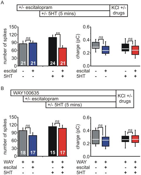
Adrenal chromaffin cells (ACCs), the neuroendocrine arm of the sympathetic nervous system, secrete catecholamines to mediate the physiological response to stress. Although ACCs do not synthesize 5-HT, they express the serotonin transporter (SERT). Genetic variations in SERT are linked to several CNS disorders but the role(s) of SERT/5-HT in ACCs has remained unclear. Adrenal glands from wild-type mice contained 5-HT at ≈ 750 fold lower abundance than adrenaline, and in SERT(-/-) mice this was reduced by ≈80% with no change in catecholamines. Carbon fibre amperometry showed that SERT modulated the ability of 5-HT1A receptors to inhibit exocytosis. 5-HT reduced the number of amperometric spikes (vesicular fusion events) evoked by KCl in SERT(-/-) cells and wild-type cells treated with escitalopram, a SERT antagonist. The 5-HT1A receptor antagonist WAY100635 blocked the inhibition by 5-HT which was mimicked by the 5-HT1A agonist 8-OH-DPAT but not the 5-HT1B agonist CP93129. There was no effect on voltage-gated Ca(2+) channels, K(+) channels, or intracellular [Ca(2+)] handling, showing the 5-HT receptors recruit an atypical inhibitory mechanism. Spike charge and kinetics were not altered by 5-HT receptors but were reduced in SERT(-/-) cells compared to wild-type cells. Our data reveal a novel role for SERT and suggest that adrenal chromaffin cells might be a previously unrecognized hub for serotonergic control of the sympathetic stress response.

Keywords: 5-HT receptor; 8-OH-DPAT (PubChem CID: 6917794); Adrenal chromaffin cell; Amperometry; CP93129 (PubChem CID: 124007); Calcium channel; Calcium imaging; Catecholamine; Escitalopram (PubChem CID: 146571); Exocytosis; GPCR; Gallein (PubChem CID: 73685); Serotonin hydrochloride (PubChem CID: 160436); Serotonin transporter; WAY100635 (PubChem CID: 11957721).


Figures

Figure 1
Figure 1. 5-HT was dramatically reduced but catecholamines were unaltered in the adrenal gland of SERT−/− mice
The monoamine content of whole adrenal glands from wild-type (WT; n = 9 mice) or SERT−/− mice (KO; n = 9) was determined using HPLC. A) 5-HT, adrenaline (Adr), noradrenaline (NA), and dopamine content expressed as nmoles / mg total protein (box denotes interquartile range, line denotes median, whiskersdenote standard deviation). The amounts of 5-HT and dopamine were much lower than adrenaline / noradrenaline (note change in scale). Glands from SERT−/− mice had dramatically reduced 5-HT but no change in catecholamines (*** P < 0.0001; ns = not significantly different: Adr, P = 0.39; NA, P = 0.65; dopamine, P = 0.07; unpaired t-tests). B) The molar ratio of each monoamine relative to adrenaline (mean ± sem). Note the change in scale due to the much lower relative abundance of dopamine and 5-HT (ns, P = 0.84; ** P = 0.004; *** P < 0.0001; unpaired t-tests).
Figure 2
Figure 2. The number of amperometric spikes evoked by KCl was modulated by 5-HT in a SERT-dependent manner
A) Chromaffin cells isolated from wild-type or SERT−/− mice were pretreated for 5 minutes in the absence (ctl) or presence of 5-HT (25nM or 1μM) before secretion was evoked by 30mM KCl (60 s stimulation). Amperometry recordings are shown from a representative wild-type (left) and SERT −/− chromaffin cell (right trace), both in the presence of 1μM 5-HT. Some examples of individual spikes from a wild-type cell are shown on an expanded time scale. B) The total number of spikes evoked by KCl in wild-type cells was not altered by 5-HT (F = 0.32, P = 0.73. one-way ANOVA). C) Time-course of secretion (cumulative number of amperometric spikes grouped by 10s bins) from wild-type cells in the absence (ctl) or presence of 5-HT (1μM) (mean ± sem) (for 5-HT, F = 0.69, P = 0.41, two-way ANOVA). D) The total number of spikes evoked by KCl in SERT −/− cells was significantly reduced by 5-HT (F = 4.43, P = 0.01. one-way ANOVA; “ns” denotes not significant, ** P < 0.01, Bonferroni multiple comparison test compared to control). E) Time-course of secretion from SERT −/− cells in the absence (ctl) or presence of 5-HT (1μM) (for 5-HT, F = 8.17, P = 0.006, two-way ANOVA).
Figure 3
Figure 3. SERT genotype, but not the presence of 5-HT, altered the charge and kinetics of amperometric spikes
Amperometry spike parameters were analyzed from the same cells as figure 2 (control conditions or in the presence of 1μM 5-HT). The median spike parameters for each cell were calculated and shown in the box plots (25%, median, 75%; whiskers denote 10% and 90%). Statistical comparison was performed using two-way ANOVA (genotype and 5-HT) with pairwise comparison using Bonferroni post-tests. The number of cells in each group was: wild-type control = 24; wild-type 5-HT = 22; SERT−/− control = 35; SERT−/− 5-HT = 27. A) Spike charge was smaller in SERT−/− cells than in wild-type cells, but was not altered by 5-HT (two-way ANOVA: for genotype F = 42.2, *** P < 0.0001; for 5-HT F = 0.01, P = 0.94) (“ns” not significant, Bonferroni post-test). B) Spike amplitude was smaller in SERT−/− cells than in wild-type cells, but was not influenced by 5-HT (two-way ANOVA: for genotype F = 19.5, *** P < 0.0001; for 5-HT F = 0.77, P = 0.38) (“ns” not significant, Bonferroni post-test). C) The half-width (spike duration at half maximal amplitude) was shorter in SERT−/− cells than in wild-type cells and 5-HT had a modest influence (two-way ANOVA: for genotype F = 16.48, *** P < 0.0001; for 5-HT F = 4.55, P = 0.04). Pairwise comparison using Bonferroni post-tests found no statistically significant difference between control and 5-HT treated cells in either genotype (“ns” not significant, Bonferroni post-test). D) Spike slope was not significantly altered by genotype or 5-HT (two-way ANOVA: for genotype F = 0.28, P = 0.60; for 5-HT F = 2.81, P = 0.10) (“ns” not significant, Bonferroni post-test).
Figure 4
Figure 4. SERT function opposed a 5-HT receptor-mediated inhibition of secretion in wild-type chromaffin cells
The experimental paradigm is depicted at the top of each panel. A) Left Panel: Escitalopram (1μM) alone had no effect on the number of amperometric spikes evoked by KCl, but combined with 5-HT (25nM) produced a significant inhibition (F = 3.42, P = 0.02, one-way ANOVA; “ns” not significant, ** P < 0.01, Bonferroni post-tests). Right Panel: Spike charge was not significantly altered by drug treatments (P = 0.22, Kruskal-Wallis test; “ns” not significant, Dunn’s multiple comparison post-test). B) The same experiment was repeated in the presence of the 5-HT1A receptor antagonist WAY100635 (25nM). Left Panel: WAY100635 prevented the inhibition of spike number produced by escitalopram + 5-HT (F = 1.55, P = 0.21, one-way ANOVA; “ns” not significant, Bonferroni post-test). Right Panel: Spike charge was not significantly altered by drug treatments in the presence of WAY100635 (P = 0.61, Kruskal-Wallis test; “ns” not significant, Dunn’s multiple comparison post-test).
Figure 5
Figure 5. The 5-HT1A receptor agonist 8-OH-DPAT inhibited the number of evoked amperometric spikes
The upper panel depicts the experimental paradigm. Cells were perfused with NaCl-based extracellular solution in the absence (control) or presence of 8-OH-DPAT (a 5-HT1A receptor agonist) or CP93129 (5-HT1B receptor agonist). Secretion was then evoked by perfusing the cells for 60s with KCl in the continued absence (controls) or presence of the agonists. A) The 5-HT1A receptor agonist 8-OH-DPAT (300 nM) significantly reduced the number of amperometry spikes evoked by KCl in wild-type cells, but the 5-HT1B receptor agonist CP93129 (300 nM) had no effect (F = 6.22, P = 0.003, one-way ANOVA) (“n.s” not significant; ** P < 0.01 for multiple pairwise comparisons to control cells using Dunnett’s post-test). B) 8-OH-DPAT significantly reduced the number of KCl-evoked spikes in SERT−/− chromaffin cells, (* P = 0.024, unpaired t-test). C) Spike charge was not significantly altered by 8-OH-DPAT or CP93129 in wild-type cells (P = 0.70, Kruskal-Wallis test; “ns” not significant, Dunn’s multiple comparison post-test). D) Spike charge was not significantly altered by 8-OH-DPAT in SERT−/− cells (P = 0.26, Mann-Whitney test).
Figure 6
Figure 6. Voltage-gated Ca2+channel or K+ channel currents were not modulated by 5-HT or the 5-HT1A receptor agonist 8-OH-DPAT
A) Amphotericin perforated whole-cell patch clamp recording of voltage-gated Ca2+ channel currents (ICa). Peak amplitude of ICa is plotted against time from a representative wild-type cell. The inset shows ICa corresponding to the numbered points. 8-OH-DPAT (DPAT) (300 nM) had no effect on ICa, but ATP (100μM) (a P2Y purinergic receptor agonist) reversibly inhibited ICa. B) Pooled data (mean ± sem) showing the fractional change in ICa amplitude for wild-type cells (n = 6) and SERT−/− cells (n = 5). 8-OH-DPAT had no effect while ATP significantly inhibited ICa in wild-type cells (F = 30.47, P < 0.0001, repeated measures ANOVA; “ns”, not significant, *** P < 0.0001 using Bonferroni post-test) and SERT−/− cells (F = 20.47, P < 0.0001, repeated measures ANOVA; “ns”, not significant, *** P < 0.0001 using Bonferroni post-test). C) Pooled data (mean ± sem) showing the fractional change in whole-cell ICa amplitude for wild-type cells (WT) exposed to 1μM 5-HT (n = 5) or 5-HT + escitalopram (1μ M) (n = 4) and SERT−/− cells exposed to 1μ M 5-HT(n = 8). None of the drugs produced a significant change in ICa amplitude (ns, not significant compared to within cell controls, paired t-test). D) Representative amphotericin perforated whole-cell patch clamp recording of K+ channel currents (IK) from a wild-type chromaffin cell. IK were evoked by steps to +80mV in the absence (control) and then in the presence of 8-OH-DPAT (300 nM). E) Current density plotted against voltage (inset shows stimulus protocol) showing no effect of 8-OH-DPAT (two-way repeated measures ANOVA; for 8-OH-DPAT treatment F = 0.11, P = 0.75; n = 6).
Figure 7
Figure 7. 5-HT did not alter resting or KCl-evoked intracellular [Ca2+]
Time-course showing intracellular [Ca2+] monitored in FURA-2 loaded chromaffin cells isolated from wild-type mice (mean ± sem; n = 61 cells). The experimental paradigm mimicked that used to evoke catecholamine secretion in previous amperometry experiments (60s stimulation with 30mM KCl after 5 min exposure to 1μM 5-HT). Labels: “a” = baseline before application of 5-HT; “b” = baseline at the end of the 5 min drug exposure; “peak” = maximal response to KCl; “end” = response at the end of KCl stimulation. B) Left panel: 5-HT had no effect on baseline [Ca2+]. Intracellular [Ca2+] before (“a”) and during (“b”) 5-HT exposure for three experimental groups (see labels “a” and “b” on panel-A): controls (0μM 5-HT; n = 53; “ns” denotes P = 0.24, paired t-test); 1μM 5-HT (n = 61; “ns” denotes P = 0.39, paired t-test); 10μM 5-HT (n = 28; “ns” denotes P = 0.06, paired t-test). Right panel: 5-HT did not significantly change [Ca2+] at the “peak” or “end” of the KCl response (“ns” denotes not significant, one-way ANOVA: “Peak” response, F = 0.56, P = 0.58; “End” response, F = 2.93, P = 0.06). C) The same experimental paradigm as in panels A and B except cells were exposed to 1μM escitalopram (escital n = 30) or a combination of 1μM escitalopram + 1μM 5-HT (escit + 5-HT, n = 38) for 5 minutes prior to KCl. Left panel: Baseline [Ca2+] was not altered by escitalopram (P = 0.62, paired t-test) or by escitalopram + 5-HT (P = 0.99, paired t-test). Right panel: Intracellular [Ca2+] at the “peak” and “end” of the KCl response in the presence of escitalopram alone or escitalopram ± 5-HT (“ns”, no significant difference, paired t-test; peak, P = 0.06; end, P = 0.22).
Figure 8
Figure 8. The 5-HT receptor-mediated inhibition of secretion was prevented by gallein, an antagonist of Gβγ signaling
A) Depicts the experimental paradigm. Chromaffin cells from wild-type mice were pretreated with DMSO (vehicle controls) or gallein (10 μ M) and stimulated with 30mM KCl in the absence or presence of 8-OH-DPAT (300 nM). Secretion was detected using carbon fiber amperometry. B) In DMSO treated cells, 8-OH-DPAT (DPAT) significantly inhibited the number of amperometric spikes evoked by KCl compared to matched controls (ctl) (* P = 0.01, unpaired t-test). There was no effect on spike charge (“ns” P = 0.57, Mann-Whitney test). C) Gallein treatment prevented the inhibition of spike number by 8-OH-DPAT (“ns”, P = 0.19, unpaired t-test). There was no change in spike charge (“ns” P = 0.56, Mann-Whitney test).

Similar articles

Cited by

References

    1. Albillos A, Gandia L, Michelena P, Gilabert JA, del Valle M, Carbone E, Garcia AG. The mechanism of calcium channel facilitation in bovine chromaffin cells. J Physiol. 1996;494(3):687–695. - PMC - PubMed
    1. Baganz NL, Blakely RD. A dialogue between the immune system and brain, spoken in the language of serotonin. ACS Chem Neurosci. 2013;4:48–63. - PMC - PubMed
    1. Bayliss DA, Li YW, Talley EM. Effects of serotonin on caudal raphe neurons: activation of an inwardly rectifying potassium conductance. J Neurophysiol. 1997a;77:1349–1361. - PubMed
    1. Bayliss DA, Li YW, Talley EM. Effects of serotonin on caudal raphe neurons: inhibition of N- and P/Q-type calcium channels and the afterhyperpolarization. J Neurophysiol. 1997b;77:1362–1374. - PubMed
    1. Bengel D, Murphy DL, Andrews AM, Wichems CH, Feltner D, Heils A, Mossner R, Westphal H, Lesch KP. Altered brain serotonin homeostasis and locomotor insensitivity to 3, 4-methylenedioxymethamphetamine ("Ecstasy") in serotonin transporter-deficient mice. Mol Pharmacol. 1998;53:649–655. - PubMed

Publication types

MeSH terms