

Acral Melanoma in Chinese: A Clinicopathological and Prognostic Study of 142 cases
- PMID: 27545198
- PMCID: PMC4992860
- DOI: 10.1038/srep31432
Acral Melanoma in Chinese: A Clinicopathological and Prognostic Study of 142 cases
Abstract
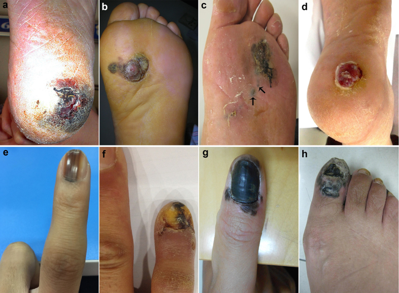
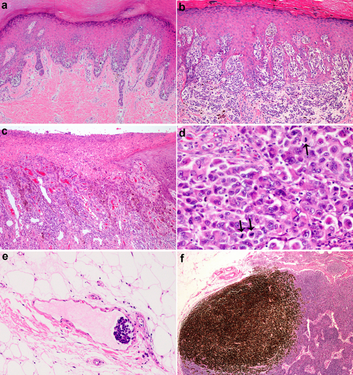
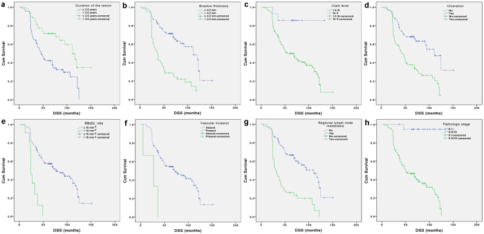
Acral melanoma (AM), as a peculiar subgroup of melanoma, is rare in Caucasians but has higher incidence in Asians. Large series of study on AM with clinicopathological features and prognostic factors is still limited, especially in Asian population. We retrospectively collected clinical, pathological and follow-up data of 142 AM cases. All patients were Chinese, with the age ranging from 24 to 87 years (mean 62.0; median 62.0). The Breslow thickness of primary lesions ranged from 0.6 to 16.3 mm (mean 4.9; median 3.7). 85.9% of the patients had acral lentiginous histologic subtype. Plantar was the most frequently involved site, followed by heels. Statistically, duration of the lesion before diagnosis (≤2.5 years), Breslow thickness >4.0 mm (T4), high mitotic index (>15 mm(-2)), presence of vascular invasion, regional lymph node metastasis at diagnosis and pathologic stage (II/III/IV) were found to be independent prognostic factors in both univariate and multivariate analyses. The prognosis of AM in Chinese is extremely poor. Our 5- and 10-year disease-specific survival (DSS) rates were 53.3% and 27.4%, respectively. Therefore, AM in Asians represents a more biologically aggressive melanoma subtype and is thought to carry a worse prognosis when compared with other races or cutaneous melanomas in other anatomic sites.
Figures
References
-
- Dai B. et al.. Analysis of KIT expression and gene mutation in human acral melanoma: with a comparison between primary tumors and corresponding metastases/recurrences. Human pathology 44, 1472–1478 (2013). - PubMed
-
- Cormier J. N. et al.. Ethnic differences among patients with cutaneous melanoma. Archives of internal medicine 166, 1907–1914 (2006). - PubMed
-
- Shaw J. H. & Koea J. B. Acral (volar-subungual) melanoma in Auckland, New Zealand. The British journal of surgery 75, 69–72 (1988). - PubMed
-
- Cascinelli N. et al.. Acral lentiginous melanoma. A histological type without prognostic significance. The Journal of dermatologic surgery and oncology 20, 817–822 (1994). - PubMed
-
- Ridgeway C. A., Hieken T. J., Ronan S. G., Kim D. K. & Das Gupta T. K. Acral lentiginous melanoma. Archives of surgery 130, 88–92 (1995). - PubMed
Publication types
MeSH terms
LinkOut - more resources
Full Text Sources
Other Literature Sources
Medical
