. 2016 Aug 25;166(5):1176-1187.e14.
doi: 10.1016/j.cell.2016.08.003. Epub 2016 Aug 18.

Cellular Cholesterol Directly Activates Smoothened in Hedgehog Signaling

Affiliations

Cellular Cholesterol Directly Activates Smoothened in Hedgehog Signaling

Pengxiang Huang et al. Cell. .

Abstract

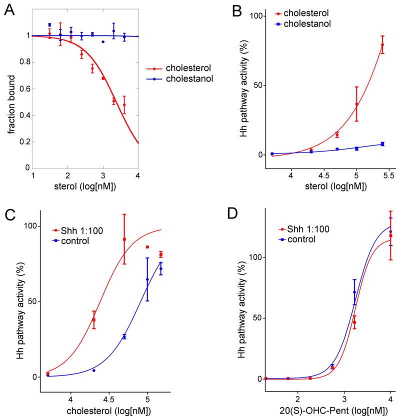
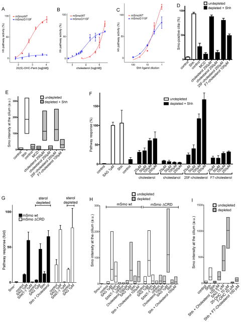
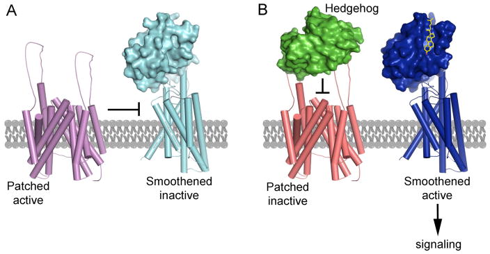
In vertebrates, sterols are necessary for Hedgehog signaling, a pathway critical in embryogenesis and cancer. Sterols activate the membrane protein Smoothened by binding its extracellular, cysteine-rich domain (CRD). Major unanswered questions concern the nature of the endogenous, activating sterol and the mechanism by which it regulates Smoothened. We report crystal structures of CRD complexed with sterols and alone, revealing that sterols induce a dramatic conformational change of the binding site, which is sufficient for Smoothened activation and is unique among CRD-containing receptors. We demonstrate that Hedgehog signaling requires sterol binding to Smoothened and define key residues for sterol recognition and activity. We also show that cholesterol itself binds and activates Smoothened. Furthermore, the effect of oxysterols is abolished in Smoothened mutants that retain activation by cholesterol and Hedgehog. We propose that the endogenous Smoothened activator is cholesterol, not oxysterols, and that vertebrate Hedgehog signaling controls Smoothened by regulating its access to cholesterol.


Figures

Figure 1
Figure 1
Structure of a SmoCRD-oxysterol complex. (A) Ribbon model showing overall structure of XSmoCRD (navy) in complex with 20(S)-OHC (yellow). Disulfide bonds (green) and the four helices are numbered. (B) Contacts mediating 20(S)-OHC recognition. Hydrogen bonds involving the two hydroxyls of oxysterol (3β-OH bonded to D68 and 20-OH bonded to E133) are shown as dashed lines. See also fig. S2D–E for all Smo-sterol contacts. (C) Shape complementarity of Smo sterol-binding site to 20(S)-OHC. Electron density of 20(S)-OHC (yellow oxysterol surrounded by red mesh, Fo-Fc omit map contoured at 3s) is shown with the molecular surface of the sterol-binding site (blue). See also fig. S2F and S2H for binding site volume. (D) Comparison between recognition of 20(S)-OHC by XSmoCRD and recognition of Xenopus Wnt8 palmitoyl moiety by mouse Fz8CRD (PDB ID 4F0A). Close up view of sterol-binding site of XSmoCRD (navy), superimposed on palmitate-binding site of mFz8CRD (cyan). 20(S)-OHC (yellow) and palmitate (orange) occupy topologically equivalent sites. Two bulky residues in Helix 3 of mFz8, Y125 and F127, cause the palmitate-binding groove to be significantly narrower than the sterol-binding groove in XSmo. Additionally, the two charged residues responsible for polar recognition of oxysterols by XSmo, D68 and E133, are absent in mFz8. See also fig. S2F–I for comparison of ligand binding site volume between XSmoCRD and mFz8CRD.
Figure 2
Figure 2
Oxysterol recognition by Smo. (A) D99 and E164, the two mSmo residues that hydrogen bond with 3β-OH and 20(S)-OH, are critical for oxysterol binding. Full-length proteins expressed in 293T cells were assayed by binding to 20(S)-OHC affinity matrix (Nedelcu et al., 2013), in the absence or presence of free 20(S)-OHC competitor. (B) Smo-null cells rescued with mSmoD99A do not respond to 20(S)-OHC-Pent, in contrast to cells rescued with wild type mSmo. Hh pathway activity was assayed by qPCR for endogenous Gli1, and is shown normalized to activation elicited by saturating levels of SAG (0.5 μM). Error bars indicate standard deviation (n=3). (C) As in (A), but treating cells with various concentrations of Shh. MSmoD99A does not respond to stimulation by Shh. (D) As in (A), but with Smo-null cells rescued with mSmoE164L. The mutant has reduced responsiveness to oxysterols. (E) mSmoG115S has reversed distereospecificity, responding stronger to 20(R)-OHC-Pent (EC50=1.3 μM) than to 20(S)-OHC-Pent (EC50=23.4 μM). See also oxysterol binding assays in fig. S3. (F) In contrast, wild-type mSmo is activated preferentially by 20(S)-OHC-Pent (EC50=0.84 μM) compared to 20(R)-OHC-Pent (EC50=26.3 μM). Experiment in panels E-I were performed in parallel, and the curve for wild-type mSmo is shown in E, G and I.
Figure 3
Figure 3
Sterol binding induces SmoCRD conformational change. (A) Close-up view of the sterol-binding site in unliganded XSmoCRD. The indicated amino acids are involved in sterol binding. See also fig. S4C for overall structure of unliganded XSmoCRD. (B) As in (A), but with unliganded XSmoCRD (cyan) superimposed on 20(S)-OHC-bound XSmoCRD (navy). In absence of sterol, the binding site is in “open” state. 20(S)-OHC-induced conformational change results in “closed” state of the binding site. (C) Ribbon diagram showing overall structure of unliganded XSmoCRD (cyan), superimposed on 20(S)-OHC-bound XSmoCRD (navy). Binding of 20(S)-OHC (yellow) induces inward rotation of helix H3’ (top right panel) and dramatic positional swap of residues in following loop (bottom right panel). See also fig. S4D for view from different angle.
Figure 4
Figure 4
Sterol-induced conformational change activates Smo. (A) Cyclopamine bound in XSmoCRD sterol-binding site. Cyclopamine OH group is recognized by the same network of hydrogen bonds (dashed lines) as 3β-OH group of 20(S)-OHC. See also fig. S5A for overall structure. (B) Superimposition of cyclopamine-bound (green) and 20(S)-OHC-bound (navy) XSmoCRD. Cyclopamine (orange) induces a protein conformation very similar to that induced by 20(S)-OHC (yellow). See also fig. S5B for a close-up view of binding site. (C) Cyclopamine (100 μM, as soluble complex with methyl-β-cyclodextrin (MCD)) binding to CRD activates mSmoD477G/E522K, a mutant in which the cyclopamine-binding site in 7TM is destroyed. MSmoD477G/E522K also responds to 20(S)-OHC-Pent (10 μM), cholesterol (250 μM, as MCD complex), and Shh, but not SAG (0.5 μM), which binds the 7TM site. Error bars indicate standard deviation (n=3).
Figure 5
Figure 5
Cholesterol binds Smo and activates Hh signaling. (A) Fluorescence polarization assay showing cholesterol competes binding of BODIPY-cyclopamine to purified XSmo ectodomain, in dose-dependent manner (IC50=2.4 μM). In contrast, the saturated analog, cholestanol, does not bind Smo. See also fig. S6 for additional assays demonstrating binding of cholesterol to XSmoCRD. (B) Cholesterol, but not cholestanol, activates Hh signaling in Smo-null cells rescued with wild-type mSmo. Cholesterol and cholestanol were delivered as MCD complexes. (C) Cholesterol (added as MCD complex) synergizes with Shh to activate Hh signaling in NIH-3T3 cells. See also fig. S7B showing synergy of Shh with cholesterol, but not cholestanol. Error bars indicate standard deviation (n=3). (D) Unlike cholesterol, the oxysterol 20(S)-OHC-Pent does not synergize with Shh in NIH-3T3 cells.
Figure 6
Figure 6
Evidence that cholesterol is endogenous activator of Smo in vertebrate Hh signaling. (A) 20(S)-OHC-Pent does not activate Hh signaling in Smo-null cells rescued with mSmoG115F. In contrast, mSmoG115F responds to cholesterol (B) and to Shh (C). This experiment was performed in parallel with the one in fig. 1E. See fig. S7C–E for similar behavior of mSmoG115S and mSmoG115S/E164L. (D) The indicated sterols (see fig. S1 for structures) were added back as MCD complexes to sterol-depleted NIH-3T3 cells, followed by incubation with Shh. Hh signaling was assayed by measuring ciliary recruitment of endogenous Smo. The graph shows fraction of Smo-positive cilia. Sterol depletion blocks Smo recruitment to cilia in response to Shh, which is rescued by cholesterol, 25F-cholesterol and F7-cholesterol, but not by cholestanol. The MCD control shows effect of adding empty MCD instead of MCD-sterol. Between 300 and 600 cilia were measured per condition. (E) As in (D), but Smo intensity at cilia is displayed as box plots representing the distribution between 25th and 75th percentile, with horizontal line indicating median intensity. (F) As in (D), but Hh signaling was measured by luciferase reporter assay in Shh Light II cells. Error bars indicate standard deviation (n=4 independent experiments). Hh signaling in sterol-depleted cells is rescued by cholesterol, 25F-cholesterol and F7-cholesterol, but not by cholestanol. Cholesterol hydroxylation at C-25 or C-27 is thus not absolutely required for Hh signaling. (G) Smo-null cells, rescued with mSmo or mSmoΔCRD, were sterol-depleted, followed by incubation with indicated agents. Hh pathway activity was measured by qPCR for Gli1 mRNA. Error bars indicate standard deviation (n=3 independent experiments). Signaling by mSmoΔCRD is not inhibited by sterol depletion, in contrast to mSmo. Both mSmo and mSmoΔCRD are inhibited by SANT1, and are activated by SAG, irrespective of sterol depletion. Cholesterol add-back (100 μM MCD complex) rescues mSmo activation by Shh, demonstrating efficiency of sterol depletion. (H) As in (G), but measuring ciliary localization of mSmo and mSmoΔCRD. Shh recruits mSmo to cilia in sterol-dependent manner; in contrast, Shh has no effect on ciliary levels of mSmoΔCRD. Both mSmo and mSmoΔCRD accumulate in cilia in response to SAG, which is unaffected by sterol depletion. Ciliary levels of both proteins are reduced by SANT1. At least 300 cilia were measured per condition. (I) Ciliary localization of endogenous Smo was measured in sterol-depleted NIH-3T3 cells, as in (E). SAG and 20-OHC strongly recruit Smo to cilia in sterol-depleted cells, in contrast to Shh. The inactive oxysterol, 7-hydroxycholesterol (7-OHC), serves as negative control. Between 80 and 140 cilia were measured per condition.
Figure 7
Figure 7
Model for Smo regulation during vertebrate Hh signaling. (A) In the absence of Hh signaling, active Ptch1 maintains Smo inhibited, with its CRD sterol-binding site empty. (B) Upon Shh binding, Ptch1 is inhibited, and cholesterol (yellow) gains access to SmoCRD. Cholesterol-induced conformational change in CRD causes allosteric activation of the 7TM domain of Smo, which, in turn, signals to the cytoplasm. These events occur in primary cilia of vertebrate cells. For simplicity, other ciliary proteins required for Smo signaling, such as EVC proteins (Dorn et al., 2012), are not shown.

References

    1. Aanstad P, Santos N, Corbit KC, Scherz PJ, Trinh le A, Salvenmoser W, Huisken J, Reiter JF, Stainier DY. The extracellular domain of Smoothened regulates ciliary localization and is required for high-level Hh signaling. Current biology : CB. 2009;19:1034–1039. - PMC - PubMed
    1. Alcedo J, Ayzenzon M, Von Ohlen T, Noll M, Hooper JE. The Drosophila smoothened gene encodes a seven-pass membrane protein, a putative receptor for the hedgehog signal. Cell. 1996;86:221–232. - PubMed
    1. Bazan JF, de Sauvage FJ. Structural ties between cholesterol transport and morphogen signaling. Cell. 2009;138:1055–1056. - PubMed
    1. Braberg H, Webb BM, Tjioe E, Pieper U, Sali A, Madhusudhan MS. SALIGN: a web server for alignment of multiple protein sequences and structures. Bioinformatics. 2012;28:2072–2073. - PMC - PubMed
    1. Carroll JN, Pinkerton FD, Su X, Gerst N, Wilson WK, Schroepfer GJ., Jr Sterol synthesis. Synthesis of 3 beta-hydroxy-25,26,26,26,27,27,27-heptafluorocholest-5-en-7-one and its effects on HMG-CoA reductase activity in Chinese hamster ovary cells, on ACAT activity in rat jejunal microsomes, and serum cholesterol levels in rats. Chemistry and physics of lipids. 1998;94:209–225. - PubMed

Publication types