. 2016 Sep 6;24(9):1537-49.
doi: 10.1016/j.str.2016.07.007. Epub 2016 Aug 18.

ALS Mutations Disrupt Phase Separation Mediated by α-Helical Structure in the TDP-43 Low-Complexity C-Terminal Domain

Affiliations

ALS Mutations Disrupt Phase Separation Mediated by α-Helical Structure in the TDP-43 Low-Complexity C-Terminal Domain

Alexander E Conicella et al. Structure. .

Abstract

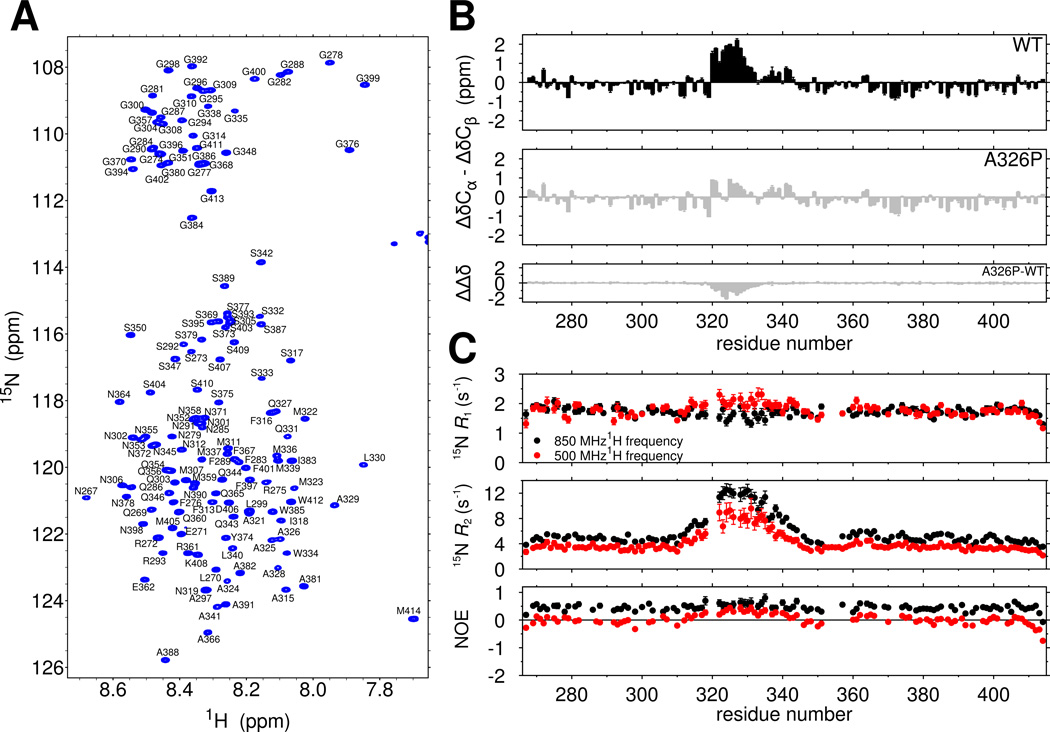
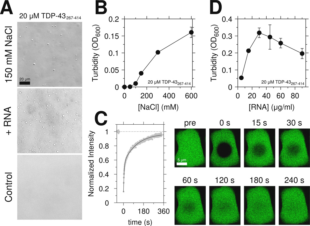
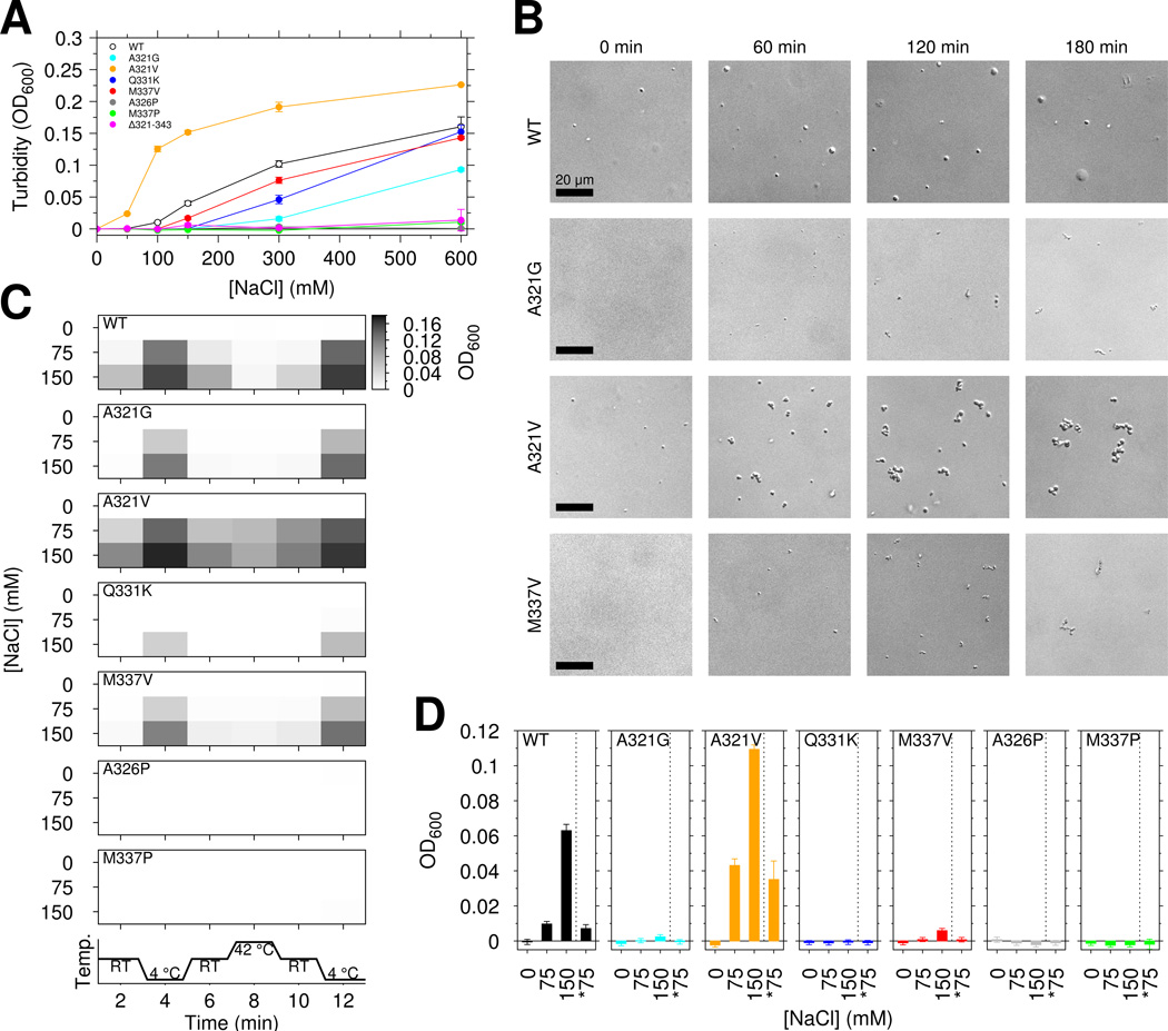
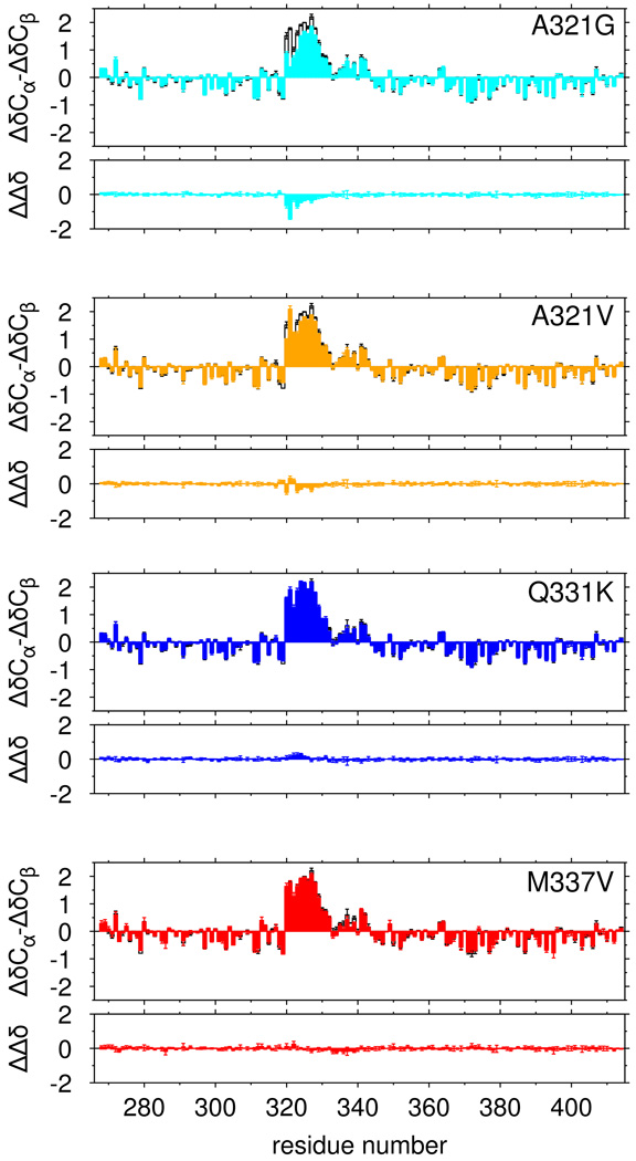
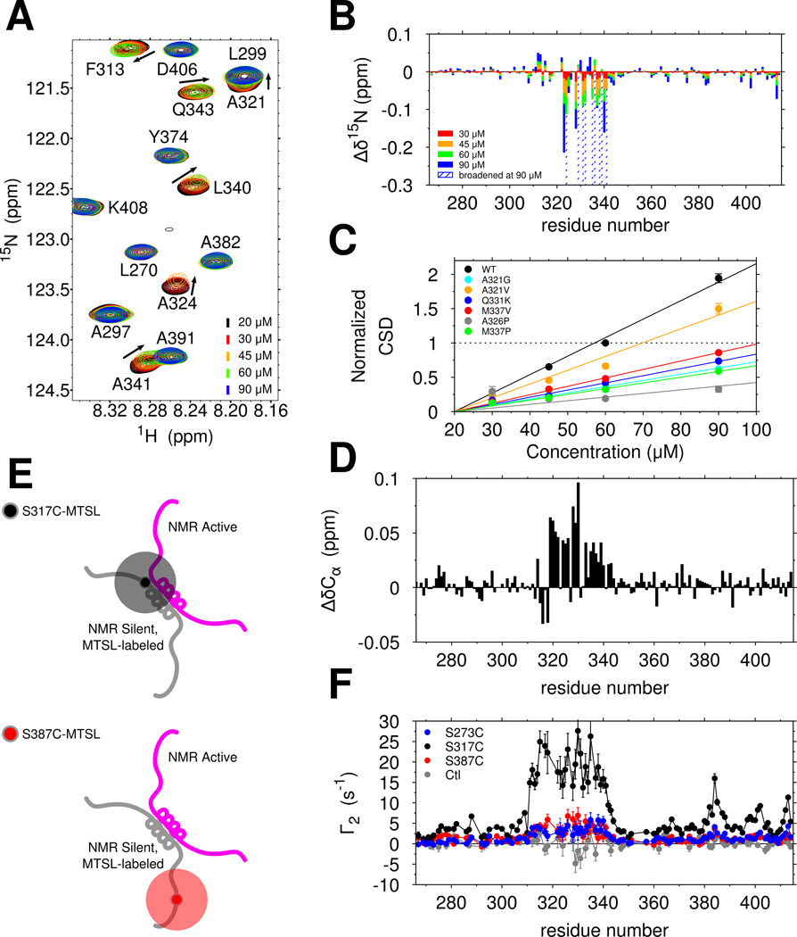
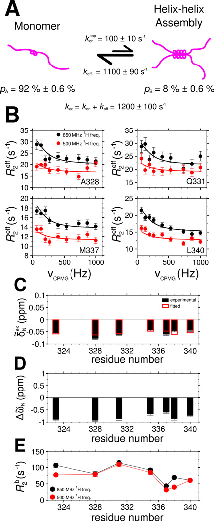
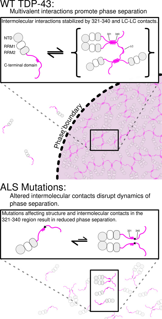
RNA-binding protein TDP-43 mediates essential RNA processing but forms cytoplasmic neuronal inclusions via its C-terminal domain (CTD) in amyotrophic lateral sclerosis (ALS). It remains unclear if aggregated TDP-43 is neurotoxic and if ∼50 ALS-associated missense mutations in TDP-43 CTD promote aggregation, or if loss of normal function plays a role in disease. Recent work points to the ability of related proteins to assemble into functional phase-separated ribonucleoprotein granules via their structurally disordered prion-like domains. Here, we provide atomic details on the structure and assembly of the low-complexity CTD of TDP-43 into liquid-liquid phase-separated in vitro granules and demonstrate that ALS-associated variants disrupt interactions within granules. Using nuclear magnetic resonance spectroscopy, simulation, and microscopy, we find that a subregion cooperatively but transiently folds into a helix that mediates TDP-43 phase separation. ALS-associated mutations disrupt phase separation by inhibiting interaction and helical stabilization. Therefore, ALS-associated mutations can disrupt TDP-43 interactions, affecting function beyond encouraging aggregation.


Figures

Figure 1
Figure 1
The TDP-43 C-terminal domain contains a cooperatively formed α-helix. See also Figure S1. (A) The narrow assigned 1H-15N HSQC of TDP-43267–414 suggests the domain is primarily intrinsically disordered. (B) Secondary shifts (ΔδCα-ΔδCβ) confirm the location of a helical region from 321–330 that is disrupted by a single helix-breaking mutation, A326P, suggesting cooperative secondary structure formation. The difference in secondary shifts for A326P relative to WT is represented as ΔΔδ. (C) 15N spin relaxation parameters suggest slowed reorientation motions for the 321–330 region. Data are plotted as mean ± SD.
Figure 2
Figure 2
Details of the TDP-43 helical subdomain monomeric structure by molecular simulation. See also Figure S2. (A) Correspondence between chemical shift deviations from random coil reference values (top) and 3JHNHα scalar coupling constants predicted from the simulation ensemble of TDP-43310–350 (red) and measured by experiment (black, open circles) demonstrate that the simulation faithfully captures structural character of the helical subdomain. (B) Based on regions of continuous backbone dihedral angles (ϕ,ψ), α-helical structure of TDP-43 peptide shows extended helical structure primarily in the 321 to 334 region. (i–v) Ensemble members and their total population (labeled %) representing populated regions of the α-helix map based on structural clustering. (C) Secondary structure sampled by each position in the TDP-43 simulated ensemble based on DSSP in three classes. Data are plotted as mean ± SEM for simulation data.
Figure 3
Figure 3
The region spanning residues 321 to 340 mediates TDP-43 C-terminal domain liquid phase separation. See also Figure S3 and Movie S1. (A) DIC micrographs of 20 µM TDP-43 CTD liquid-liquid phase separation in the presence of salt (150 mM NaCl, top) or yeast RNA extract (middle). No phase-separation is observed in the control condition. Scale bar is 20 µm. (B) The extent of phase separation increases with increasing salt concentration as monitored by turbidity. (C) Fluorescence recovery curve (and corresponding images) measured for a large TDP-43 C-terminal domain liquid droplet at a total sample concentration of 50 µM. Scale bar is 5 µm. Data are plotted as mean±SD.
Figure 4
Figure 4
ALS mutations affect TDP-43 liquid-liquid phase separation, reversibility, and aggregation. See also Figure S4. (A) TDP-43 CTD ALS variants decrease the salt-dependent turbidity with respect to wild type, except increased turbidity for A321V. Data for wild type are repeated from Figure 3B with open circles for clarity. (B) DIC micrographs from aliquots taken at time points after addition of 150 mM salt to 20 µM CTD suggest that over time CTD ALS variants form assemblies that are morphologically distinct from liquid droplets. Scale bar is 20 µm. (C) Temperature cycling of wild type and mutant CTD immediately after addition of salt modulates sample turbidity, highlighting the dynamic, reversible character of CTD liquid phase separation. (D) CTD liquid-liquid phase separation is also reversible upon dilution of salt. Turbidity of 20 µM CTD was measured for samples prepared in 0, 75, and 150 mM NaCl as well samples prepared at 150 mM NaCl and then diluted to 75 mM NaCl at constant protein concentration (*75, e.g. 1:1 dilution of 20 µM CTD in 150 mM NaCl with addition of 20 µM CTD in 0 mM NaCl). Data are plotted as mean ± SD.
Figure 5
Figure 5
Only select ALS-associated mutations disrupt α-helical structure of TDP-43. Secondary shifts (ΔδCα-ΔδCβ), and the difference in secondary shift relative to WT (ΔΔδ) for ALS-associated mutations A321G (cyan) and A321V (orange) show minor local disruption of helical signatures compared to wild type (black, open bars) while Q331K (blue), and M337V (red) show no significant change in helical structure. Data are plotted as mean ± SD.
Figure 6
Figure 6
The TDP-43 C-terminal domain self-assembles via helix-helix contacts that are disrupted by ALS-associated mutations. See also Figure S4. (A) Overlay of 1H-15N HSQC spectra of TDP-43 C-terminal domain at increasing concentrations show chemical shift differences (arrows) and intensity reduction associated with intermolecular interactions. (B) Interactions between copies of TDP-43 C-terminal domain as probed by large 15N chemical shift differences (Δδ15N) are localized to region 321 to 340. (C) Normalized 15N chemical shift differences (CSDs) are fit to a line and demonstrate that ALS-associated mutations and structure-breaking variants disrupt domain interactions. (D) Positive Cα chemical shift differences (ΔδCα) between 60 µM and 20 µM wild type, consistent with enhanced helical structure, localize to the 321 to 340 region. (E) Labeling scheme for probing transient, intermolecular interactions by paramagnetic relaxation enhancement (PRE). (F) Intermolecular PRE values (Γ2) measured for mixtures of WT TDP-43 with MTSL-labeled S273C (blue), S317C (black) and S387C (red) confirm that 321 to 340 primarily interacts with itself and weakly interacts with other regions. A control experiment (gray, unconjugated MTSL mixed with 15N CTD) confirms that MTSL does directly interact with the 310–340 region.
Figure 7
Figure 7
Characterization of TDP-43 C-terminal domain interaction dynamics by 15N CPMG relaxation dispersion. See also Figure S5. (A) A two-state model for TDP-43 C-terminal domain interaction at 45 µM in which a population of monomers (pA) is in equilibrium with a multimeric assembled state (pB) governed by a global exchange rate kex. (B) Relaxation dispersion profiles and (C) exchange-induced 15N chemical shifts measured (circles, B; filled boxes, C) and calculated (lines, B; open boxes, C) by simultaneous optimization. Best-fit values of model parameters: (D) 15N chemical shift differences (ΔῶN) between the assembled and monomeric state and (E) 15N transverse relaxation in the assembled state (R2b). Data are plotted as mean ± SD.
Figure 8
Figure 8. Model for TDP-43 phase separation driven by the C-terminal domain
In the context of WT TDP-43 (top), the C-terminal domain mediates multimerization via intermolecular contacts in the 321–340 region that taken on additional α-helical structure upon assembly. These intermolecular contacts cooperate with additional interactions between the remainder of the aromatic-rich “prion-like” low-complexity (LC) domain to promote liquid phase separation of TDP-43 via multivalent (helix-helix and LC-LC) contacts. Additional interaction valency stimulating phase separation may be added by LC domain RNA-binding and, in full-length TDP-43, by N-terminal domain multimerization (gray circles) and RNA-binding by tandem RNA recognition motifs (RRM1 and RRM2, gray/white squares). ALS mutations (bottom) that affect structure or intermolecular contacts in the 321–340 region disrupt multivalent interactions and result in altered phase separation. Only the A321V mutation increases phase separation.

Comment in

  • A New Phase in ALS Research.
    Chong PA, Forman-Kay JD. Chong PA, et al. Structure. 2016 Sep 6;24(9):1435-6. doi: 10.1016/j.str.2016.08.003. Structure. 2016. PMID: 27602988

References

    1. Alami NH, Smith RB, Carrasco MA, Williams LA, Winborn CS, Han SS, Kiskinis E, Winborn B, Freibaum BD, Kanagaraj A, et al. Axonal transport of TDP-43 mRNA granules is impaired by ALS-causing mutations. Neuron. 2014;81:536–543. - PMC - PubMed
    1. Amador-Ortiz C, Lin WL, Ahmed Z, Personett D, Davies P, Duara R, Graff-Radford NR, Hutton ML, Dickson DW. TDP-43 immunoreactivity in hippocampal sclerosis and Alzheimer's disease. Ann Neurol. 2007;61:435–445. - PMC - PubMed
    1. Arnold ES, Ling SC, Huelga SC, Lagier-Tourenne C, Polymenidou M, Ditsworth D, Kordasiewicz HB, McAlonis-Downes M, Platoshyn O, Parone PA, et al. ALS-linked TDP-43 mutations produce aberrant RNA splicing and adult-onset motor neuron disease without aggregation or loss of nuclear TDP-43. Proc Natl Acad Sci U S A. 2013 - PMC - PubMed
    1. Ash PE, Zhang YJ, Roberts CM, Saldi T, Hutter H, Buratti E, Petrucelli L, Link CD. Neurotoxic effects of TDP-43 overexpression in C. elegans. Hum Mol Genet. 2010;19:3206–3218. - PMC - PubMed
    1. Ayala YM, Pantano S, D'Ambrogio A, Buratti E, Brindisi A, Marchetti C, Romano M, Baralle FE. Human, Drosophila, and C.elegans TDP43: nucleic acid binding properties and splicing regulatory function. J Mol Biol. 2005;348:575–588. - PubMed

MeSH terms