. 2016 Sep 1;99(3):683-694.
doi: 10.1016/j.ajhg.2016.06.020. Epub 2016 Aug 18.

Biallelic Variants in UBA5 Link Dysfunctional UFM1 Ubiquitin-like Modifier Pathway to Severe Infantile-Onset Encephalopathy

Affiliations

Biallelic Variants in UBA5 Link Dysfunctional UFM1 Ubiquitin-like Modifier Pathway to Severe Infantile-Onset Encephalopathy

Mikko Muona et al. Am J Hum Genet. .

Abstract

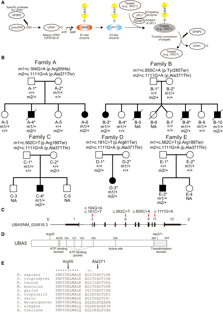
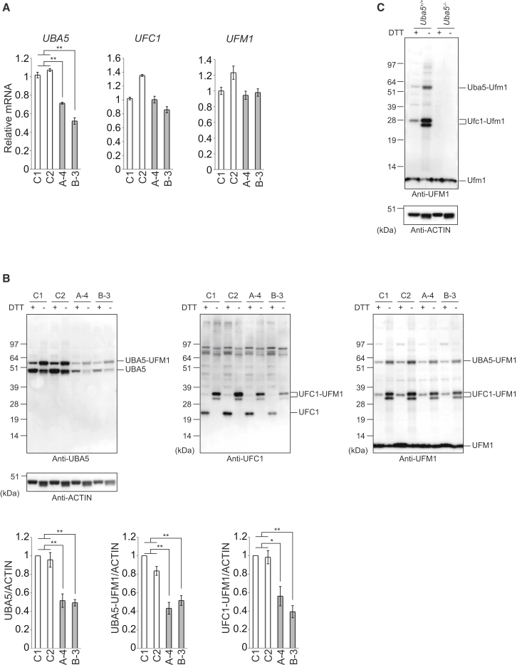
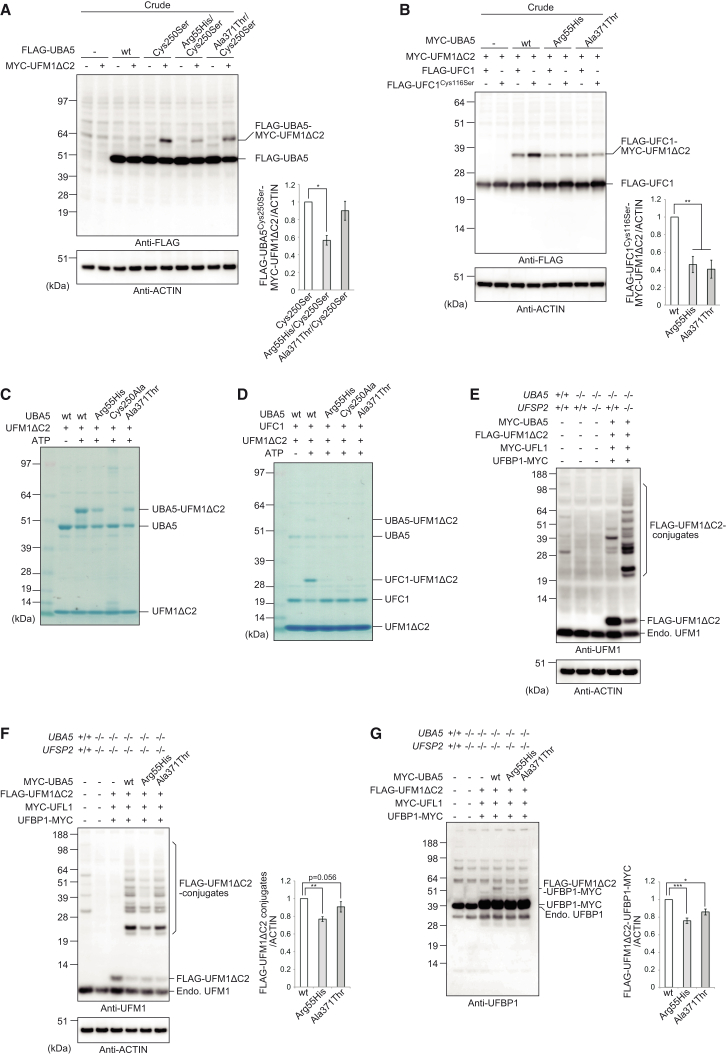
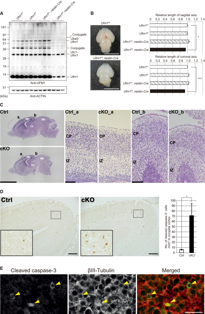
The ubiquitin fold modifier 1 (UFM1) cascade is a recently identified evolutionarily conserved ubiquitin-like modification system whose function and link to human disease have remained largely uncharacterized. By using exome sequencing in Finnish individuals with severe epileptic syndromes, we identified pathogenic compound heterozygous variants in UBA5, encoding an activating enzyme for UFM1, in two unrelated families. Two additional individuals with biallelic UBA5 variants were identified from the UK-based Deciphering Developmental Disorders study and one from the Northern Finland Intellectual Disability cohort. The affected individuals (n = 9) presented in early infancy with severe irritability, followed by dystonia and stagnation of development. Furthermore, the majority of individuals display postnatal microcephaly and epilepsy and develop spasticity. The affected individuals were compound heterozygous for a missense substitution, c.1111G>A (p.Ala371Thr; allele frequency of 0.28% in Europeans), and a nonsense variant or c.164G>A that encodes an amino acid substitution p.Arg55His, but also affects splicing by facilitating exon 2 skipping, thus also being in effect a loss-of-function allele. Using an in vitro thioester formation assay and cellular analyses, we show that the p.Ala371Thr variant is hypomorphic with attenuated ability to transfer the activated UFM1 to UFC1. Finally, we show that the CNS-specific knockout of Ufm1 in mice causes neonatal death accompanied by microcephaly and apoptosis in specific neurons, further suggesting that the UFM1 system is essential for CNS development and function. Taken together, our data imply that the combination of a hypomorphic p.Ala371Thr variant in trans with a loss-of-function allele in UBA5 underlies a severe infantile-onset encephalopathy.


Figures

Figure 1
Figure 1
Compound Heterozygous Variants in UBA5 (A) A schematic of the UFM1 ubiquitin-like modifier cascade. UFM1 is synthesized in a precursor form and cleaved at the C terminus by specific protease, UFSP2. The E1-like enzyme UBA5 activates mature UFM1 (UFM1ΔC2), forming a high-energy thioester bond. The activated UFM1 is then transferred to an E2-like (conjugating) enzyme, UFC1, through a similar thioester linkage. Finally, UFM1 is covalently conjugated (ufmylated) with cellular proteins such as UFM1-binding protein 1 (UFBP1, official symbol DDRGK1) and a nuclear receptor coactivator, ASC1 (official symbol TRIP4) via UFL1 (E3-ligating enzyme)., The conjugates are cleaved by UFSP2, implying the reversibility of the UFM1 conjugating system. (B) Pedigrees of five families with biallelic variants in UBA5. Variants present in each family are shown above the pedigrees. Exome-sequenced individuals are marked with asterisks. Plus sign (+) indicates wild-type. (C) A schematic of the exon structure of UBA5 showing the locations of the variants. (D) A schematic of the domain structure of UBA5 protein. (E) ClustalX alignment of the Arg55 and Ala371 residues of UBA5 in metazoa and plants. Asterisks () and colons (:) indicate fully conserved and highly conserved residues, respectively.
Figure 2
Figure 2
Defective E1 Activity of UBA5 in Fibroblasts Derived from Subjects with Pathogenic Biallelic UBA5 Variants (A) Quantitative real-time PCR analyses of UBA5, UFC1 (MIM: 610554) and UFM1 (MIM: 610553) in case (A-4 and B-3) and control (C1: female, age 26; C2: female, age 43) primary skin fibroblasts. Using a Transcriptor First Strand cDNA Synthesis Kit (Roche Applied Science), cDNA was synthesized from 1 μg of total RNA extracted from indicated fibroblasts. Quantitative PCR was performed using LightCycler 480 Probes Master (Roche Applied Science) in a LightCycler 480 (Roche Applied Science). Signals were assessed relative to that of GAPDH (MIM: 138400). Values were normalized to the amount of mRNA in control C1. The experiments were performed three times. The sequences of the primers are shown in Table S7. Statistical analysis was performed using the unpaired t test (Welch test). Data are means ± SE. ∗∗p < 0.01. (B and C) Immunoblot analysis of UBA5, UFC1, and UFM1 with reducing and nonreducing samples that were prepared from fibroblasts of affected individuals and human controls (B) and mouse embryonic fibroblasts (C). Indicated fibroblasts were lysed with ice-cold TNE buffer (10 mM Tris-HCl [pH 7.5], 1% Nonidet P-40, 150 mM NaCl, 1 mM ethylenediaminetetraacetic acid [EDTA], and protease inhibitors). Samples were prepared with NuPAGE-loading buffer in presence or absence of DTT, separated using a NuPAGE system (Life Technologies) on 4%–12% Bis-Tris gels in MOPS-SDS buffer, and then transferred to a polyvinylidene difluoride (PVDF) membrane. Mouse monoclonal anti-actin antibody (Chemicon International cat# MAB1501R), rabbit monoclonal anti-UFM1 antibody (Abcam cat# ab109305, RRID: AB_10864675), anti-UBA5 antibody, and anti-UFC1 antibody were used for immunodetection. The immunoreactive bands were detected by LAS-4000 (GE Healthcare UK). In the cases of samples prepared without DTT, the intermediates corresponding to UBA5-UFM1 and UFC1-UFM1 were clearly detected. Bar graphs indicate the quantitative densitometric analyses using Multi Gauge Version 3.2 Image software (Fuji Film) of UBA5, UBA5-UFM1, and UFC1-UFM1 intermediates relative to ACTIN. Statistical analysis was performed using the unpaired t test (Welch test). The data represent the means ± SE of five separate experiments. p < 0.05 and ∗∗p < 0.01.
Figure 3
Figure 3
Impaired Function of UBA5 Mutants (A and B) Immunoblot assay of UBA5 mutant p.Cys250Ser and double mutants p.Arg55His/p.Cys250Ser and p.Ala371Thr/p.Cys250Ser in UBA5−/− HEK293T cells. Indicated constructs (0.1 μg for UBA5, 0.5 μg for UFC1, and 2 μg for UFM1ΔC2) were expressed in UBA5-deficient HEK293T cells. 24 hr after transfection, the cell lysates were subjected to immunoblot analysis with indicated antibodies as described in Figure 2B. Bar graphs indicate the quantitative densitometric analyses of UBA5-UFM1 and UFC1-UFM1 intermediates relative to ACTIN. Statistical analysis was performed using the unpaired t test (Welch test). The data represent the means ± SE of four separate experiments. p < 0.05 and ∗∗p < 0.01. (C and D) In vitro thioester formation assay of UFM1 by UBA5 (C) and of UFM1 by UFC1 (D). Recombinant GST-UFM1ΔC2, GST-UFC1, and GST-UBA5, as well as UBA5 mutants p.Arg55His (GST-UBA5Arg55His), p.Ala371Thr (GST-UBA5Ala371Thr), and p.Cys250Ala (negative control; GST-UBA5Cys250Ala) were produced in E. coli and the recombinant proteins were purified by chromatography on Glutathione Sepharose 4B (GE Healthcare UK). After digestion of GST by PreScission Protease (GE Healthcare UK), the recombinant proteins were dialyzed against 50 mM BisTris (pH 6.5), 100 mM NaCl, 10 mM MgCl2, and 0.1 mM DTT (reaction buffer). Most thioester formation reactions contained reaction buffer with 0.8 μg UFM1ΔC2 and some of the following: 5 mM ATP, 0.08 (for UFC1-UFM1 thioester formation assay) or 0.8 (for UBA5-UFM1 thioester formation assay) μg UBA5 or UBA5 mutants, and 0.8 μg UFC1. Reactions were incubated for 5 min at 25°C and stopped by the addition of NuPAGE-loading buffer lacking reducing agent, followed by 10 min incubation at 37°C, NuPAGE (4%–12% acrylamide gradient), and Coomassie brilliant blue staining. Data shown are representative of three separate experiments. (E) Immunoblot assay to detect UFM1 conjugates. MYC-UBA5 (0.1 μg) was expressed in combination with indicated constructs (each 1 μg) in UBA5-deficient or UBA5-UFSP2 double-deficient HEK293T cells. Cells were lysed by 200 μL of TNE, and the lysate was then centrifuged at 10,000 × g for 10 min at 4°C to remove debris. The supernatant was subjected to immunoblot analyses with indicated antibodies. (F) Immunoblot assay to study the effect of UBA5 mutants on UFM1 conjugate formation. MYC-UBA5 or MYC-UBA5 mutants (0.1 μg) were expressed in combination with indicated constructs (each 1 μg) in UBA5-UFSP2 double-deficient HEK293T cells. Cells were lysed by 200 μL of TNE, and the lysate was then centrifuged at 10,000 × g for 10 min at 4°C to remove debris. The supernatant was subjected to immunoblot analyses with indicated antibodies. Bar graph indicates the quantitative densitometric analyses of FLAG-UFM1 conjugates relative to ACTIN. Statistical analysis was performed using the unpaired t test (Welch test). The data represent the means ± SE of six separate experiments. p < 0.05. (G) Immunoblot assay to study the effect of UBA5 mutants on UFM1-UFBP1 conjugate formation. Transfection and subsequent immunoblot analysis were conducted as shown in (F). Bar graph indicates the quantitative densitometric analyses of FLAG-UFM1-UFBP1-MYC relative to ACTIN. Rabbit polyclonal anti-UFBP1 antibody was used for immunodetection. Statistical analysis was performed using the unpaired t test (Welch test). The data represent the means ± SE of six separate experiments. p < 0.05 and ∗∗∗p < 0.001.
Figure 4
Figure 4
Loss of UFM1 in Central Nervous System Causes Microcephaly (A) Immunoblot analysis of UFM1 in mice with indicated genotypes. Mice were delivered by caesarean section at E18.5, and then mouse brains were homogenized in 0.25 M sucrose, 10 mM 2-[4-(2-hydroxyethyl)-1-piperazinyl]ethanesulfonic acid (HEPES) (pH 7.4), and 1 mM dithiothreitol (DTT). The homogenates were subjected to immunoblot analysis with indicated antibodies. Samples prepared from three mice with indicated genotype were loaded. (B) A dorsal view of brains of Ufm1f/f and Ufm1f/f;nestin-Cre mice delivered by Caesarean section at E18.5. Graphs show axial distance (from the anterior edge of cerebrum to posterior edge of mid brain) and maximal lateral distance of brains of indicated genotype mice. Data presented as mean ± SE of Ufm1f/+ (n = 4), Ufm1f/f (n = 4), Ufm1f/+;nestin-Cre (n = 5), and Ufm1f/f;nestin-Cre (n = 6). Statistical analysis was performed using the unpaired t test. p < 0.05 and ∗∗∗p < 0.001. (C) Histological analyses of brains of Ufm1f/+;nestin-Cre (Ctrl) and Ufm1f/f;nestin-Cre (cKO) mice. Embryos at E18.5 were delivered by Caesarean section, and their heads were fixed by immersion in 0.1 M phosphate buffer (pH 7.4) containing 4% paraformaldehyde and 4% sucrose. Each brain was carefully dissected and processed for paraffin embedding, and then 3 μm sagittal sections were prepared for haematoxylin and eosin staining. Images were captured with BZ-9000 (Keyence) and BX51 microscopes (Olympus). Boxed regions a and b in the neopallium are magnified and shown on the right as indicated. Note that the occipital region (b) of neopallium in the mutant brain is thinner than that in control, while the difference in the parietal region (a) is less apparent. Scale bars are 2 mm and 0.1 mm. Abbreviations are as follows: Np, neopallium; Mb, midbrain; Th, thalamus; IZ, intermediate zone; CP, cortical plate. (D) Apoptotic cells in the occipital region of neopallium of Ufm1f/+;nestin-Cre (Ctrl) and Ufm1f/f; nestin-Cre (cKO) mice at E18.5. Sections prepared as described in (C) were immunostained by rabbit polyclonal anti-cleaved caspase-3 antibody (Cell Signaling Technology [CST] cat# 9661, RRID: AB_2314091; 1:500) as described previously. Images were captured with BX53 microscope (Olympus). Each inset is a magnified image. Scale bars represent 100 μm. For quantification, the number of cleaved caspase-3-positive cells per unit area was calculated in each occipital cortex, which was defined as the cerebral cortex located posterior to the hippocampus. Statistical analysis was performed using the unpaired t test (n = 3 animals for each group). Data represent the means ± SE. p < 0.05. The area was measured by NIH Image/ImageJ. (E) Double-immunofluorescence analysis. Section of cKO brain (occipital region of neopallium) prepared as described in (C) was double-immunostained with anti-cleaved-caspase-3, mouse monoclonal anti-βIII Tubulin antibody (clone 5G8, Promega, 1:1,000), goat anti-mouse Alexa Fluor 594, and goat anti-rabbit Alexa Fluor 488 (Molecular Probes, 1:1,000). Images were captured with confocal FV1200 microscope (Olympus). Scale bar represents 20 μm.

Similar articles

Cited by

References

    1. van der Veen A.G., Ploegh H.L. Ubiquitin-like proteins. Annu. Rev. Biochem. 2012;81:323–357. - PubMed
    1. Schulman B.A., Harper J.W. Ubiquitin-like protein activation by E1 enzymes: the apex for downstream signalling pathways. Nat. Rev. Mol. Cell Biol. 2009;10:319–331. - PMC - PubMed
    1. Kang S.H., Kim G.R., Seong M., Baek S.H., Seol J.H., Bang O.S., Ovaa H., Tatsumi K., Komatsu M., Tanaka K., Chung C.H. Two novel ubiquitin-fold modifier 1 (Ufm1)-specific proteases, UfSP1 and UfSP2. J. Biol. Chem. 2007;282:5256–5262. - PubMed
    1. Komatsu M., Chiba T., Tatsumi K., Iemura S., Tanida I., Okazaki N., Ueno T., Kominami E., Natsume T., Tanaka K. A novel protein-conjugating system for Ufm1, a ubiquitin-fold modifier. EMBO J. 2004;23:1977–1986. - PMC - PubMed
    1. Yoo H.M., Kang S.H., Kim J.Y., Lee J.E., Seong M.W., Lee S.W., Ka S.H., Sou Y.S., Komatsu M., Tanaka K. Modification of ASC1 by UFM1 is crucial for ERα transactivation and breast cancer development. Mol. Cell. 2014;56:261–274. - PubMed

MeSH terms