. 2016 Sep 1;99(3):695-703.
doi: 10.1016/j.ajhg.2016.06.030. Epub 2016 Aug 18.

Biallelic Variants in UBA5 Reveal that Disruption of the UFM1 Cascade Can Result in Early-Onset Encephalopathy

Collaborators, Affiliations

Biallelic Variants in UBA5 Reveal that Disruption of the UFM1 Cascade Can Result in Early-Onset Encephalopathy

Estelle Colin et al. Am J Hum Genet. .

Abstract

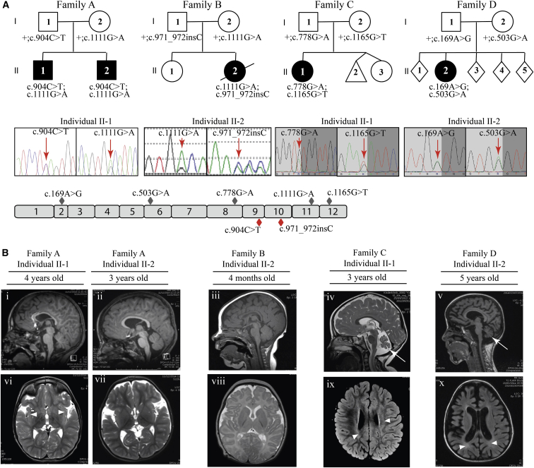
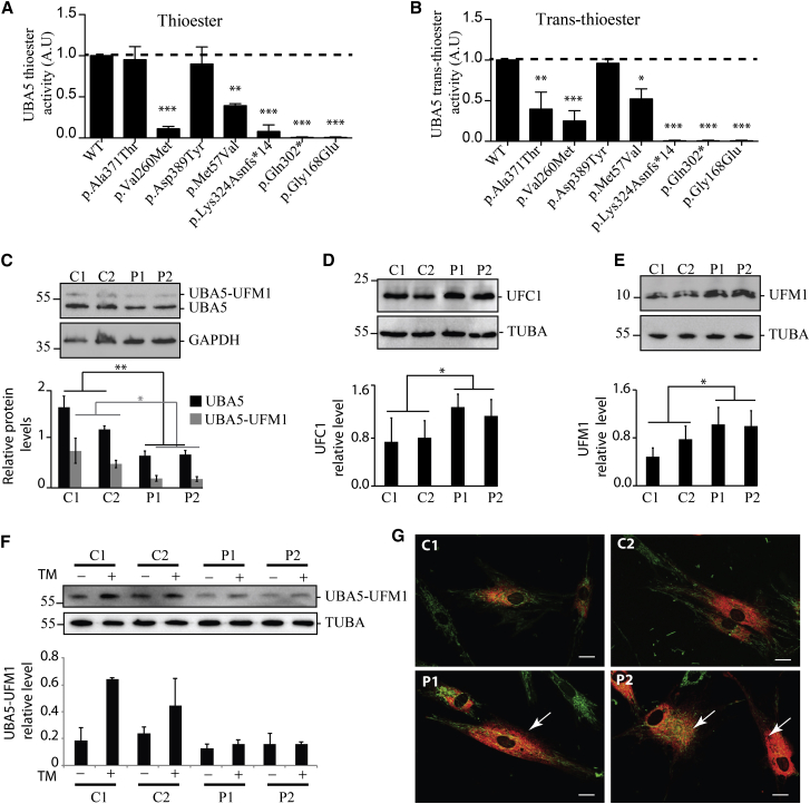
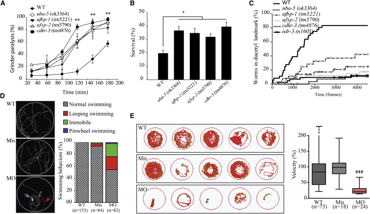
Via whole-exome sequencing, we identified rare autosomal-recessive variants in UBA5 in five children from four unrelated families affected with a similar pattern of severe intellectual deficiency, microcephaly, movement disorders, and/or early-onset intractable epilepsy. UBA5 encodes the E1-activating enzyme of ubiquitin-fold modifier 1 (UFM1), a recently identified ubiquitin-like protein. Biochemical studies of mutant UBA5 proteins and studies in fibroblasts from affected individuals revealed that UBA5 mutations impair the process of ufmylation, resulting in an abnormal endoplasmic reticulum structure. In Caenorhabditis elegans, knockout of uba-5 and of human orthologous genes in the UFM1 cascade alter cholinergic, but not glutamatergic, neurotransmission. In addition, uba5 silencing in zebrafish decreased motility while inducing abnormal movements suggestive of seizures. These clinical, biochemical, and experimental findings support our finding of UBA5 mutations as a pathophysiological cause for early-onset encephalopathies due to abnormal protein ufmylation.


Figures

Figure 1
Figure 1
Identification and Segregation of UBA5 Mutations and Brain MRI of the Five Affected Individuals (A) Families with mutations in UBA5. Filled black symbols represent the affected individuals. Allelic status is given below each tested individual. Representative chromatograms show the compound heterozygous mutations for each family (family A: c.904C>T, c.1111G>A; family B: c.1111G>A, c.971_972insC; family C: c.778G>A, c.1165G>T; family D: c.169A>G, c.503G>A). Red arrows indicate the position of the nucleotide change. Schematic overview of the 12 exons (gray boxes) of human UBA5, with missense (gray diamonds) and nonsense (red diamonds) mutations. (B) Brain MRI of the five affected individuals. Sagittal T1 (i, ii, iii, and v) and sagittal T2 (iv) images are shown. A thin corpus callosum in individuals II-1 from family A, II-2 from family B, and II-2 from family D (i, iii, v), cortical atrophy in individual II-2 from family D (v), and cerebellar atrophy (arrows) without brainstem anomalies in individuals II-1 from family C and II-2 from family D (iv, v) are observed. Axial T2 images (vi, vii, and viii) and T2 Flair (ix, x) are shown. White matter hyperintensities (arrowheads) in insula subcortical white matter (individual II-1 in family A [vi]), periventricular region (individuals II-1 in family C and II-2 in family D [ix, x]), and delayed myelination (individual II-2 in family B) are observed (viii). Widening of sylvian fissures (individuals II-1 and II-2 in family A [vi, vii]) and global cortical atrophy with ventricular dilation (individual II-2 in family D [x]) are observed.
Figure 2
Figure 2
Biochemical and Cellular Characterization of the UBA5 Disease-Causing Variants (A and B) UBA5, UFM1, and UFC1 recombinant proteins were produced in E. coli, purified, and used for in vitro UFM1 thioester activation and trans-thioester conjugation reactions, according to published protocols. (A) In vitro assessment of UFM1 thioester activation by the UBA5-Glutathion-S-Transferase (GST) fusion proteins, each carrying one of the mutations of the affected families. The thioester formations were monitored after 2 min of incubation. (B) Assessment of UFM1 trans-thioester conjugation to UFC1 by the UBA5-GST fusion proteins carrying each one of the mutations of the affected families. The trans-thioester formations were monitored after 3 min of incubation. Three independent experiments were used for quantification and statistical analyses by one-way ANOVA, Bonferroni’s multiple comparison test, p < 0.05, ∗∗p < 0.01, ∗∗∗p < 0.001. (C–G) Skin fibroblasts from the two affected individuals from family A (P1 equate to II-1 and P2 equate to II-2) and from two control subjects were grown in DMEM-F12 for 24 hr and used for assessing the UFM1 cascade. (C) Representative western blots (top) of free (lower bands) and ufmylated (upper bands) UBA5 (Proteintech cat# 12093-1-AP, RRID: AB_2211743) and GAPDH (GeneTex cat# GTX100118, RRID: AB_1080976) proteins in control (C1, C2) and UBA5 mutant (P1, P2) fibroblast protein extracts, and quantification (bottom) of the relative levels of UBA5/GAPDH and UBA5-UFM1/GAPDH. (D) Representative western blots (top) of UFC1 (Abcam cat# ab189251) and quantification (bottom) of UFC1 expression level related to TUBA (Sigma-Aldrich cat# T9026, RRID: AB_477593). (E) Representative western blots (top) in non-reducing conditions (using Optiblot LDS Sample buffer, Abcam) of free UFM1 (Abcam cat# ab109305, RRID: AB_10864) and quantification (bottom) of UFM1 expression level related to TUBA. (F) Representative western blots (top) of UBA5 ufmylation with (+) and without (−) 10 μg/mL of tunicamycin (TM, BML-CC104, Enzo Life Sciences) treatment for 24 hr and quantification (bottom) related to TUBA. Three independent experiments were used for quantification and statistical analyses using Student’s t test, p < 0.05; ∗∗p < 0.01. (G) Confocal live images of WT (C1, C2) and UBA5 mutant (P1, P2) fibroblasts transduced by an RFP-tagged ER red probe (C10591, CellLight ER-RFP, BacMam 2.0, Thermo Fischer Scientific), and stained with MitoTracker-green (M7514, Thermofisher) to reveal the ER and mitochondria. Arrows highlight expanded ER network in UBA5 mutant fibroblasts. Scale bar represents 10 μm.
Figure 3
Figure 3
Characterization of C. elegans Strains Deleted for Genes Involved in Ufmylation and Zebrafish Silenced for uba5 (A–C) C. elegans UFM-1 cascade deletion strains uba-5(ok3364), ufbp-1(tm5221), ufsp-2(tm5790), and cdkr-3(tm4876) were used for the following experiments and compared to the wild-type (WT) strain (N2 Bristol). (A) Assessment of grinder paralysis of the UFM-1 cascade deletion strains treated with 0.5 mM aldicarb for 60 min reveal that the mutants display a highly significant increase in grinder paralysis, witnessing failure to negatively regulate acetylcholine release (ANOVA, ∗∗p < 0.01). (B) Assessment of survival of the UFM-1 cascade deletion strains, after an 18 hr treatment with the ER-stressor dithiothreitol (DTT, 11 mM), show that mutants are resistant to ER stress, compared to WT animals (ANOVA, p < 0.05). (C) Assessment of chemotaxis toward the attractant diacetyl of the UFM-1 cascade deletion strains shows that the percentage of animals entering the landmark containing diacetyl (1/1,000) was significantly lower for the UFM-1 cascade deletion mutants compared to WT (Kaplan-Meier curve, p < 0.01). Negative control odr-3(n1605) worms were unable to sense diacetyl. Trajectories of the worms were recorded for 7.5 min with 10 frames per second via the FTIR-based Imaging Method (FIM). (D and E) Zebrafish from the AB genetic background were maintained at 28°C on a 14 hr light and 10 hr dark cycle, and fertilized eggs were injected with the antisense uba5 morpholino oligonucleotide (MO) and the mismatch oligonucleotide (Mis) at a concentration of 0.35 pM. (D) Assessment of 72 hpf larvae motility, after a slight mechanical stimulation on the tail. The motion of individual larvae was examined (left) and scored (right) as normal swimming, looping swimming (red arrow), pinwheel swimming (blue arrow), or immobile and revealed a significant and specific alteration of swimming in uba5-depleted animals (MO), compared to larvae injected with the mismatch morpholino (Mis) and control larvae (WT). Quantification of the swimming behavior showed a normal motility for WT and Mis-injected larvae, whereas 45.1% of the MO showed impaired motility: 22% were immobile, 20.7% exhibited looping swimming, and 2.4% showed pinwheel swimming. (E) Spontaneous motility of zebrafish monitored at 5 days post fertilization (dpf) using the Zebrabox recording system (Viewpoint). The cumulative movement of representative WT, Mis, and MO larvae is represented (left). The quantification of the velocity (right, mm/s), expressed as percentages of the WT values, revealed a significant decreased motility in uba5-depleted zebrafish (∗∗∗p < 0.001 between WT/MO and between Mis/MO but not significant between WT/Mis; statistical significance was inferred with the non-parametric Mann-Whitney test). Horizontal bars in the boxes represent the median values, horizontal bars outside the boxes represent the maximal and minimal individual values (in the WT, the maximal individual value was 676%).

Similar articles

Cited by

References

    1. van der Veen A.G., Ploegh H.L. Ubiquitin-like proteins. Annu. Rev. Biochem. 2012;81:323–357. - PubMed
    1. Komatsu M., Chiba T., Tatsumi K., Iemura S., Tanida I., Okazaki N., Ueno T., Kominami E., Natsume T., Tanaka K. A novel protein-conjugating system for Ufm1, a ubiquitin-fold modifier. EMBO J. 2004;23:1977–1986. - PMC - PubMed
    1. Daniel J., Liebau E. The ufm1 cascade. Cells. 2014;3:627–638. - PMC - PubMed
    1. Zhang M., Zhu X., Zhang Y., Cai Y., Chen J., Sivaprakasam S., Gurav A., Pi W., Makala L., Wu J. RCAD/Ufl1, a Ufm1 E3 ligase, is essential for hematopoietic stem cell function and murine hematopoiesis. Cell Death Differ. 2015;22:1922–1934. - PMC - PubMed
    1. Lemaire K., Moura R.F., Granvik M., Igoillo-Esteve M., Hohmeier H.E., Hendrickx N., Newgard C.B., Waelkens E., Cnop M., Schuit F. Ubiquitin fold modifier 1 (UFM1) and its target UFBP1 protect pancreatic beta cells from ER stress-induced apoptosis. PLoS ONE. 2011;6:e18517. - PMC - PubMed

MeSH terms