. 2016 Sep 7;91(5):975-987.
doi: 10.1016/j.neuron.2016.07.036. Epub 2016 Aug 18.

High-Throughput Mapping of Single-Neuron Projections by Sequencing of Barcoded RNA

Affiliations

High-Throughput Mapping of Single-Neuron Projections by Sequencing of Barcoded RNA

Justus M Kebschull et al. Neuron. .

Abstract

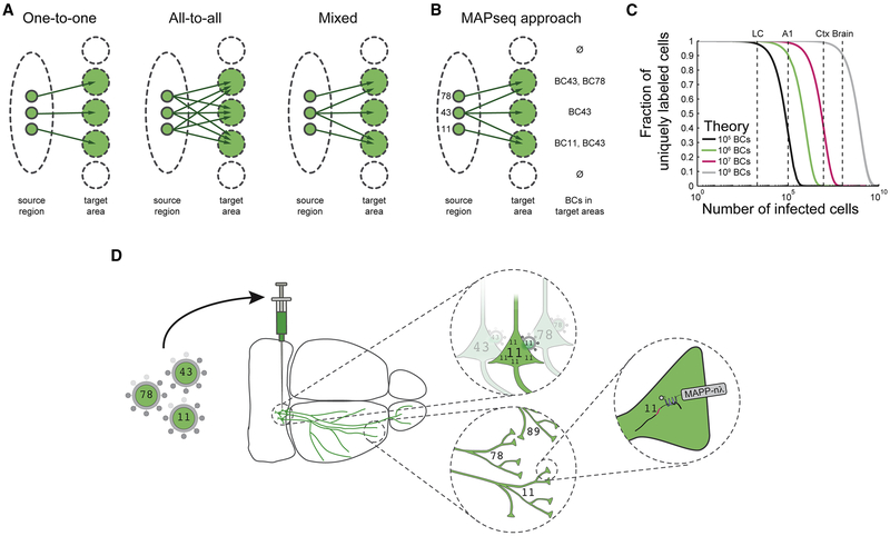
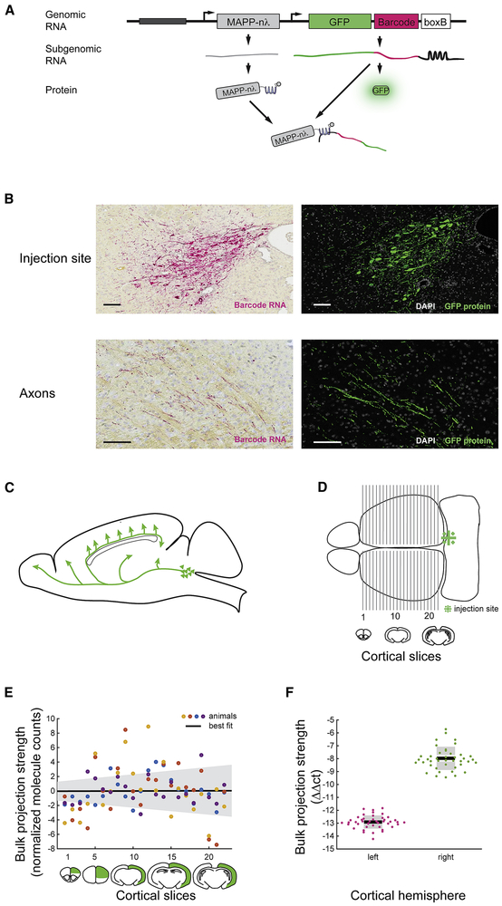
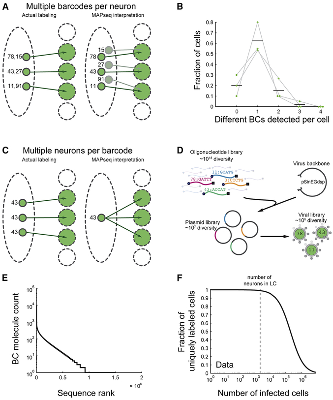
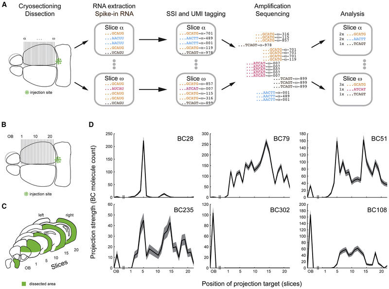
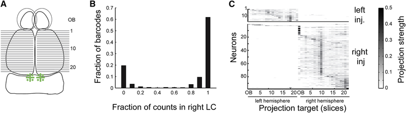
Neurons transmit information to distant brain regions via long-range axonal projections. In the mouse, area-to-area connections have only been systematically mapped using bulk labeling techniques, which obscure the diverse projections of intermingled single neurons. Here we describe MAPseq (Multiplexed Analysis of Projections by Sequencing), a technique that can map the projections of thousands or even millions of single neurons by labeling large sets of neurons with random RNA sequences ("barcodes"). Axons are filled with barcode mRNA, each putative projection area is dissected, and the barcode mRNA is extracted and sequenced. Applying MAPseq to the locus coeruleus (LC), we find that individual LC neurons have preferred cortical targets. By recasting neuroanatomy, which is traditionally viewed as a problem of microscopy, as a problem of sequencing, MAPseq harnesses advances in sequencing technology to permit high-throughput interrogation of brain circuits.


Conflict of interest statement

Conflict of interests

The authors declare no conflict of interests.

Figures

Figure 1:
Figure 1:
Barcoding allows high-throughput single neuron tracing. (a) Identical bulk mapping results can arise from different underlying projection patterns. (b) Single neuron resolution can be achieved by randomly labeling neurons with barcodes and reading out barcodes in target areas. (c) The expected fraction of uniquely labeled cells is given by F=(1-1/N)(k-1), where N is the number of barcodes and k is the number of infected cells, assuming a uniform distribution of barcodes. The number of neurons for various mouse brain areas are indicated according to refs (Herculano-Houzel et al., 2006; Schüz and Palm, 1989) (A1= primary auditory cortex; Ctx = neocortex). (d) In MAPseq, neurons are infected at low multiplicity of infection (MOI) with a barcoded virus library. Barcode mRNA is expressed, trafficked and can be extracted from distal sites as a measure of single neuron projections.
Figure 2:
Figure 2:
Barcoded Sindbis virus can be used for projection mapping. (a) A dual promoter Sindbis virus was used to deliver barcodes to neurons. The virus encoded GFP, barcodes and MAPP-nλ. (b) Barcode mRNA labeling of LC neurons is comparable to GFP labeling of these neurons in an adjacent 6μm slice both at the injection site (top) and in the axon tract (bottom). Scale bar = 100μm. Representative data from 3 animals. (c) Axons from LC project rostrally from the cell body, before changing direction and innervating cortex. LC axons that project to frontal cortices have thus traveled only about half as long as axons innervating visual cortex. (d) We injected right LC with MAPseq virus and dissected cortex along the anterior-posterior axis as shown. (e) Bulk projection strength of LC to ipsilateral cortex as measured by barcode mRNA is independent of the anterior-posterior position of the cortical slice, suggesting a uniform RNA fill of LC axons. N=4. (f) qPCR for barcode mRNA shows approximately 30× stronger LC projections to ipsi- than to contralateral cortex. N=2 animals and 21 cortical slices per animal. BC=barcodes. The y-axis displays ΔΔct values, which are equivalent to the log2(foldchange of barcode mRNA per sample) normalized to β-actin levels in each sample and to the amount of barcode mRNA in the injection site of each animal (Livak and Schmittgen, 2001).
Figure 3:
Figure 3:
Random labeling of neurons with a barcoded virus library can achieve unique labeling of many neurons. (a) When single neurons are labeled with several barcodes, MAPseq will overestimate of the number of neurons identified, but will not distort the projection patterns recorded for individual neurons. (b) Single cell isolation of GFP-positive, barcoded neurons, followed by sequencing of their barcode complement reveals a low chance of double infection. We interpret neurons for which no barcodes were recovered as technical failures of cell isolation, rather than biological phenomena. N=3 animals. Mean and individual data points are plotted. (c) When several neurons share the same barcode, MAPseq misinterprets this as a single neuron whose projection pattern is given by the union of the projection patterns of the two infected neurons. (d) High diversity Sindbis virus libraries are produced by shotgun cloning random oligonucleotides into a plasmid followed by virus production. (e) The virus library used in this work has a diversity of ~106 different barcodes (BC), but the distribution was non-uniform. The sequence rank is a number that ranges from 1 to the total number of barcodes, where 1 corresponds to the most abundant sequence, 2 to the second most abundant and so on. (f) Based on the empirically observed non-uniform barcode distribution, we determined that the virus library used is sufficiently diverse to uniquely label all of LC with low error rate.
Figure 4:
Figure 4:
MAPseq reveals large diversity of projections from LC. (a) Barcode mRNAs from target areas are sequenced as described (SSI = slice specific identifier, UMI = unique molecular identifier). (b,c) Barcodes from ipsilateral olfactory bulb and cortex show projection patterns (d) with single or multiple peaks in cortex and/or olfactory bulb. The shaded area indicates Poisson error bars given by the square root of barcode (BC) counts per slice.
Figure 5:
Figure 5:
MAPseq provides a robust readout of single neuron projection patterns. (a) Two representative pairs of barcodes with projection patterns more similar than expected by chance for two distinct neurons, likely the result of double infection of a single neuron. The close agreement between the two barcode profiles indicates that MAPseq provides a reliable measure of projection patterns. The closest match across animals is indicated in grey for comparison. (b) Cumulative distribution of distances between the best barcode pairs within one animal and across animals. The shift in the within animal distribution reflects the higher fraction of closely matched projection profiles, consistent with double infection. Representative data from one animal.
Figure 6:
Figure 6:
LC neurons tile cortex with their maximum projections, but innervate large areas of cortex at a low level. (a) A heatmap of all 995 projection patterns from 4 animals shows a strong diagonal component after sorting by maximum projection site. Barcode abundances are normalized to sum to one across target areas and are color-coded as indicated. (b) Average cortical drop-off rate from maximum for all barcodes shows a rapid drop-off and a structure that is different from the drop-off after randomly shuffling slices for all neurons. N=4. (c) Cumulative distribution of cortical projection widths indicates a broad low intensity innervation of cortex by individual LC neurons. BC = barcode.
Figure 7:
Figure 7:
MAPseq can be multiplexed to several injection sites. (a) Following bilateral injection of barcoded Sindbis virus into LC, left and right olfactory bulb and cortex were dissected as before. (b) Histogram of the fraction of barcode counts in the right vs. left injection site across barcodes. Barcodes show strong abundance differences in the left and right injection sites allowing them to be assigned to one of the two sites. (c) Bilateral injections produce the projection pattern expected from unilateral injections. Differences in the number of neurons traced from the left and right LC arise from injection variability.

References

    1. Aston-Jones G, and Cohen JD (2005). An integrative theory of locus coeruleus-norepinephrine function: adaptive gain and optimal performance. Annu. Rev. Neurosci. 28, 403–450. - PubMed
    1. Bredenbeek PJ, Frolov I, Rice CM, and Schlesinger S (1993). Sindbis virus expression vectors: packaging of RNA replicons by using defective helper RNAs. Journal of Virology 67, 6439–6446. - PMC - PubMed
    1. Cetin A, Komai S, Eliava M, Seeburg PH, and Osten P (2007). Stereotaxic gene delivery in the rodent brain. Nat Protoc 1, 3166–3173. - PubMed
    1. Chandler DJ, Gao WJ, and Waterhouse BD (2014). Heterogeneous organization of the locus coeruleus projections to prefrontal and motor cortices. Proceedings of the National Academy of Sciences 111, 6816–6821. - PMC - PubMed
    1. Chandler D, and Waterhouse BD (2012). Evidence for Broad Versus Segregated Projections from Cholinergic and Noradrenergic Nuclei to Functionally and Anatomically Discrete Subregions of Prefrontal Cortex. Front. Behav. Neurosci. 6. - PMC - PubMed