

Type-dependent differences in Fas expression and phagocytes distribution in rat corpora lutea during natural regression: an immunohistochemical evidence
- PMID: 27546215
- PMCID: PMC5240753
- DOI: 10.1292/jvms.16-0199
Type-dependent differences in Fas expression and phagocytes distribution in rat corpora lutea during natural regression: an immunohistochemical evidence
Abstract
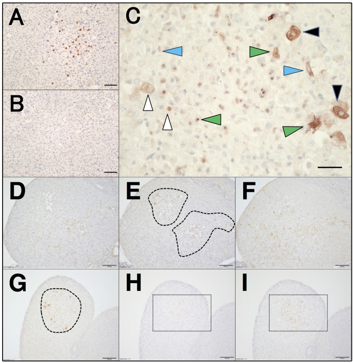
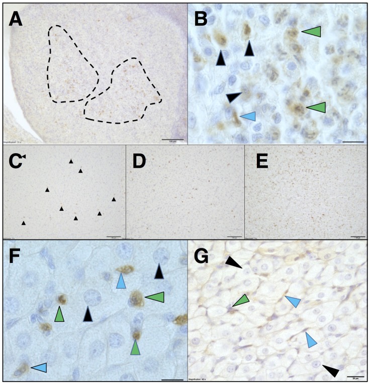
Though Fas/Fas ligand (FasL) system-dependent apoptosis is considered to be the primary form of cell death in regressing corpus luteum (CL), the cellular identity and regulation of expression of the ligand and receptor molecules are not fully understood. Here, we focused on immunohistochemical determination of Fas expression during natural regression with comparison of three different types of rat CLs. Detected Fas was in good spatial association with cleaved caspase-3 and FasL proteins and with macrophages and neutrophils. In CLs of the cycle and pseudopregnancy, Fas-positive cell types included large and small luteal (steroidogenic) cells and capillary endothelial cells mainly, and blood-derived immune cells occasionally. Fas signals were abundant at multiple focal inflammatory-like sites. In contrast, Fas signals in CL of pregnancy did not localize in steroidogenic cells, but almost exclusively in endothelial cells and granulocytes. The signals scattered evenly throughout the CL tissue as phagocytes also did. In all CLs types, the numbers of Fas-expressing cells increased transiently after functional inactivation and at the early phase of structural regression. This observation revealed spatio-temporally regulated expression of Fas that was highly associated with apoptotic and phagocytotic systems and type-dependent differences in Fas expression and phagocytes dynamics in naturally regressing CL of rats.
Figures
Similar articles
-
Is FAS/Fas ligand system involved in equine corpus luteum functional regression?Biol Reprod. 2010 Dec;83(6):901-8. doi: 10.1095/biolreprod.110.084699. Epub 2010 Aug 18. Biol Reprod. 2010. PMID: 20720169
-
Changes in localization of immune cells and cytokines in corpora lutea during luteolysis in murine ovaries.J Exp Zool A Comp Exp Biol. 2003 Apr 1;296(2):152-9. doi: 10.1002/jez.a.10246. J Exp Zool A Comp Exp Biol. 2003. PMID: 12658721
-
Autotaxin as a novel, tissue-remodeling-related factor in regressing corpora lutea of cycling rats.FEBS J. 2013 Dec;280(24):6600-12. doi: 10.1111/febs.12565. Epub 2013 Nov 1. FEBS J. 2013. PMID: 24128332
-
Luteal regression vs. prepartum luteolysis: regulatory mechanisms governing canine corpus luteum function.Reprod Biol. 2014 Apr;14(2):89-102. doi: 10.1016/j.repbio.2013.11.004. Epub 2013 Dec 21. Reprod Biol. 2014. PMID: 24856467 Review.
-
Multiple roles of TNF super family members in corpus luteum function.Reprod Biol Endocrinol. 2003 Nov 10;1:95. doi: 10.1186/1477-7827-1-95. Reprod Biol Endocrinol. 2003. PMID: 14613529 Free PMC article. Review.
Cited by
-
Autophagy in the corpus luteum correlates with tissue growth in pregnant rats.J Reprod Dev. 2024 Oct 1;70(5):286-295. doi: 10.1262/jrd.2024-019. Epub 2024 Jul 7. J Reprod Dev. 2024. PMID: 38972734 Free PMC article.
References
-
- Carambula S. F., Pru J. K., Lynch M. P., Matikainen T., Gonçalves P. B. D., Flavell R. A., Tilly J. L., Rueda B. R.2003. Prostaglandin F2α- and FAS-activating antibody-induced regression of the corpus luteum involves caspase-8 and is defective in caspase-3 deficient mice. Reprod. Biol. Endocrinol. 1: 15. doi: 10.1186/1477-7827-1-15 - DOI - PMC - PubMed
-
- Chen X., Gao H., Wei P., Zhang Z., Liu Y.2003. Expression of apoptosis-related genes Fas/FasL, Bax/Bcl-2 and Caspase-3 in rat corpus luteum during luteal regression. Sci. China C Life Sci. 46: 273–285. - PubMed
MeSH terms
Substances
LinkOut - more resources
Full Text Sources
Other Literature Sources
Research Materials
Miscellaneous