. 2016 Sep 13;7(3):454-470.
doi: 10.1016/j.stemcr.2016.06.012. Epub 2016 Aug 18.

G9a and ZNF644 Physically Associate to Suppress Progenitor Gene Expression during Neurogenesis

Affiliations

G9a and ZNF644 Physically Associate to Suppress Progenitor Gene Expression during Neurogenesis

Jonathan B Olsen et al. Stem Cell Reports. .

Abstract

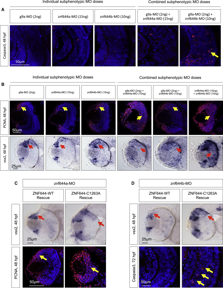
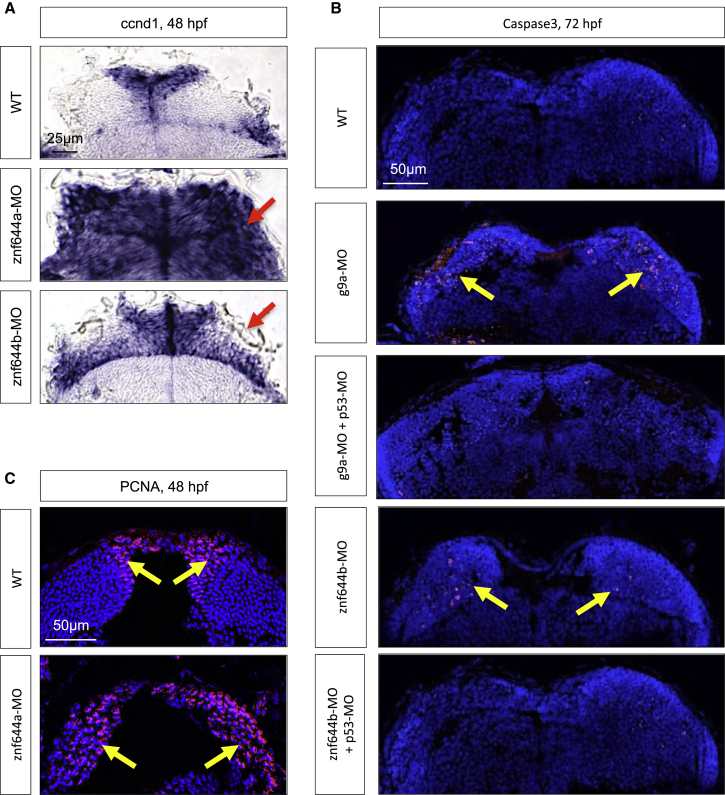
Proliferating progenitor cells undergo changes in competence to give rise to post-mitotic progeny of specialized function. These cell-fate transitions typically involve dynamic regulation of gene expression by histone methyltransferase (HMT) complexes. However, the composition, roles, and regulation of these assemblies in regulating cell-fate decisions in vivo are poorly understood. Using unbiased affinity purification and mass spectrometry, we identified the uncharacterized C2H2-like zinc finger protein ZNF644 as a G9a/GLP-interacting protein and co-regulator of histone methylation. In zebrafish, functional characterization of ZNF644 orthologs, znf644a and znf644b, revealed complementary roles in regulating G9a/H3K9me2-mediated gene silencing during neurogenesis. The non-overlapping requirements for znf644a and znf644b during retinal differentiation demarcate critical aspects of retinal differentiation programs regulated by differential G9a-ZNF644 associations, such as transitioning proliferating progenitor cells toward differentiation. Collectively, our data point to ZNF644 as a critical co-regulator of G9a/H3K9me2-mediated gene silencing during neuronal differentiation.


Figures

Figure 1
Figure 1
ZNF644 Is a G9a/GLP-Interacting Protein and Co-regulator of H3K9me2 (A) Physical interaction map of histone methylation-related complexes. Nodes represent proteins identified by AP-MS, black edges represent high-confidence interactions, and known interactions are in green. (B) Hierarchical clustering of bait interaction profile similarity. Colored boxes indicate proteins known to reside in the same complex. (C) (Top) Protein domain architecture of WIZ and ZNF644. (Bottom) Sequence alignment of atypical C2H2-like ZF motifs. Putative zinc ion-chelating residues are circled. (D) Summary of AP-MS analyses showing average peptide spectral counts of GFP-tagged forms of full-length WT ZNF644, the C1263A or H1283A point mutants, or the ZF8-containing C-terminal region only. (E) Western blot analysis of pulldown assays using GST-tagged WT, C1263A, or H1283A versions of the ZF8 domain of ZNF644 with the His-tagged G9a SET domain. (F) Western blot assays monitoring G9a, H3K9me2, H3K9me3, histone H3, and GAPDH levels in HEK293 cell lines expressing shRNAs targeting a non-silencing control, G9a, or ZNF644.
Figure 2
Figure 2
Zebrafish znf644 Paralogs Are Specifically Expressed In and Regulate the Development of Retinal and Midbrain Progenitor Cells (A) Conservation of C2H2-like ZF motifs between human and zebrafish ZNF644 genes. (B) Representative lateral views, retinal cross-sections, and midbrain cross-sections of whole-mount in situ hybridization (WISH) assays monitoring znf644a or znf644b expression at 24 hpf (n > 15 in each group). Arrows denote the inner part of the central retinal epithelium. (C) Lateral views of g9a (20/32), znf644a (10/18), or znf644b (14/21) morphant embryos compared with WT (21/21). Dashed circles denote 2D area of the WT retina. Arrowheads denote the dorsal hindbrain region. (D) Quantitation (mean ± SD) of retinal 2D cross-sectional area (μm2) of znf644a, znf644b, or g9a morphants relative to WT embryos. Error bars represent SD. p < 0.005, Student's t test. (E) RT-PCR assays monitoring unspliced (unspl) and spliced (spl) znf644a or znf644b transcripts at 24 hpf.
Figure 3
Figure 3
znf644a and znf644b Are Required for H3K9me2-Mediated Gene Silencing in the Forming Retina (A) Frequency and distance from TSS at which H3K9me2 peaks were identified in WT, znf644a, and znf644b morphants. (B) Venn diagram illustrating the number and overlap of H3K9me2 peaks at 48 hpf in znf644a or znf644b morphants or WT embryos. (C) ChIP-PCR assays monitoring H3K9me2 levels near the TSSs of vsx2 or ccnd1 genes at 48 hpf. (D) WISH assays monitoring expression of progenitor or proneural genes in WT, znf644a, or znf644b morphant retinas. Arrows highlight expression domains.
Figure 4
Figure 4
znf644a and znf644b Morphant Retinas Are Composed of Distinct Populations of Retinal Cells with Differing Characteristics (A) Immunostaining of retinal cross-sections monitoring BrdU incorporation and PCNA+ cells at 48 hpf in WT, znf644a, and znf644b morphants. (B) Quantitation of BrdU+ cells/μm2 in central retina of WT, znf644a, or znf644b morphants at 48 hpf (n = 3 for each group). Error bars represent SD. p < 0.05, Student's t test. (C–E) Immunostaining of pH3 expression in retinal cross-sections at 48 and 72 hpf in WT, and znf644a morphants (C). Quantitation of pH3+ cells/μm2 in the central retina of WT, znf644a, or znf644b morphants at (D) 48 hpf (n = 5 for each group) and (E) 72 hpf (n = 2 for each group). Quantitation represented as mean ± SD. (F) Immunostaining monitoring marker protein expression in retinal cross-sections. At 72 hpf, amacrine cells express GABA, bipolar neurons express PKC, and cone photoreceptors express Zpr1. White arrows highlight populations of proliferative or differentiated cells.
Figure 5
Figure 5
znf644a and znf644b Morphant Retinas Exhibit Distinct Cellular Defects (A) Immunostaining of retinal cross-sections of Vsx2 (red), Pax6 (red), Isl2b (green), or H3K9me2 (red) in WT or znf644a morphant embryos at indicated time points. Vsx2 expression at 48 hpf marks proliferating RPCs in the CMZ (gray arrows) and in a subpopulation of bipolar neurons in the central retina (yellow arrows). Pax6 expression marks amacrine cells and a subset of RGCs, and Isl2b expression marks a subset of RGCs. Differentiated retinal neurons with H3K9me2+ nuclei are highlighted. (B and C) Immunostaining monitoring cleaved Caspase3 (red) in retinal cross-sections from WT, g9a, or znf644b morphant embryos at 48 or 72 hpf (B). Yellow arrows highlight Caspase3+ apoptotic cells. (C) Quantitation of Caspase3+ cells in WT, znf644a morphant, or znf644b morphant retinas with or without co-injection of p53-MO at 72 hpf. Quantitation represented as mean ± SD (n=3 for each group). p < 0.05, ∗∗p < 0.005, ∗∗∗p < 0.0005, Student's t test.
Figure 6
Figure 6
The Retinal Functions of znf644a and znf644b Are Dependent on Functional and Physical Interactions with g9a (A) Immunostaining of cleaved Caspase3 (red) in retinal cross-sections of embryos injected with individual or combined subphenotypic doses of g9a-MO, znf644a-MO, or znf644b-MO (n = 3 in each group). Yellow arrows highlight apoptotic cells. (B) (Top) Immunostaining of PCNA (n = 6 in each group) in retinal cross-sections from embryos injected with individual and combined subphenotypic doses of MOs (n = 6 in each group). Yellow arrows denote the position of the PCNA+ population. (Bottom) WISH assays monitoring the expression of vsx2 in retinal cross-sections of embryos injected with individual subphenotypic doses (n = 10 in each group) and combined subphenotypic co-injection of g9a-MO and znf644a-MO (n = 10), g9a-MO and znf644b-MO (n = 13), and znf644a-MO and znf644b-MO (n = 10). Red arrows denote the position of the vsx2 expressing cells. (C) (Top) WISH assays in retinal cross-sections monitoring vsx2 expression at 48 hpf in znf644a morphant embryos rescued by co-injection of either WT human ZNF644 mRNA (n = 12) or C1263A mutant mRNA (n = 9). (Bottom) Immunostaining of retinal cross-sections monitoring PCNA expression at 48 hpf showing rescue of znf644b morphants by co-injection of WT human ZNF644 mRNA (n = 3) but not C1263A mutants (n = 3). (D) (Top) WISH assays in retinal cross-sections at 48 hpf monitoring vsx2 expression from znf644b morphant embryos rescued by co-injection of human WT ZNF644 mRNA (n = 14) or C1263A ZNF644 mutant mRNA (n = 9). (Bottom) Immunostaining of retinal cross-sections monitoring cleaved Caspase3 at 72 hpf in znf644b morphant embryos rescued by co-injection of WT human ZNF644 mRNA (n = 3) or a C1263A mutant (n = 3). Red arrows highlight vsx2 expression domains, and yellow arrows highlight PCNA+ cell populations (C) or apoptotic cells (D).
Figure 7
Figure 7
The Retinal Defects of g9a, znf644a, and znf644b Morphants Are Recapitulated in the Midbrain (A) WISH assays monitoring ccnd1 expression in WT, znf644a, or znf644b morphant midbrain cross-sections at 48 hpf. Arrows indicate mislocalized ccnd1 expression. (B) Immunostaining of cleaved Caspase3 in g9a or znf644b morphant midbrain cross-sections at 72 hpf and rescue by p53-MO (n = 3 in each group). Arrows denote the position of Caspase3+ cells. (C) Immunostaining of PCNA in WT or znf644a morphant midbrain cross-sections (n = 3 in each group). Arrows denote the position of PCNA+ cells.

Comment in

References

    1. Agathocleous M., Harris W.A. From progenitors to differentiated cells in the vertebrate retina. Annu. Rev. Cell Dev. Biol. 2009;25:45–69. - PubMed
    1. Balemans M.C., Huibers M.M., Eikelenboom N.W., Kuipers A.J., van Summeren R.C., Pijpers M.M., Tachibana M., Shinkai Y., van Bokhoven H., Van der Zee C.E. Reduced exploration, increased anxiety, and altered social behavior: autistic-like features of euchromatin histone methyltransferase 1 heterozygous knockout mice. Behav. Brain Res. 2010;208:47–55. - PubMed
    1. Balemans M.C., Ansar M., Oudakker A.R., van Caam A.P., Bakker B., Vitters E.L., van der Kraan P.M., de Bruijn D.R., Janssen S.M., Kuipers A.J. Reduced Euchromatin histone methyltransferase 1 causes developmental delay, hypotonia, and cranial abnormalities associated with increased bone gene expression in Kleefstra syndrome mice. Dev. Biol. 2014;386:395–407. - PubMed
    1. Barton K.M., Levine E.M. Expression patterns and cell cycle profiles of PCNA, MCM6, cyclin D1, cyclin A2, cyclin B1, and phosphorylated histone H3 in the developing mouse retina. Dev. Dyn. 2008;237:672–682. - PubMed
    1. Bassett E.A., Wallace V.A. Cell fate determination in the vertebrate retina. Trends Neurosci. 2012;35:565–573. - PubMed

Publication types

MeSH terms

Grants and funding