. 2016 Sep 1;63(5):739-52.
doi: 10.1016/j.molcel.2016.07.012. Epub 2016 Aug 18.

Members of the Hsp70 Family Recognize Distinct Types of Sequences to Execute ER Quality Control

Affiliations

Members of the Hsp70 Family Recognize Distinct Types of Sequences to Execute ER Quality Control

Julia Behnke et al. Mol Cell. .

Abstract

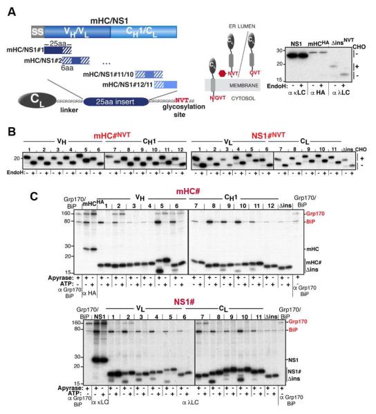
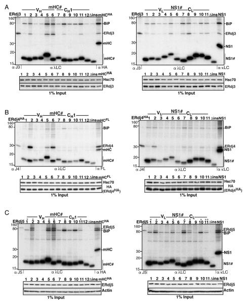
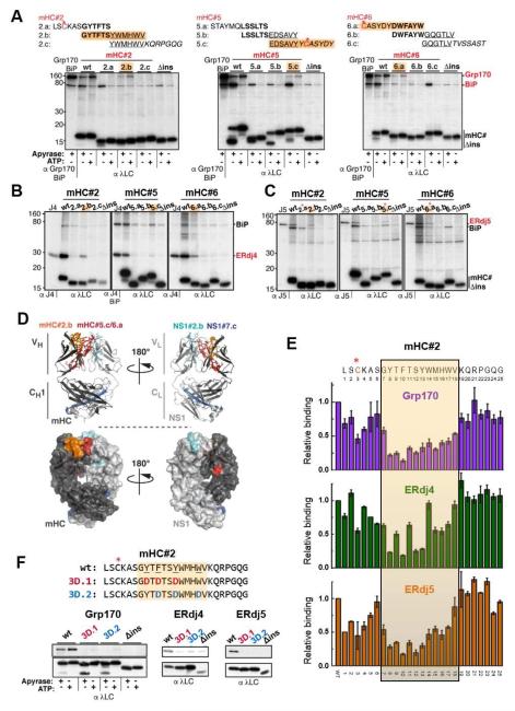
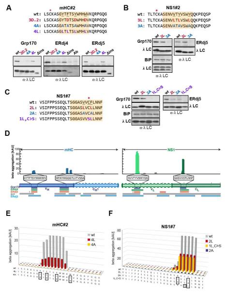
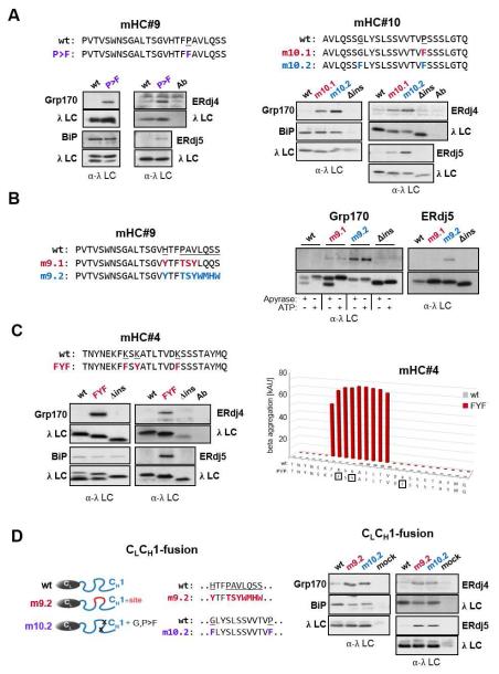
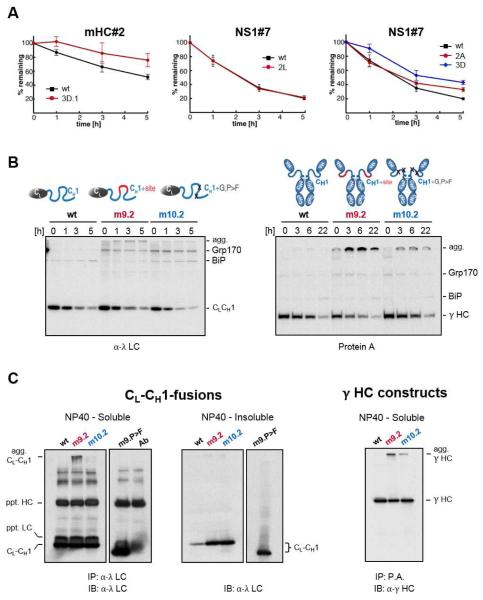
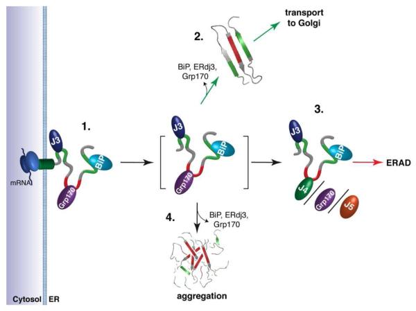
Protein maturation in the endoplasmic reticulum is controlled by multiple chaperones, but how they recognize and determine the fate of their clients remains unclear. We developed an in vivo peptide library covering substrates of the ER Hsp70 system: BiP, Grp170, and three of BiP's DnaJ-family co-factors (ERdj3, ERdj4, and ERdj5). In vivo binding studies revealed that sites for pro-folding chaperones BiP and ERdj3 were frequent and dispersed throughout the clients, whereas Grp170, ERdj4, and ERdj5 specifically recognized a distinct type of rarer sequence with a high predicted aggregation potential. Mutational analyses provided insights into sequence recognition characteristics for these pro-degradation chaperones, which could be readily introduced or disrupted, allowing the consequences for client fates to be determined. Our data reveal unanticipated diversity in recognition sequences for chaperones; establish a sequence-encoded interplay between protein folding, aggregation, and degradation; and highlight the ability of clients to co-evolve with chaperones, ensuring quality control.


Figures

Figure 1
Figure 1. An in vivo peptide library reveals distinct substrate binding patterns for BiP and Grp170
(A) A series of 25 amino acid overlapping peptides corresponding to the mHC and NS1 were inserted into a flexible GS-linker downstream of an ER-targeted λ LC constant domain (CL) ending with a C-terminal NVT glycosylation site or QVT. Only upon ER entry is the NVT site modified (red hexagon). EndoH resistance confirmed that NS1 and mHC are glycoproteins (right). (B) After transfection into COS-1 cells, metabolically labeled peptide constructs were immunoprecipitated with anti-λ antiserum, treated with (+) or without (−) EndoH and analyzed by SDS-PAGE. (C) COS-1 cells co-expressing Grp170, BiP, and the indicated peptide constructs were metabolically labeled, lysed with ATP or apyrase added. Peptide constructs were isolated with anti-λ and separated on SDS gels. Cells transfected with only Grp170 and BiP serve as markers and the full-length client for each set of peptides is included as a reference for chaperone binding.
Figure 2
Figure 2. ERdj3 binds frequently throughout mHC and NS1, while ERdj4 and ERdj5 possess fewer, mostly shared binding motifs
(A) COS-1 cells were co-transfected with ERdj3 and BiP together with the indicated peptide constructs and metabolically labeled, DSP-crosslinked and lysed in RIPA buffer. Peptide constructs were immunoprecipitated with anti-λ antiserum and analyzed by reducing SDS-PAGE. Cells transfected with ERdj3 and BiP alone were immunoprecipitated with antiserum specific for ERdj3 as molecular weight marker, and full-length clients were included as a reference for chaperone binding. Levels of ERdj3 expression in each sample by immunoblotting are shown under the panel. (B) Binding of mHC and NS1 peptides to ERdj4 was performed as in (A), except that HA-tagged ERdj4 was co-transfected and cells were lysed directly in NP40-lysis buffer. (C) Binding of ERdj5 to mHC and NS1 peptides was performed as in (B), except that ERdj5 was co-transfected and the lysis/washing buffers contained 20 mM NEM.
Figure 3
Figure 3. Grp170/ERdj4/ERdj5 recognize shared motifs that are buried after protein folding or subunit assembly
(A) The three 25 amino acid peptides in the mHC recognized by Grp170 were further subdivided into 12 amino acid overlapping peptides as shown above the gel images. COS-1 cells were co-transfected with Grp170 and BiP along with the indicated reporter constructs and analyzed as in Figure 1C. Peptides recognized by Grp170 are highlighted in orange, and cysteines are marked by red asterisks. (B-C) The same subdivided peptides were examined for ERdj4 (B) or ERdj5 (C) binding as described in Figure 2. Interacting peptides are indicated in orange, and in (C) cysteine containing peptides are marked with a red asterisk. (D) Binding sites shared by Grp170, ERdj4 and ERdj5 were mapped on a model of a mHC-NS1 heterodimer (top: ribbon representation, bottom: surface representation). The mHC is shown in dark grey with mHC#2.b in orange and #5.c/#6.a in red. NS1 is shown in light grey with NS1#2.b in turquois and #7.c in blue. (E) Each amino acid in the mHC#2 peptide was individually mutated to Asp. The subdivided peptide that bound all three (co-)chaperones is indicated with an orange box. COS-1 cells transfected with Grp170, ERdj4 or ERdj5 together with BiP and each of the constructs were metabolically labeled and analyzed as before. The levels of the co-precipitated (co-)chaperones were quantified as described in Extended Experimental Procedures, and are shown ±SEM (n≥3). The value obtained for the parent mHC#2 (wt) was set to 1. (F) (Co-)chaperone binding to the mHC#2 wild-type and mutant constructs was determined by immunoprecipitation with α-λ and blotted with the appropriate (co-)chaperone (top) or λ (bottom) antiserum.
Figure 4
Figure 4. Grp170, ERdj4 and ERdj5 binding sites correspond to aggregation-prone regions in their clients
(A-C) Cells expressing the indicated (co-)chaperones together with mHC#2 (A), NS1#2 (B) or NS1#7 (C) peptide constructs were analyzed by immunoprecipitation-coupled western blotting as described in Figure 3F. The 12-aa fragments identified in Figure 3 and S3 are indicated with orange boxes. The top panel in each set shows anti-(co-)chaperone western blots and the bottom anti-λ blots. For ERdj4, a separate lane with the anti-λ antibody was included and samples were run under-non-reducing conditions to reduce background signal from the precipitating antibody. (D) Sequences for the mini-HC (blue) and NS1 LC (green) were subjected to analysis by the TANGO algorithm, which identifies regions that are predicted to be prone to β-aggregate formation. Wavy segments indicate unfolded domains in each client (when expressed in isolation). The core binding peptides for Grp170 (purple), ERdj4 (green), and ERdj5 (orange) are indicated below, as are binding sites for BiP (grey) and ERdj3 (blue). (E-F) The TANGO algorithm was used to calculate the β-aggregation potential for the indicated mHC#2 (E) and NS1#7 (F) constructs. Each mutant is color-coded with sequences indicated below and mutated residues boxed.
Figure 5
Figure 5. Mutations predicted to increase aggregation lead to (co-)chaperone binding and can result in aggregation of full-length clients
(A) Putative (co-)chaperone binding sites were introduced into mHC#9 and mHC#10 with the indicated substitutions and analyzed by immunoprecipitation-coupled western blotting for (co-)chaperone binding as before. (B) The indicated substitutions were made in mHC#9 to create a partial (m9.1) or the complete (m9.2) binding site identified in mHC#2. Constructs were analyzed as in (A). In the case of Grp170 binding samples were lysed with apyrase or ATP added. (C) The TANGO algorithm was used to predict amino acid changes that would lead to an aggregation-prone region in mHC#4, which was negative for both BiP and (co-)chaperone binding. The wild-type and mutant constructs were examained for BiP and (co-)chaperone binding as above. (D) Mutations that led to (co-)chaperone binding in either mHC#9 or #10 were engineered into the corresponding region of the CH1 domain in a chimeric protein composed of the ER targeted λ CL domain and the γ1 CH1 domain. (Co-)chaperone interactions and BiP binding were detected by immunoprecipitation-coupled western blotting.
Figure 6
Figure 6. Introduction or disruption of (co-)chaperone binding sites alters the fate of client proteins in vivo
(A) COS-1 cells expressing the indicated mHC#2 and NS1#7 constructs were pulse-labeled and chased for the indicated times. Immunoprecipitated peptide constructs were analyzed by SDS-PAGE, signals were quantified by phosphorimaging and the amount of remaining constructs over time was calculated ±SEM (n≥3). (B) Grp170 and BiP were co-expressed with the wild-type CL-CH1-fusion protein (left) and with the corresponding mutants in the full-length γ HC (right). Cells were pulse-labeled and chased for the indicated times. Immunoprecipitated material was analyzed by reducing SDS-PAGE and migration of clients and chaperones are marked. Slower migrating aggregated clients are indicated (agg). (C) COS-1 cells were co-transfected with Grp170, wild-type (D), and the indicated CL-CH1-fusion proteins. Cells were lysed in NP40 lysis buffer and soluble material from the indicated transfectants was immunoprecipitated with anti-λ (CL-CH1-fusion) and blotted with anti-λ antisera. The antibody control reveals bands present in the antiserum used for immunoprecipation, which are recognized when the same anti-λ serum is used for blotting. Equal amounts of NP40-insoluble material was directly analyzed by western blotting and the images of soluble and insoluble for each mutant are from the same exposure.
Figure 7
Figure 7. A model predicting how members of the ER Hsp70 chaperone system might interact to control the fate of client proteins
(1.) Proteins entering the ER can encounter the (co-)chaperones ERdj3, BiP, and Grp170, which reside in proximity to the translocon. Grp170 recognizes sequences (indicated in red) that are distinct from BiP and ERdj3 binding sequences (indicated in green). (2.) As folding procedes, these (co-)chaperones are released allowing the properly matured client to be transported to the Golgi. Grp170/ERdj4/ERdj5 binding sites (indicated in red) become buried during folding, protecting them from aggregation. (3.) If folding fails, the sites recognized by Grp170/ERdj4/ERdj5 remain exposed, leading the client to be targeted for ERAD. (4.) Release of BiP, ERdj3, and Grp170 in the absence of folding can lead to aggregation. Thus, either rapid burial of the Grp170/ERdj4/ERdj5 recognition sites upon folding or their detection for the purpose of ERAD is critical, as these sites possess high aggregation propensity.

Comment in

References

    1. Ahmed AB, Kajava AV. Breaking the amyloidogenicity code: methods to predict amyloids from amino acid sequence. FEBS Lett. 2013;587:1089–1095. - PubMed
    1. Behnke J, Hendershot LM. The large hsp70 grp170 binds to unfolded protein substrates in vivo with a regulation distinct from conventional hsp70s. J Biol. Chem. 2014;289:2899–2907. - PMC - PubMed
    1. Blond-Elguindi S, Cwirla SE, Dower WJ, Lipshutz RJ, Sprang SR, Sambrook JF, Gething MJ. Affinity panning of a library of peptides displayed on bacteriophages reveals the binding specificity of BiP. Cell. 1993;75:717–728. - PubMed
    1. Bole DG, Hendershot LM, Kearney JF. Posttranslational association of immunoglobulin heavy chain binding protein with nascent heavy chains in nonsecreting and secreting hybridomas. J. Cell. Biol. 1986;102:1558–1566. - PMC - PubMed
    1. Braakman I, Hebert DN. Protein folding in the endoplasmic reticulum. Cold Spring Harb. Perspect. Biol. 2013;5:a013201. - PMC - PubMed

MeSH terms