Review
. 2016 Sep;39(9):625-637.
doi: 10.1016/j.tins.2016.07.002. Epub 2016 Aug 18.

Apolipoprotein E and Sex Bias in Cerebrovascular Aging of Men and Mice

Affiliations
Review

Apolipoprotein E and Sex Bias in Cerebrovascular Aging of Men and Mice

Caleb E Finch et al. Trends Neurosci. 2016 Sep.

Abstract

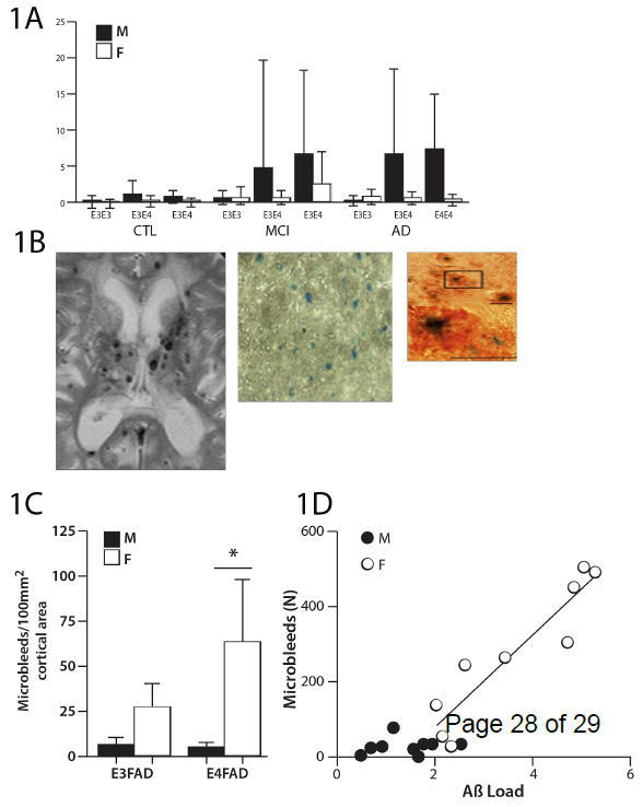
Alzheimer disease (AD) research has mainly focused on neurodegenerative processes associated with the classic neuropathologic markers of senile plaques and neurofibrillary tangles. Additionally, cerebrovascular contributions to dementia are increasingly recognized, particularly from cerebral small vessel disease (SVD). Remarkably, in AD brains, the apolipoprotein E (ApoE) ɛ4 allele shows male excess for cerebral microbleeds (CMBs), a marker of SVD, which is opposite to the female excess of plaques and tangles. Mouse transgenic models add further complexities to sex-ApoE ɛ4 allele interactions, with female excess of both CMBs and brain amyloid. We conclude that brain aging and AD pathogenesis cannot be understood in humans without addressing major gaps in the extent of sex differences in cerebrovascular pathology.

Keywords: apolipoprotein E; cerebral amyloid angiopathy; cerebral microbleeds; cerebrovasculature; magnetic resonance imaging; sex; small vessel disease.


Figures

Figure 1
Figure 1. Cerebral microbleeds
Figures are redrawn from [4] except for B panel 1. A) Human microbleed frequencies analyzed by apoE ε4 alleles in cohorts in two large memory clinics: ADNI (Alzheimer Disease Neuroimaging Initiative) and KIDS (Karolinska Imaging Dementia Study). Error bars denote the standard error of the mean. Male AD patients show a statistically significant excess of CMBs over female patients (P<0.001); the male CMB excess is further exacerbated by ε4(P<0.001), as tested by the Mann-Whitney U-test. AD, Alzheimer disease (ε3/ ε3, n= M42/F33; ε3/ ε4, n=M46/F60; ε4/ ε4, n=M25/F36); CMBs, cerebral microbleeds; F= Female; M= Male; MCI, mild cognitive impairment (ε3/ ε3, n= M144/F133; ε3/ ε4, n=M120/F88; ε4/ ε4, n=M37/F33); CTL, controls (ε3/ ε3, n= M84/F98; ε3/ ε4, n=M43/F66; ε4/ ε4, n=M5/F6). B) CMB images: panel 1, male AD patient, with multiple CMBs on a T2* weighted MR image; panel 2, female EFAD-mouse at three magnifications showing whole field stained for hemosiderin by Prussian Blue and individual microbleeds Prussian Blue and co-immunostained for Aβ (orange). The majority (67%) of hemosiderin puncta resided within Aβ deposits. C) EFAD mice: CMB frequencies show female excess with additive effect of ε4. D) EFAD mice: The number of CMBs regressed against the Aβ load in the cerebral cortex of individual mice showed linear increase above a threshold level of 1.7 Aβ units; this relationship holds for females, but not males. Both apoE alleles were included.
Figure 2
Figure 2
MR imaging markers of small vessel disease: A) Cerebral microbleeds (black arrows) are seen as dark round hypointensities on the gradient echo T2* MR image. Note the larger bleed in the right thalamus. The size of microbleeds on the image slightly exaggerates their size in vivo. B) Cortical superficial siderosis (black arrows) are seen on MR as characteristically dark, linear, and gyriform, and are considered a sensitive marker of CAA [110]. C) Enlarged perivascular spaces in the centrum semiovale on the T2-weighted MRI sequence. Perivascular spaces in the centrum semiovale and basal ganglia are thought to represent CAA, and hypertensive arteriopathy respectively. D) White matter hyperintensities are seen best on FLAIR MRI. White matter hyperintensities are thought to be multifactorial, and a marker of SVD. E) Lacune (arrow). Seen as CSF-signal on T1 and T2, sometimes with a surrounding hyperintense rim. F) Cortical microinfarct (arrow), seen as <5 mm, hypointensities, perpendicular to the cortex. Images are from the KIDS cohort[10].

References

    1. Mahley RW, et al. Apolipoprotein E: structure determines function, from atherosclerosis to Alzheimer’s disease to AIDS. J Lipid Res. 2009;50(Suppl):S183–188. - PMC - PubMed
    1. Sing CF, Davignon J. Role of the apolipoprotein E polymorphism in determining normal plasma lipid and lipoprotein variation. Am J Hum Genet. 1985;37:268–285. - PMC - PubMed
    1. Fullerton SM, et al. Apolipoprotein E Variation at the Sequence Haplotype Level: Implications for the Origin and Maintenance of a Major Human Polymorphism. Am J Hum Genet. 2000;67:881–900. - PMC - PubMed
    1. Leduc V, et al. APOE and cholesterol homeostasis in Alzheimer’s disease. Trends Mol Med. 2010;16:469–477. - PubMed
    1. Dey AK, et al. Pathoconnectomics of cognitive impairment in small vessel disease: A systematic review. Alzheimers Dement J Alzheimers Assoc. 2016 doi: 10.1016/j.jalz.2016.01.007. - DOI - PubMed

Publication types