Review
. 2016 Aug 7;22(29):6595-609.
doi: 10.3748/wjg.v22.i29.6595.

Management of a large mucosal defect after duodenal endoscopic resection

Affiliations
Review

Management of a large mucosal defect after duodenal endoscopic resection

Shintaro Fujihara et al. World J Gastroenterol. .

Abstract

Duodenal endoscopic resection is the most difficult type of endoscopic treatment in the gastrointestinal tract (GI) and is technically challenging because of anatomical specificities. In addition to these technical difficulties, this procedure is associated with a significantly higher rate of complication than endoscopic treatment in other parts of the GI tract. Postoperative delayed perforation and bleeding are hazardous complications, and emergency surgical intervention is sometimes required. Therefore, it is urgently necessary to establish a management protocol for preventing serious complications. For instance, the prophylactic closure of large mucosal defects after endoscopic resection may reduce the risk of hazardous complications. However, the size of mucosal defects after endoscopic submucosal dissection (ESD) is relatively large compared with the size after endoscopic mucosal resection, making it impossible to achieve complete closure using only conventional clips. The over-the-scope clip and polyglycolic acid sheets with fibrin gel make it possible to close large mucosal defects after duodenal ESD. In addition to the combination of laparoscopic surgery and endoscopic resection, endoscopic full-thickness resection holds therapeutic potential for difficult duodenal lesions and may overcome the disadvantages of endoscopic resection in the near future. This review aims to summarize the complications and closure techniques of large mucosal defects and to highlight some directions for management after duodenal endoscopic treatment.

Keywords: Bleeding; Clip; Closure; Complication; Duodenum; Endoscopic full-thickness resection; Endoscopic mucosal resection; Endoscopic submucosal dissection; Over-the-scope clip; Perforation.


Figures

Figure 1
Figure 1
Schema of endoscopic closure with through-the-scope-clips and over-the scope-clip. TTSC: Through-the-scope-clips; OTSC: Over-the scope-clip.
Figure 2
Figure 2
Immediate perforation after duodenal endoscopic mucosal resection with circumferential mucosal incision. A: An adenoma, 20 mm in size with an ulcer scar, was located in the inferior wall of the duodenal bulb; B: After duodenal EMR, immediate perforation occurred; C, D: The perforation was closed using multiple hemoclips. EMR: Endoscopic mucosal resection.
Figure 3
Figure 3
Delayed bleeding after duodenal endoscopic submucosal dissection. A: An adenoma, 20 mm in size, was located in the posterior wall of the second part of the duodenum; B: Prophylactic closure with a hemoclip was performed after ESD; C: The patient complained of melena at post-ESD day 13, and esophagoduodenoscopy revealed bleeding at the base of the ulcer because several clips had fallen off; D: The large mucosal defect was closed with multiple clips and Endoloops. ESD: Endoscopic submucosal dissection.
Figure 4
Figure 4
Prophylactic closure after duodenal endoscopic mucosal resection. A: Chromoendoscopy with indigo carmine was performed before endoscopic resection; B: A small-sized mucosal defect was detected; C, D: Prophylactic closure was performed with a hemoclip.
Figure 5
Figure 5
Diagram of a closure technique with the additional use of a loop clip, 8-ring loop, and small incision around the mucosal defect using a needle-type knife.
Figure 6
Figure 6
Diagram of a closure technique with the additional use of endoloop and a ring-shaped thread.
Figure 7
Figure 7
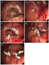
Methods for endoscopic sliding closure after endoscopic submucosal dissection. A: A mucosal defect was identified after ESD; B, C: The ring thread was clipped at two points across the maximal diameter of the mucosal defect; D: A third clip was placed across to the edge of the mucosal defect, and after pulling the first and second clips, the mucosal defect area slid together; E: The mucosal defect was smaller, and closing it with additional clips was easier. ESD: Endoscopic submucosal dissection.

References

    1. Kakushima N, Kanemoto H, Tanaka M, Takizawa K, Ono H. Treatment for superficial non-ampullary duodenal epithelial tumors. World J Gastroenterol. 2014;20:12501–12508. - PMC - PubMed
    1. Matsumoto S, Miyatani H, Yoshida Y. Future directions of duodenal endoscopic submucosal dissection. World J Gastrointest Endosc. 2015;7:389–395. - PMC - PubMed
    1. Gaspar JP, Stelow EB, Wang AY. Approach to the endoscopic resection of duodenal lesions. World J Gastroenterol. 2016;22:600–617. - PMC - PubMed
    1. Yamamoto Y, Yoshizawa N, Tomida H, Fujisaki J, Igarashi M. Therapeutic outcomes of endoscopic resection for superficial non-ampullary duodenal tumor. Dig Endosc. 2014;26 Suppl 2:50–56. - PubMed
    1. Matsumoto S, Yoshida Y. Selection of appropriate endoscopic therapies for duodenal tumors: an open-label study, single-center experience. World J Gastroenterol. 2014;20:8624–8630. - PMC - PubMed

MeSH terms