

Assessing the prevalence of paranasal sinuses anatomical variants in patients with sinusitis using Cone Beam Computer Tomography
- PMID: 27547064
- PMCID: PMC4990440
- DOI: 10.15386/cjmed-598
Assessing the prevalence of paranasal sinuses anatomical variants in patients with sinusitis using Cone Beam Computer Tomography
Abstract
Background and aims: To asses, by using the Cone Beam CT (CBCT) reformatted images, the presence of anatomical variants of the sinonasal cavities and to determine the correlation of these variations with the onset of maxillary sinus inflammations.
Method: The study is a retrospective one and consists of the investigation of 130 patients with CBCT imaging, patients that were referred to the Maxillo-Facial Clinic, Radiology Department of the Iuliu Hatieganu University of Medicine and Pharmacy in Cluj-Napoca, for clinical symptoms of sinusitis within a period of 24 months. The images were analyzed for the presence of different anatomical variations and sinus inflammation. The CBCT images were obtained using a NewTom 3G scanner and the data acquired were statistically analyzed using Chi-square test, Odds ratio data and confidence intervals, with a determined p<0.05 considered to be statistically significant.
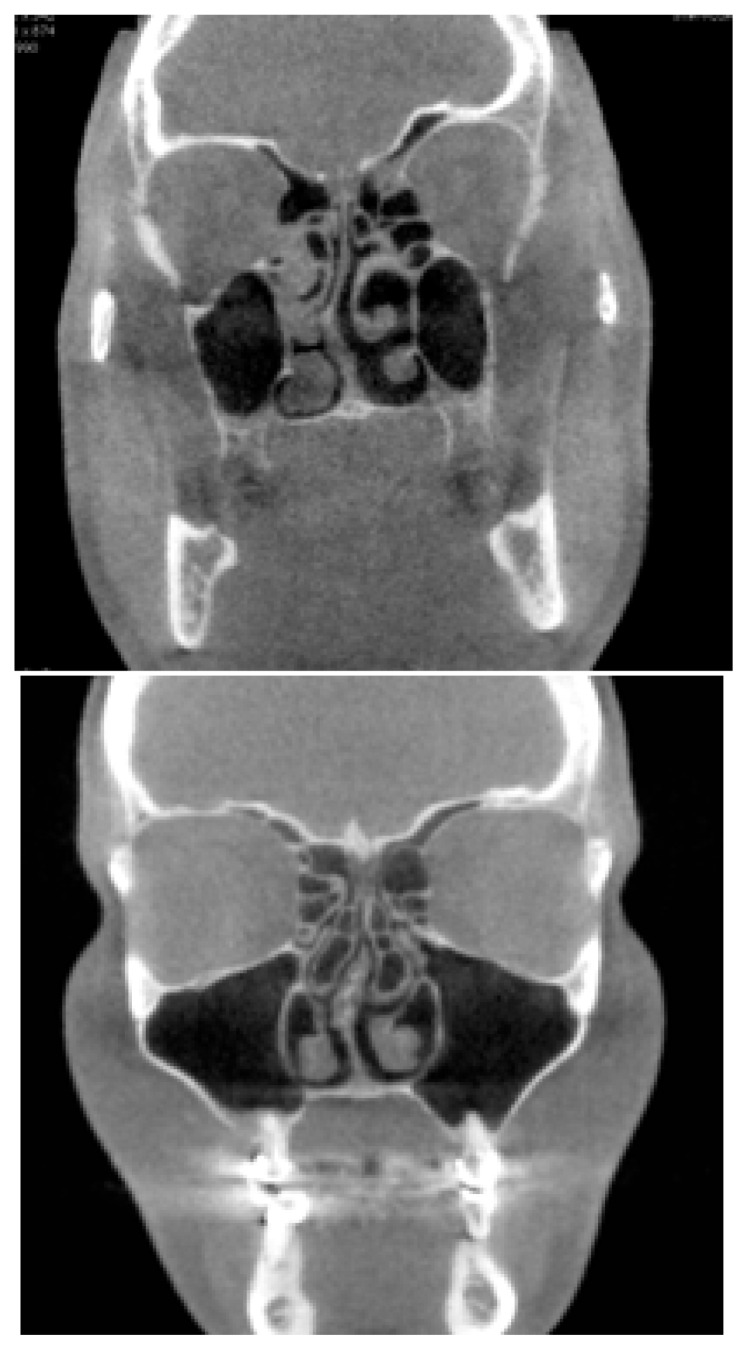
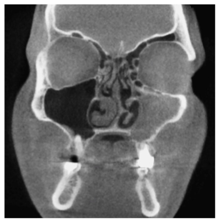
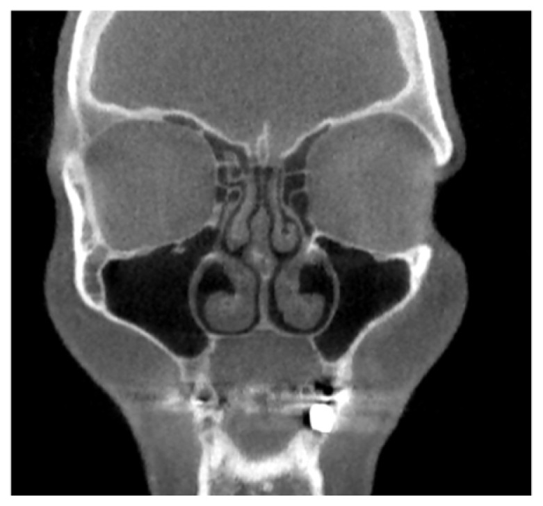
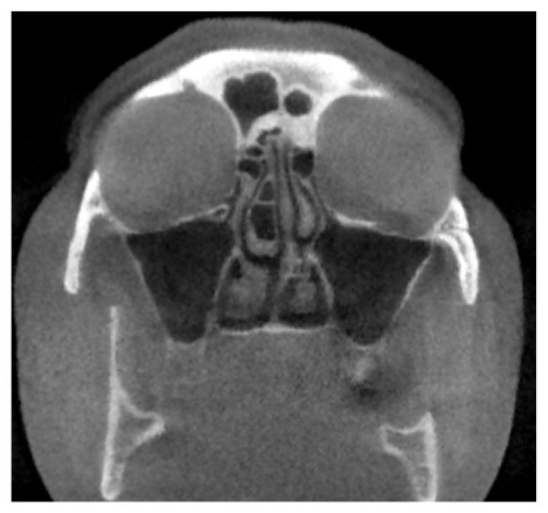
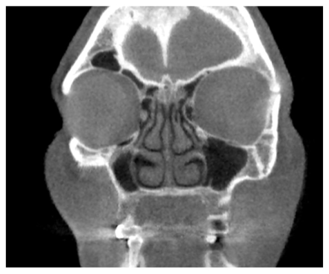
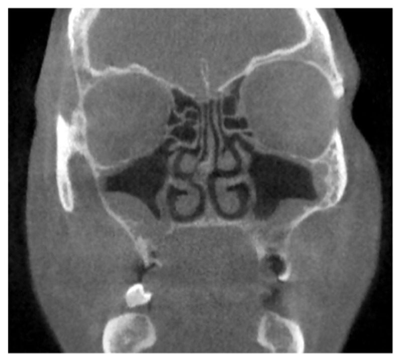
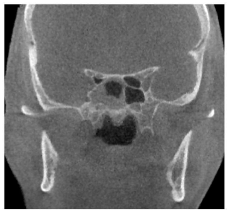
Results: The anatomical variants were detected both in the inflammation and control group. From the spectrum of variations concha bullosa, deviation of uncinate process and asymmetrical ethmoid roof presented significant association with sinusitis. The deviated position of the uncinate process appeared in more than 50% of patients in the positive group [OR=2.55] compared with a third of the control group. Concha bullosa was observed in 31% cases, 23% in the control group and 34% in the positive group [OR=1.73]. Haller cells showed a small difference between groups [OR=1.14] whereas the ethmoid roof asymmetry was evidently more prevalent in the inflammation group.
Conclusion: The anatomical variants of the paranasal sinuses are not incidental, being found in a large number of patients and may be a predisposing factor in the onset and recurrence of sinuses inflammation. The CBCT technique, due to the finest multiplanar reconstruction, permits a very good pre-therapeutic assessment of these predisposing conditions.
Keywords: CBCT; anatomical variants; maxillary sinuses.
Figures
References
-
- Nouraei SA, Elisay AR, Dimarco A, Abdi R, Majidi H, Madani SA, et al. Variations in paranasal sinus anatomy: implications for the pathophysiology of chronic rhinosinusitis and safety of endoscopic sinus surgery. J Otolaryngol Head Neck Surg. 2009;38(1):32–37. - PubMed
-
- Limeme M, Benzina M, Majerbi M, Kheireddine N, Zaghouani H, et al. Anatomical variants in the sino-nasal region: a pictorial review. EPOS. doi: 10.1594/ecr2015/C-2406. ECR 2015. C-2406. - DOI
-
- Alkire BC, Bhattacharyya N. An assessment of sinonasal anatomic variants potentially associated with recurrent acute rhinosinusitis. Laryngoscope. 2010;120:631–634. - PubMed
LinkOut - more resources
Full Text Sources
Other Literature Sources
Molecular Biology Databases