Review
. 2016 Aug 3:5:F1000 Faculty Rev-1909.
doi: 10.12688/f1000research.8881.1. eCollection 2016.

Recent advances in cohesin biology

Affiliations
Review

Recent advances in cohesin biology

Susannah Rankin et al. F1000Res. .

Abstract

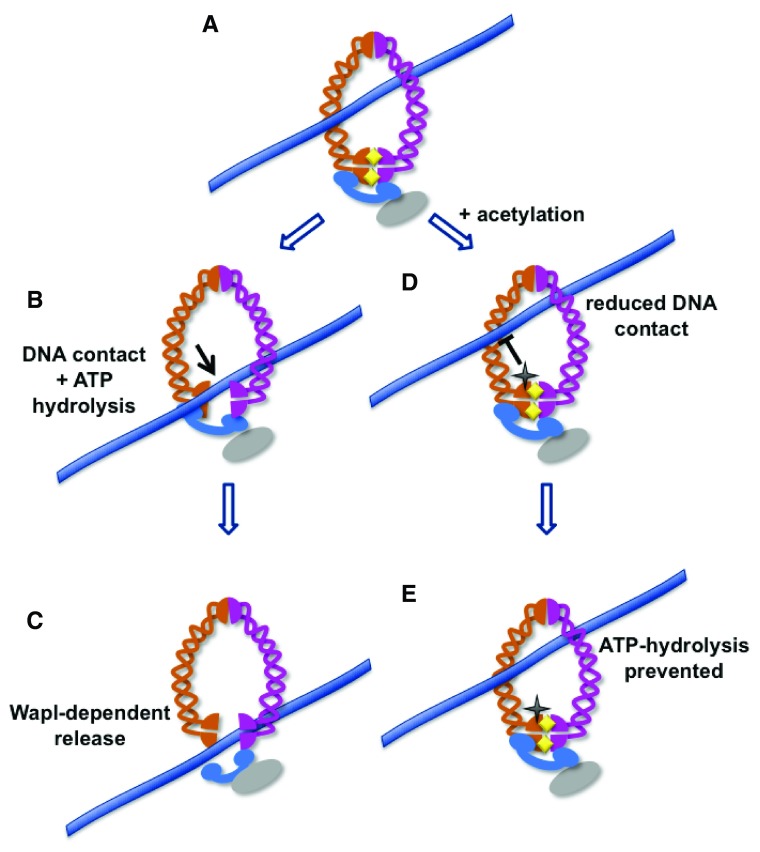
Sister chromatids are tethered together from the time they are formed in S-phase until they separate at anaphase. A protein complex called cohesin is responsible for holding the sister chromatids together and serves important roles in chromosome condensation, gene regulation, and the repair of DNA damage. Cohesin contains an open central pore and becomes topologically engaged with its DNA substrates. Entrapped DNA can be released either by the opening of a gate in the cohesin ring or by proteolytic cleavage of a component of the ring. This review summarizes recent research that provides important new insights into how DNA enters and exits the cohesin ring and how the rings behave on entrapped DNA molecules to provide functional cohesion.

Keywords: chromosome; cohesin; mitosis.


Conflict of interest statement

Competing interests: The authors declare that they have no competing interests.

No competing interests were disclosed.

Figures

Figure 1.
Figure 1.. The cohesin complex.
The four-subunit complex contains two SMC proteins, each of which forms long antiparallel coiled-coils. The SMC proteins interact in two places: where they fold back on themselves (hinge domains) and at head domains formed by interaction of their N and C termini. The interactions of the Smc1 and Smc3 head domains together form two intermolecular ATPases. ATP is indicated by yellow diamonds. Note that the Smc1 and 3 subunits, when interacting at both the hinge and the ATPase domains, would form a structure with an internal pore. The Mcd1 subunit (blue) interacts with both SMC head domains. Because the ATPase domains interact, a second pore may exist between Mcd1 and the Smc proteins. Separation of the Smc ATPase domains by ATP hydrolysis would create a single pore surrounded by Mcd1, Smc3, and Smc1. The Scc3 subunit, shown in grey, is not thought to form part of the ring.
Figure 2.
Figure 2.. Model for DNA unloading through the Smc3-Mcd1 gate.
DNA is loaded into the cohesin ring, which is closed in part through interaction of the Smc1 and Smc3 head domains ( A). Once loaded into the cohesin ring, interaction of DNA (lavender line) with the head domain of Smc3 can stimulate ATPase activity at the Smc3-Smc1 interface, resulting in partial opening of the cohesin ring ( B). This allows Wapl to open the Smc3-Rad21 interface, fully releasing the DNA ( C) . In contrast, acetylation of the Smc3 head domain, indicated by the black star, would prevent close interaction of the DNA with the head domains, preventing ATP hydrolysis ( D) and thus preventing ring opening ( E). In this model, acetylation of the Smc3 head domain might prevent both entry and exit of DNA.
Figure 3.
Figure 3.. Different models for functional interaction of cohesin with sister chromatids.
In the most basic “embrace” model ( A), both sister chromatids are entrapped together within individual cohesin rings, which are loaded either before or during DNA replication. The finding that two different non-functional alleles of individual cohesin subunits are able to promote cohesion when expressed in the same cell is consistent with the notion that higher order cohesin interactions can promote sister chromatid tethering . Examples include the handcuff ( B) and stacked cohesin ( C) models.
Figure 4.
Figure 4.. Single molecule analysis of cohesin’s interaction with DNA.
Purified cohesin incubated with tethered and extended DNA molecules shows the behaviors illustrated . The presence of the loader complex (not shown) results in the highly stable interaction of cohesin with DNA. Once loaded, cohesin appears to move freely along the length of the DNA, unless there is an obstacle of greater than ~11 nm in size. Cohesin is released from the untethered end of the DNA molecule (shown at left) and stops when it reaches the tethered end (right).

References

    1. Murayama Y, Uhlmann F: Biochemical reconstitution of topological DNA binding by the cohesin ring. Nature. 2014;505(483):367–71. 10.1038/nature12867 - DOI - PMC - PubMed
    1. Mc Intyre J, Muller EG, Weitzer S, et al. : In vivo analysis of cohesin architecture using FRET in the budding yeast Saccharomyces cerevisiae. EMBO J. 2007;26(16):3783–93. 10.1038/sj.emboj.7601793 - DOI - PMC - PubMed
    1. Peters J, Nishiyama T: Sister chromatid cohesion. Cold Spring Harb Perspect Biol. 2012;4(11): pii: a011130. 10.1101/cshperspect.a011130 - DOI - PMC - PubMed
    1. Rowland BD, Roig MB, Nishino T, et al. : Building sister chromatid cohesion: smc3 acetylation counteracts an antiestablishment activity. Mol Cell. 2009;33(6):763–74. 10.1016/j.molcel.2009.02.028 - DOI - PubMed
    1. Rolef Ben-Shahar T, Heeger S, Lehane C, et al. : Eco1-dependent cohesin acetylation during establishment of sister chromatid cohesion. Science. 2008;321(5888):563–6. 10.1126/science.1157774 - DOI - PubMed

LinkOut - more resources