. 2016 Aug 3:4:e2312.
doi: 10.7717/peerj.2312. eCollection 2016.

The pes of Australovenator wintonensis (Theropoda: Megaraptoridae): analysis of the pedal range of motion and biological restoration

Affiliations

The pes of Australovenator wintonensis (Theropoda: Megaraptoridae): analysis of the pedal range of motion and biological restoration

Matt A White et al. PeerJ. .

Abstract

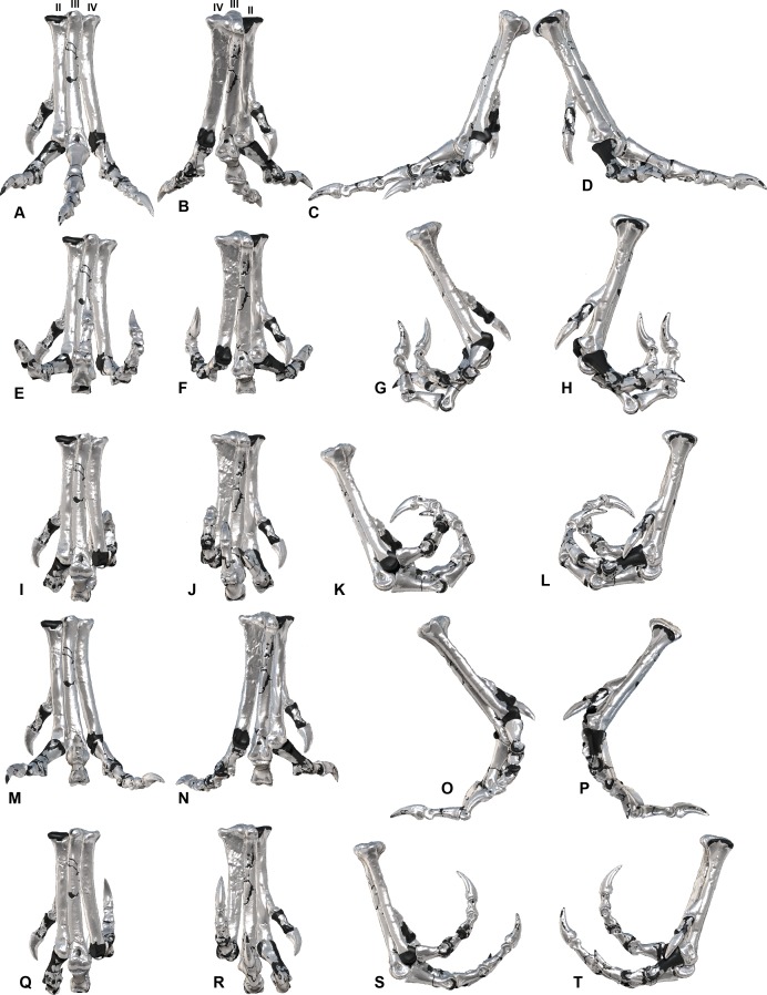
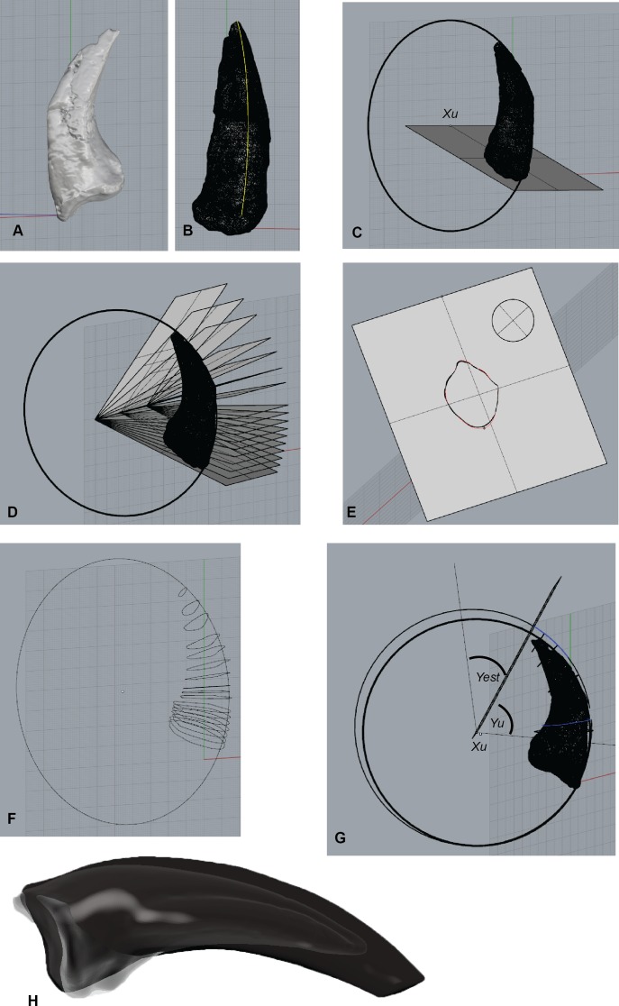
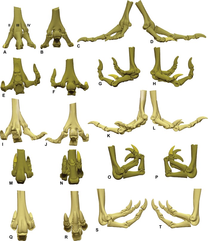
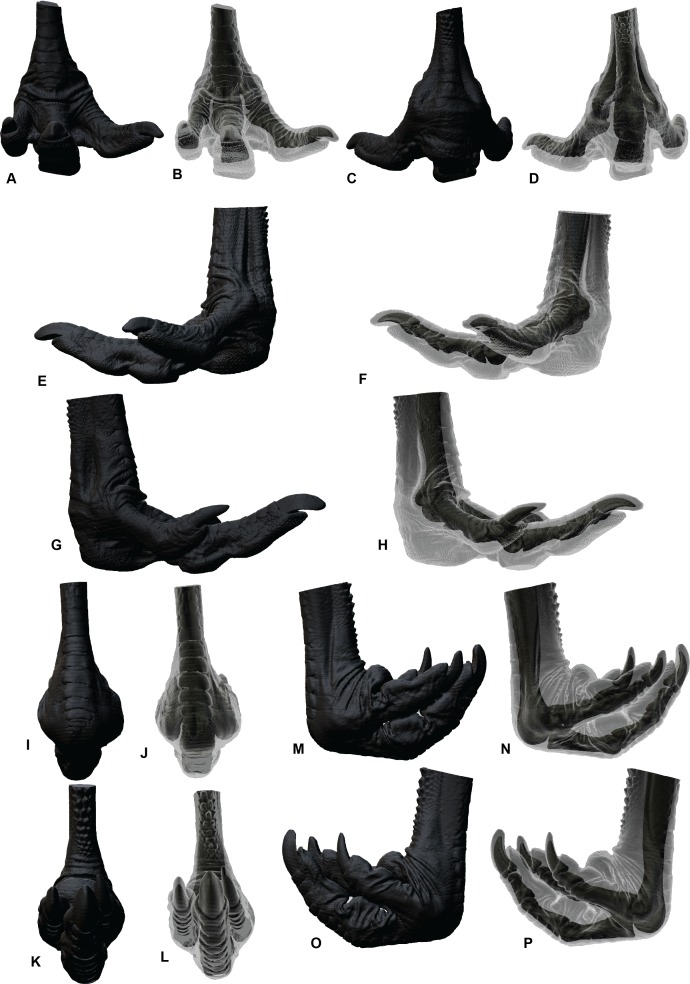
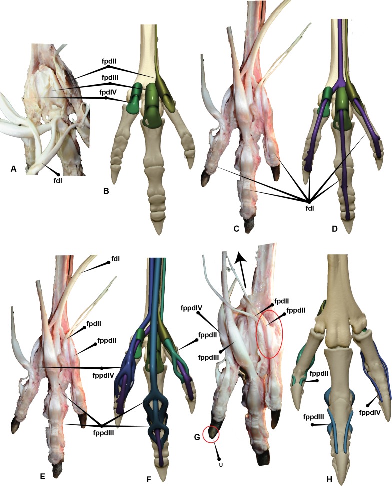
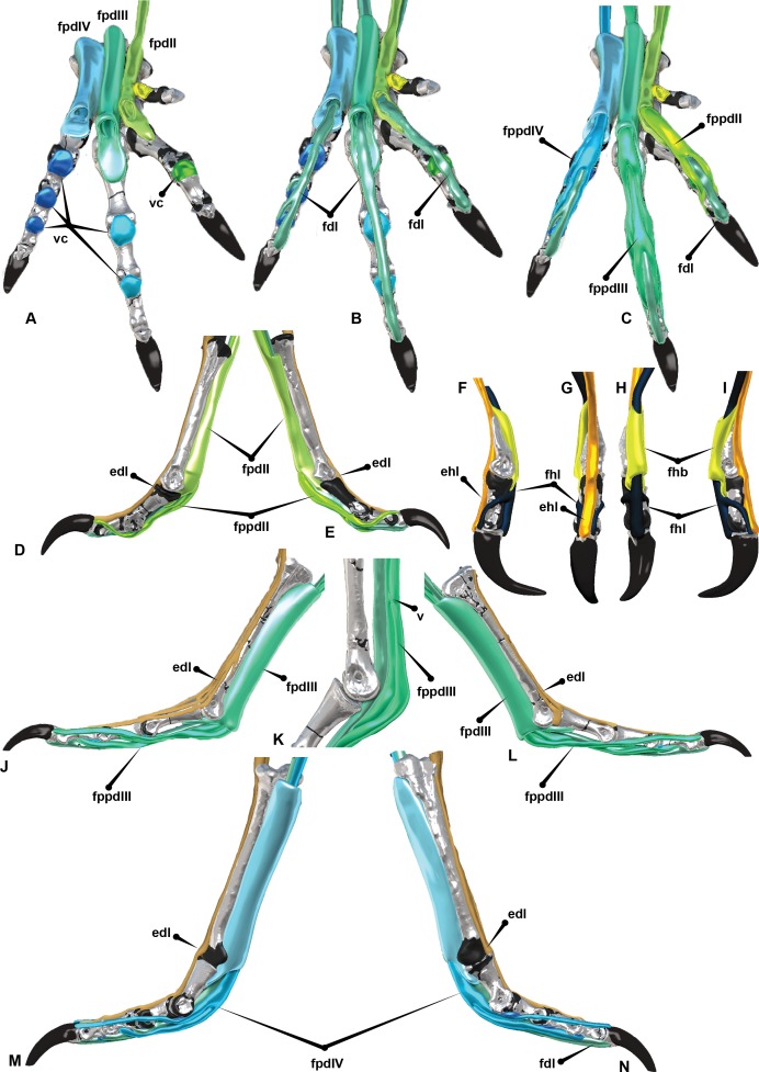
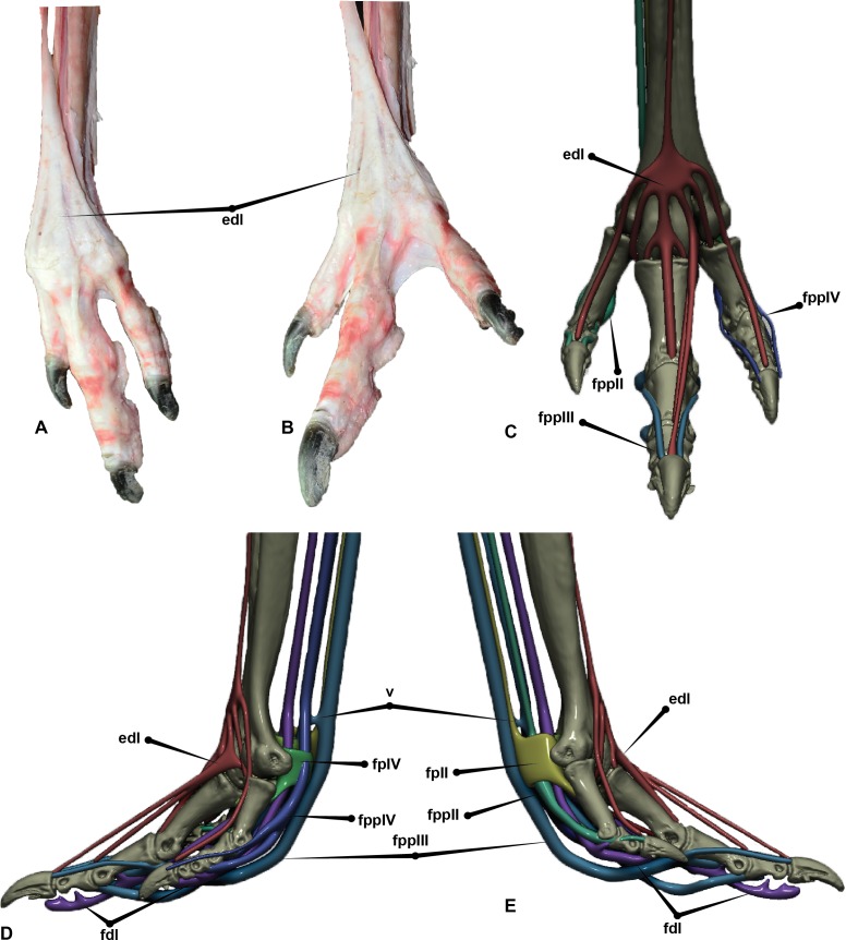
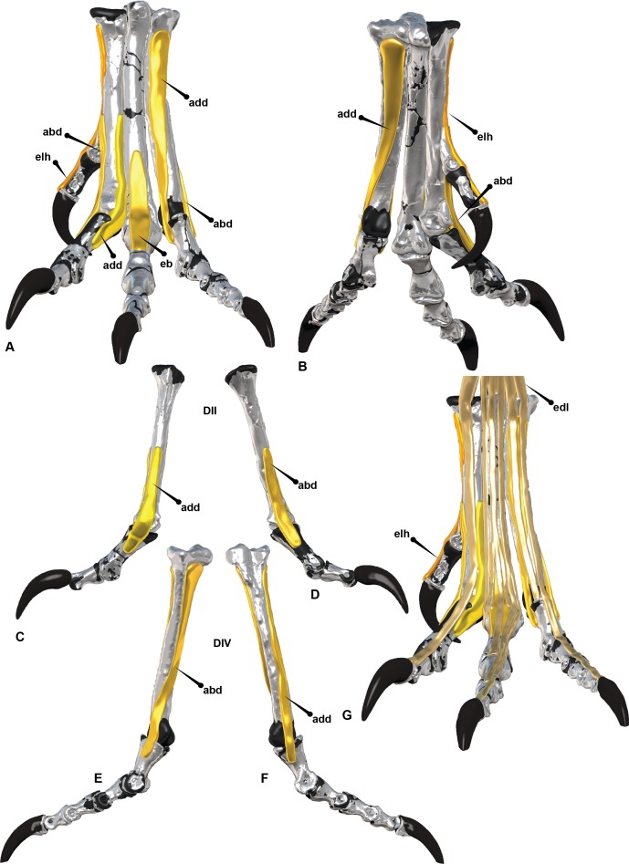
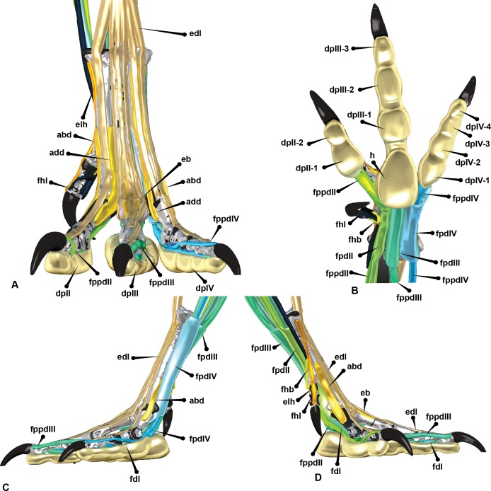
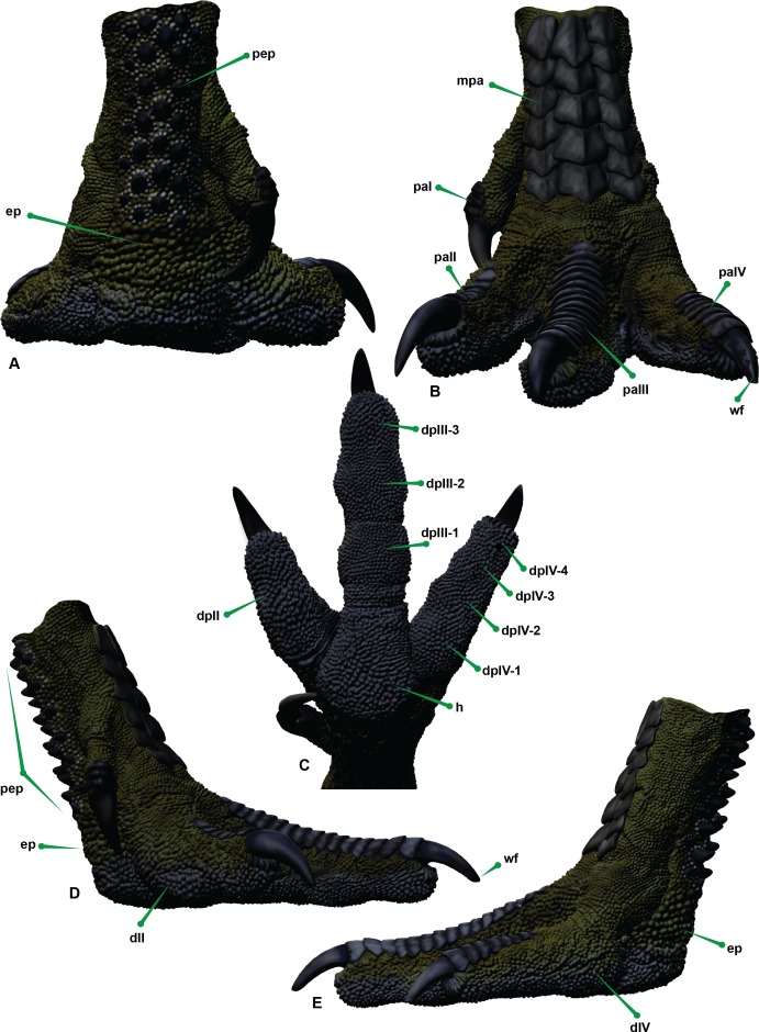
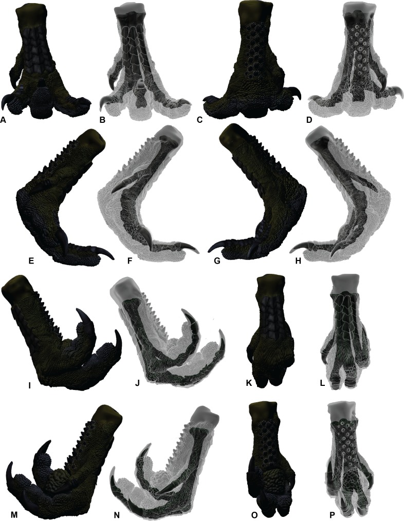
The pedal range of motion in Australovenator wintonensis is investigated to determine what influence soft tissue had on range of motion in the foot. Fortunately, the theropod pes shares a close morphology with extant large cursorial birds. Therefore, to better understand the pedal range of motion of Australovenator, the pedal range of motion of Dromaius novaehollandiae (commonly known as the emu) was analysed with and without soft tissue. We used a variety of innovative digital techniques to analyse the range of motion and biologically restore the Australovenator pes. Computed tomography scans of Dromaius pes in fully flexed and fully extended positions provided the soft tissue range of motion limits. The bone on bone range of motion of the same specimen was replicated following the removal of soft tissue. It was identified that there was an increase in range of motion potential with the removal of soft tissue. This variation provided a guide to develop the potential range of motion of a fully fleshed Australovenator pes. Additionally, the dissection of the Dromaius pes provided a guide enabling the replication of the corresponding soft tissue and keratin sheaths of the Australovenator pes.

Keywords: Australovenator; Dinosaurs; Foot print; Pes; Range of motion; Reconstruction; Theropod.


Conflict of interest statement

The authors declare there are no competing interests.

Figures

Figure 1
Figure 1. Australovenator wintonensis.
Australovenator attacking a Muttaburrasaurus. Artwork by Travis R. Tischler.
Figure 2
Figure 2. ROM of the Australovenator pedal digits with and without soft tissue.
The black coloration represents missing elements. Neutral posture: (A) Anterior; (B) Plantar; (C) Lateral; (D) Medial. Extended without soft tissue: (E) Anterior; (F) Plantar; (G) Lateral; (H) Medial. Flexed without soft tissue: (I) Anterior; (J) Plantar; (K) Lateral; (L) Medial. Extended with soft tissue: (M) Anterior; (N) Plantar; (O) Lateral; (P) Medial. Flexed with soft tissue: (Q) Anterior; (R) Plantar; (S) Lateral; (T) Medial.
Figure 3
Figure 3. Australovenator ungual and sheath restoration.
(A) Pedal ungual II-3 imported into Rhinoceros 5.0. (B) Outer surface curve drawn to dorsal ungual surface. (C) Circle attached to the best fit of outer curve of the ungual. (D) Section planes drawn from a centre point of the drawn circle. (E) Section through the ungual displaying the outer surface of the ungual. (F) Cross-section ribs of the ungual redrawn to represent unbroken sections. (G) A perfect circle was dorsally offset to the claw’s rim for sheath thickness in Rhinoceros 5.0. The offset synthesized the dorsal sheath thickness which was progressive until the sheath’s tip. (H) Wear facets on te reconstructed sheaths. Abbreviations: Centre of circle (Xu), Ungual angle (Yu), Sheath extent (Yest).
Figure 4
Figure 4. Determining the ROM using Zbrush and Rhino 5.0 using metatarsal II (MTII) and II-1 as an example.
(A) Metatarsal II and phalanx II-1 in extended and flexed positions with a sphere representing the range of motion surface of the condyle end of MTII. (B) Separate overlapping spheres drawn as attachments to the central sphere which bisecting along the axis of the distal positions of II-1. (C) Converted from sphere to a mesh. (D) The mesh exported from Zbrush 4R7 into Rhinoceros 5.0. Lines are drawn centrally dissecting the meshes so the ROM can be determined using the angle function. (E) Medial view of a soft tissue articulation angle. The soft tissue angle (Table 2) is replicated with a 3-D protractor with the joint’s corresponding soft tissue angle, which was aligned with the interphalangeal rotation point. The phalanx is re-oriented to its suspected soft tissue articulation. (F) Oblique view of soft tissue ROM adjustment. Abbreviations: bone (b) ROM; sto, soft tissue ROM angle.
Figure 5
Figure 5. ROM of the Dromaius pes with and without soft tissue.
Neutral posture: (A) Anterior; (B) Plantar; (C) Lateral; (D) Medial. Extended without soft tissue: (E) Anterior; (F) Plantar; (G) Lateral; (H) Medial. Flexed without soft tissue: (I) Anterior; (J) Plantar; (K) Lateral; (L) Medial. Extended with soft tissue: (M) Anterior; (N) Plantar; (O) Lateral; (P) Medial. Flexed with soft tissue: (Q) Anterior; (R) Plantar; (S) Lateral; (T) Medial.
Figure 6
Figure 6. Soft tissue ROM of the Dromaius pes.
Extended in: (A, B) Cranial; (C, D) Plantar; (E, F) Medial; (G, H) Lateral. Flexed in: (I, J) Cranial; (K, L) Plantar; (M, N) Medial; (O, P) Lateral.
Figure 7
Figure 7. Dromaius pes with intermediate, superficial and deep flexors.
Plantar view displaying: (A) Extension of superficial flexors for each corresponding digit that housed the deep and intermediate flexors. (B) Corresponding digital render of superficial flexors. (C) Ventral dissection identifying the superficial flexors (cartilage housing for the tendons) and the flexor digitorum longus. (D) Corresponding digital rendering of dissection. (E) Ventral dissection identifying intermediate, superficial and deep tendons. (F) Corresponding digital rendering of dissection. (G) Close up of the digit II’s superficial flexor. (H) Cranial view of the intermediate flexor attachments. Abbreviations: M. flexor digitorum longus (fdl); M. flexor hallucis brevis (fhb); M. flexor hallucis longus (fhl); M. extensor digitorum longus (edl); M. flexor perforatus digiti II (fpdII); M. flexor perforans et perforatus digiti II (fppdll); flexor perforates digiti III (fpdIII); M. flexor perforans et perforatus digiti III (fppdIII); flexor perforatus digiti IV (fpdIV).
Figure 8
Figure 8. Australovenator pedal digits I–IV with reconstructed intermediate, superficial and deep flexors.
Plantar view displaying: (A) Extension of proximo-ventral cartilage of each phalanx and superficial flexors for each corresponding digit (vc). (B) Deep flexor digitorum longus. (C) Superficial flexors, intermediate flexors and deep flexors combined. Digit II with flexor digitorum longus, flexor perforatus digiti II and flexor perforans et perforatus digiti II, (D) Lateral, (E) Medial. Digit I with flexor hallucis brevis, flexor hallucis and extensor digitorum longus, (F) Medial, (G) Lateral, (H) Plantar, (I) Cranial. Digit III with flexor digitorum longus, flexor perforatus digiti III, flexor perforans et perforatus digiti III; Flexor perforatus digiti III and flexor perforans et perforatus digiti III connected by the fibrous vinculum: (J, K) Lateral, (L) Medial. Digit IV with flexor digitorum longus and flexor perforatus digiti IV: (M) Lateral, (N) Medial. Abbreviations: M. flexor digitorum longus (fdl); M. flexor hallucis brevis (fhb); M. flexor hallucis longus (fhl); M. extensor digitorum longus (edl); M. flexor perforatus digiti II (fpdII); M. flexor perforans et perforatus digiti II (fppdll); M. flexor perforatus digiti III (fpdIII); M. flexor perforans et perforatus digiti III (fppdIII); M. flexor perforatus digiti IV (fpdIV).
Figure 9
Figure 9. Extensors of the Dromaius pes.
(A) Dromaius pes in suspended flexed position; (B) Dromaius pes in extended position; (C) Digital render of extensor digitorum longus in cranial view. Dromaius pes with dissection in exploded view; (D) Lateral (E) Medial. Abbreviations: M. flexor digitorum longus (fdl); M. flexor hallus brevis (fhb); M. flexor hallucis longus (fhl); M. extensor digitorum longus (edl); M. flexor perforans II (fpdII); M. flexores perforans et perforates digiti II (fppdll); M. flexor perforans III (fpdIII); M. flexores perforans et perforates digiti III (fppdIII); M. flexor perforans IV (fpdIV).
Figure 10
Figure 10. Adductor and abductor tendons of the Australovenator pes.
(A) Cranial; (B) Plantar; (C) Adductor digiti II; (D) Abductor digiti II; (E) Abductor digiti IV; (F) Adductor digiti IV; (G) Extensors: extensor digitorum longus (edl), extensor hallucis longus (elh). Abbreviations: adductor (add), abductor (abd), extensor brevis digiti III (eb).
Figure 11
Figure 11. Reconstructed pedal biology.
(A) Cranial. (B) Plantar. (C) Lateral. (D) Medial. Abbreviations: M. flexor digitorum longus (fdl); M. flexor hallucis brevis (fhb); M. flexor hallucis longus (fhl); M. extensor digitorum longus (edl); M. flexor perforatus digiti II (fpdII); M. flexor perforans et perforatus digiti II (fppdll); M. flexor perforatus digiti III (fpdIII); M. flexores perforans et perforatus digiti III (fppdIII); M. flexor perforatus digiti IV (fpdIV), vinculum (v).
Figure 12
Figure 12. Reconstructed left pes of Australovenator.
(A) Proximal. (B) Cranial. (C) Plantar. (D) Medial. (E) Lateral view. Abbreviations: enlarged papillae ep; digit I DI; digit II DII; digit III DIII; digit IV DIV; digit pad (dp) (II, III-1, III-2, III-3, IV-1; IV-2, IV-3, IV-4); Digit podotheca (pa); main heal pad (h); metatarsus podotheca (mpa); proximal enlarged podotheca (pep); ungual wear facet (wf).
Figure 13
Figure 13. Soft tissue ROM of the Australovenator pes.
Extended in: (A, B) Cranial; (C, D) Plantar; (E, F) Medial; (G, H) Lateral. Flexed in: (I, J) Lateral; (K, L) Cranial; (M, N) Medial; (O, P) Plantar.

References

    1. Bonnan MF, Sandrik JL, Nishiwaki T, Wilhite DR, Elsey RM, Vitore C. Calcified cartilage shape in archosaur long bones reflects overlying joint shape in stress-bearing elements: implications for nonavian dinosaur locomotion. The Anatomical Record. 2010;293:2044–2055. doi: 10.1002/ar.21266. - DOI - PubMed
    1. Brooks A. On Dendragapus Obscurus Obscurus. The Auk. 1929;46:111–113. doi: 10.2307/4075798. - DOI
    1. Brusatte SL, Benson R, Hutt S. The osteology of Neovenator salerii (Dinosauria: Theropoda) from the Wealden Group (Barremian) of the Isle of Wight. Monograph of the Palaeontographical Society. 2008;162:1–75. doi: 10.1080/02724630903416092. - DOI
    1. Bryan SE, Cook AG, Allen CM, Siegel C, Purdy DJ, Greentree JS, Uysal T. Early—mid Cretaceous tectonic evolution of eastern Gondwana: from silicic LIP to continental rupture. Episodes Special Issue, Oceania Geology. 2012;35(1):142–152.
    1. Carpenter K. Forelimb biomechanics of nonavian theropod dinosaurs in predation. Senckenbergiana Lethaea. 2002;82:59–76. doi: 10.1007/BF03043773. - DOI

LinkOut - more resources