. 2016 Aug 1;16(10):7.
doi: 10.1167/16.10.7.

Speed discrimination in the far monocular periphery: A relative advantage for interocular comparisons consistent with self-motion

Speed discrimination in the far monocular periphery: A relative advantage for interocular comparisons consistent with self-motion

Devon A Greer et al. J Vis. .

Abstract

Some animals with lateral eyes (such as bees) control their navigation through the 3D world using velocity differences between the two eyes. Other animals with frontal eyes (such as primates, including humans) can perceive 3D motion based on the different velocities that a moving object projects upon the two retinae. Although one type of 3D motion perception involves a comparison between velocities from vastly different (monocular) portions of the visual field, and the other involves a comparison within overlapping (binocular) portions of the visual field, both compare velocities across the two eyes. Here we asked whether human interocular velocity comparisons, typically studied in the context of binocularly overlapping vision, operate in the far lateral (and hence, monocular) periphery and, if so, whether these comparisons were accordant with conventional interocular motion processing. We found that speed discrimination was indeed better between the two eyes' monocular visual fields, as compared to within a single eye's (monocular) visual field, but only when the velocities were consistent with commonly encountered motion. This intriguing finding suggests that mechanisms sensitive to relative motion information on opposite sides of an animal may have been retained, or at some point independently achieved, as the eyes became frontal in some animals.


Figures
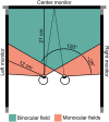
Figure 1
Figure 1
Top view schematic of the three-monitor setup, observer viewing location, and visual field locations. The observer simultaneously viewed three monitors: one at 21 cm directly in front, and one each to the left and the right, positioned such that stimuli could be presented in the center of the monocular fields (orange) at roughly 12 cm from the nearest eye. The typical observer's binocular field (teal) in this setup was measured to span approximately 120 visual degrees and monocular fields spanned approximately 28 visual degrees on each side.
Figure 2
Figure 2
Perimetry visualization from the naive observer confirmed stimuli placement exclusively in the monocular fields. Observer visual field was estimated by superimposing the left and right eye perimetry results. Teal areas show locations in which the subject reported seeing the stimulus in both left and right eye (both eyes response). Areas of blended teal and orange specify locations in which the observer provided mixed responses as to if the stimulus was seen with one eye or both. Orange areas show locations in which the observer reported seeing the stimulus in only one eye (one eye response). Lighter orange areas mark locations that the observer reported seeing the stimulus at that location for 50% of the trials in one eye. Blank areas were not reported as seen. The black circles show the locations of one of the peripheral stimulus configurations (left and right conditions), demonstrating that our monocular stimuli did indeed fall only in the truly monocular fields. The eccentricities along the vertical and horizontal meridian are specified at a few critical locations on the three-monitor setup. The stimuli were too large and bright to detect the blindspots, so the monocular field boundaries are very conservative estimates.
Figure 3
Figure 3
Speed discrimination conditions in Experiment 1 consisted of spatially separate, oppositely drifting gratings presented simultaneously. Arrows show the drift direction of the gratings. In the monocular within-field condition (A), stimuli were presented exclusively within a single monocular field of the observer. The mIOVD condition stimuli (B) were presented separately in each monocular field. Note that the radial eccentricities of the stimuli in A and B were slightly different, but not importantly so, given their large size and huge overall eccentricity. The central binocular field condition stimuli (C, D) were presented in the central area of the observer's binocular field. The stimuli in this condition are vertically (C) or horizontally (D) offset across the fixation point. Note that Figure 3 and the other similar Figures are not to scale; in the horizontally offset central binocular field condition (D), for example, the stimuli were entirely on the center monitor.
Figure 4
Figure 4
Speed discrimination thresholds for Experiment 1. (A) Sensitivity for speed differences within a single monocular field (within-field condition, blue) and for the mIOVD condition (red). Average sensitivity for the central binocular field is shown in gray. Icons illustrate stimulus configurations. (B) Sensitivity to speed differences in the central binocular field condition with vertically offset gratings (dark gray) and with horizontally offset gratings (light gray). (C) Difference in average thresholds for the peripheral monocular field and the central binocular field conditions. Monocular within-field threshold difference is shown in light blue and mIOVD threshold difference is shown in pink. The colored regions indicate the bootstrapped 68% confidence intervals.
Figure 5
Figure 5
Experiment 2 speed discrimination conditions in which simultaneously presented gratings drifted in the same relative direction (same general format as described for Figure 3). Arrows indicate the relative drift directions of the gratings (same horizontal drift direction for vertically oriented gratings in Experiment 2).
Figure 6
Figure 6
Speed discrimination thresholds for Experiment 2. (A) Weber fractions for speed differences within either the left or right monocular field (blue) and for the mIOVD stimulus (red). Averaged central binocular thresholds (gray) are shown for reference. (B) Speed discrimination thresholds for the central binocular field with vertically offset gratings (dark gray) and for the central binocular field with horizontally offset gratings (light gray). (C) The difference in average thresholds of the peripheral monocular field conditions (monocular within-field, light blue; mIOVD, pink) and the equivalent central binocular field condition. Plotting conventions are as in Figure 4.
Figure 7
Figure 7
Speed discrimination conditions in Experiment 3, in which gratings drifted orthogonally. Conventions are as in Figures 3 and 5. For each condition, one grating was oriented vertically and drifted horizontally, the second grating was oriented horizontally and drifted vertically. See text for details.
Figure 8
Figure 8
Speed discrimination thresholds for orthogonal motion. (A) Weber fractions for speed differences within a single monocular field (“within-field” blue) and for between both monocular fields (mIOVD, red). Averaged central binocular thresholds (gray) are shown for comparison. (B) Speed difference thresholds in the central binocular field with vertically offset gratings (dark gray) and for the central binocular field with horizontally offset gratings (light gray). (C) The difference in average thresholds measured the drop in performance of the peripheral monocular field conditions (monocular within-field,light blue; mIOVD, pink) after the equivalent central binocular field condition was deducted. Plotting conventions are as in Figures 4 and 6.

Similar articles

Cited by

References

    1. Bennett P. J,, Banks M. S. (1987). Sensitivity loss in odd-symmetric mechanisms and phase anomalies in peripheral vision. Nature, 326, 873–876. - PubMed
    1. Bex P. J,, Dakin S. C. (2005). Spatial interference among moving targets. Vision Research, 45, 1385–1398. - PubMed
    1. Bex P. J,, Dakin S. C,, Simmers A. J. (2003). The shape and size of crowding for moving targets. Vision Research, 43, 2895–2904. - PubMed
    1. Bhagavatula P. S,, Claudianos C,, Ibbotson M. R,, Srinivasan M. V. (2011). Optic flow cues guide flight in birds. Current Biology, 21, 1794–1799. - PubMed
    1. Brainard D. H. (1997). The Psychophysics Toolbox. Spatial Vision, 10, 433–436. - PubMed

LinkOut - more resources