

Trichostatin A Enhances the Apoptotic Potential of Palladium Nanoparticles in Human Cervical Cancer Cells
- PMID: 27548148
- PMCID: PMC5000750
- DOI: 10.3390/ijms17081354
Trichostatin A Enhances the Apoptotic Potential of Palladium Nanoparticles in Human Cervical Cancer Cells
Abstract
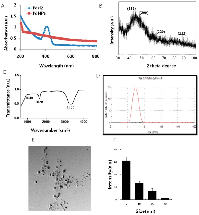
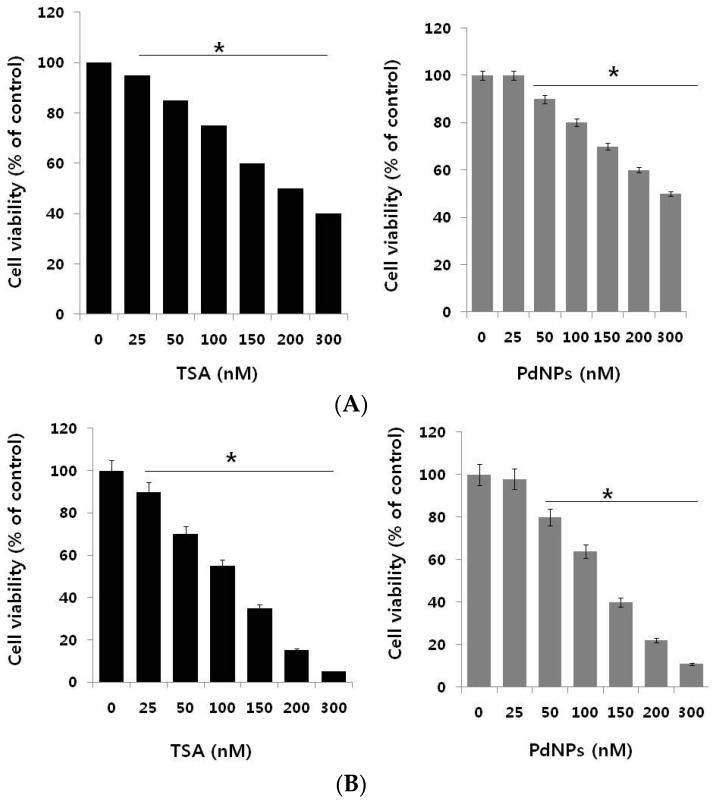
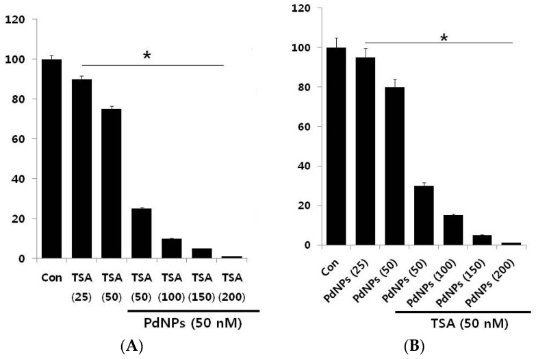
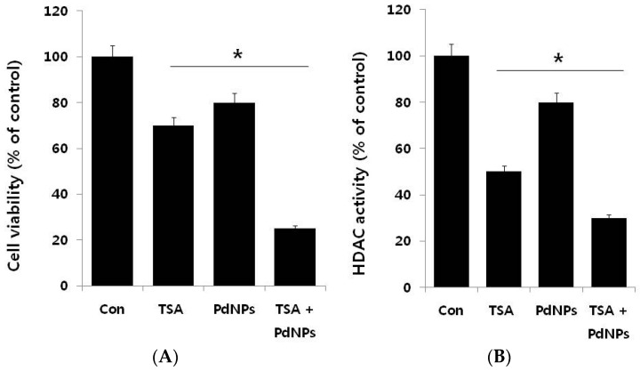
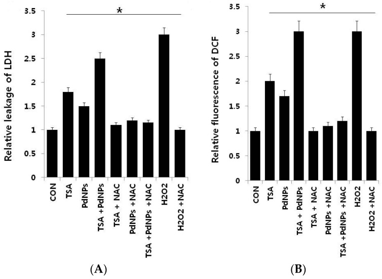
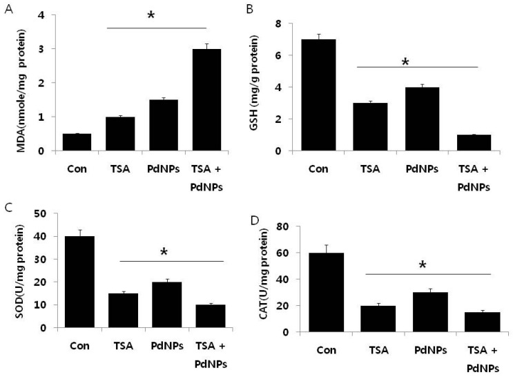
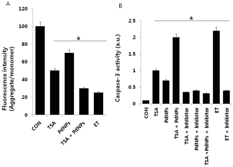
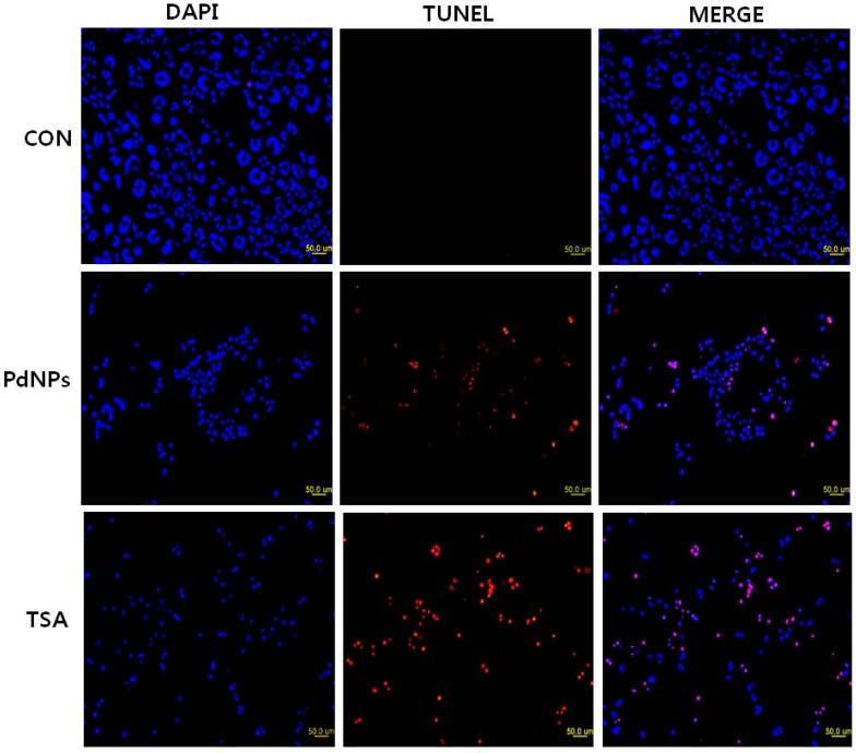
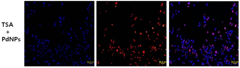
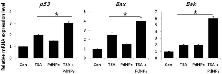
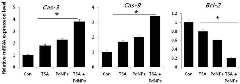
Cervical cancer ranks seventh overall among all types of cancer in women. Although several treatments, including radiation, surgery and chemotherapy, are available to eradicate or reduce the size of cancer, many cancers eventually relapse. Thus, it is essential to identify possible alternative therapeutic approaches for cancer. We sought to identify alternative and effective therapeutic approaches, by first synthesizing palladium nanoparticles (PdNPs), using a novel biomolecule called saponin. The synthesized PdNPs were characterized by several analytical techniques. They were significantly spherical in shape, with an average size of 5 nm. Recently, PdNPs gained much interest in various therapies of cancer cells. Similarly, histone deacetylase inhibitors are known to play a vital role in anti-proliferative activity, gene expression, cell cycle arrest, differentiation and apoptosis in various cancer cells. Therefore, we selected trichostatin A (TSA) and PdNPs and studied their combined effect on apoptosis in cervical cancer cells. Cells treated with either TSA or PdNPs showed a dose-dependent effect on cell viability. The combinatorial effect, tested with 50 nM TSA and 50 nMPdNPs, had a more dramatic inhibitory effect on cell viability, than either TSA or PdNPs alone. The combination of TSA and PdNPs had a more pronounced effect on cytotoxicity, oxidative stress, mitochondrial membrane potential (MMP), caspase-3/9 activity and expression of pro- and anti-apoptotic genes. Our data show a strong synergistic interaction between TSA and PdNPs in cervical cancer cells. The combinatorial treatment increased the therapeutic potential and demonstrated relevant targeted therapy for cervical cancer. Furthermore, we provide the first evidence for the combinatory effect and cytotoxicity mechanism of TSA and PdNPs in cervical cancer cells.
Keywords: apoptosis; caspases; cell viability; cervical cancer; mitochondrial membrane potential; oxidative stress; palladium nanoparticles; trichostatin A.
Figures
Similar articles
-
Combination of palladium nanoparticles and tubastatin-A potentiates apoptosis in human breast cancer cells: a novel therapeutic approach for cancer.Int J Nanomedicine. 2017 Sep 5;12:6503-6520. doi: 10.2147/IJN.S136142. eCollection 2017. Int J Nanomedicine. 2017. PMID: 28919751 Free PMC article.
-
Trichostatin A induces apoptotic cell death of HeLa cells in a Bcl-2 and oxidative stress-dependent manner.Int J Oncol. 2013 Jan;42(1):359-66. doi: 10.3892/ijo.2012.1705. Epub 2012 Nov 19. Int J Oncol. 2013. PMID: 23165748
-
Combination of salinomycin and silver nanoparticles enhances apoptosis and autophagy in human ovarian cancer cells: an effective anticancer therapy.Int J Nanomedicine. 2016 Aug 2;11:3655-75. doi: 10.2147/IJN.S111279. eCollection 2016. Int J Nanomedicine. 2016. PMID: 27536105 Free PMC article.
-
Copper and palladium nanostructures: a bacteriogenic approach.Appl Microbiol Biotechnol. 2018 Sep;102(18):7693-7701. doi: 10.1007/s00253-018-9180-5. Epub 2018 Jul 11. Appl Microbiol Biotechnol. 2018. PMID: 29998411 Review.
-
An Up-To-Date Review on Biomedical Applications of Palladium Nanoparticles.Nanomaterials (Basel). 2019 Dec 27;10(1):66. doi: 10.3390/nano10010066. Nanomaterials (Basel). 2019. PMID: 31892149 Free PMC article. Review.
Cited by
-
Cytotoxicity and Transcriptomic Analyses of Biogenic Palladium Nanoparticles in Human Ovarian Cancer Cells (SKOV3).Nanomaterials (Basel). 2019 May 22;9(5):787. doi: 10.3390/nano9050787. Nanomaterials (Basel). 2019. PMID: 31121951 Free PMC article.
-
The Effects of Apigenin-Biosynthesized Ultra-Small Platinum Nanoparticles on the Human Monocytic THP-1 Cell Line.Cells. 2019 May 10;8(5):444. doi: 10.3390/cells8050444. Cells. 2019. PMID: 31083475 Free PMC article.
-
The Emerging Role of Histone Deacetylase Inhibitors in Cervical Cancer Therapy.Cancers (Basel). 2023 Apr 10;15(8):2222. doi: 10.3390/cancers15082222. Cancers (Basel). 2023. PMID: 37190151 Free PMC article. Review.
-
Pharmacological Properties of Trichostatin A, Focusing on the Anticancer Potential: A Comprehensive Review.Pharmaceuticals (Basel). 2022 Oct 8;15(10):1235. doi: 10.3390/ph15101235. Pharmaceuticals (Basel). 2022. PMID: 36297347 Free PMC article. Review.
-
Tangeretin-Assisted Platinum Nanoparticles Enhance the Apoptotic Properties of Doxorubicin: Combination Therapy for Osteosarcoma Treatment.Nanomaterials (Basel). 2019 Jul 29;9(8):1089. doi: 10.3390/nano9081089. Nanomaterials (Basel). 2019. PMID: 31362420 Free PMC article.
References
-
- Cervical Cancer Statistics World Cancer Research Fund International GLOBOCAN Cancer Fact Sheets: Cervical Cancer—Iarc. [(accessed on 6 July 2016)]. Available online: http://www.globocan.iarc.fr.
-
- Chemotherapy for Cervical Cancer—American Cancer Society. [(accessed on 6 July 2016)]. Available online: http://www.cancer.org.
-
- Anton M., Horký M., Kuchtícková S., Vojtĕsek B., Bláha O. Immunohistochemical detection of acetylation and phosphorylation of histone H3 in cervical smears. Ceska Gynekol. 2004;69:3–6. - PubMed
-
- Zhong S., Fields C.R., Su N., Pan Y.X., Robertson K.D. Pharmacologic inhibition of epigenetic modifications, coupled with gene expression profiling, reveals novel targets of aberrant DNA methylation and histone deacetylation in lung cancer. Oncogene. 2007;26:2621–2634. doi: 10.1038/sj.onc.1210041. - DOI - PubMed
MeSH terms
Substances
LinkOut - more resources
Full Text Sources
Other Literature Sources
Research Materials
