. 2016 Aug 19;8(8):507.
doi: 10.3390/nu8080507.

Post-Intake of S-Ethyl Cysteine and S-Methyl Cysteine Improved LPS-Induced Acute Lung Injury in Mice

Affiliations

Post-Intake of S-Ethyl Cysteine and S-Methyl Cysteine Improved LPS-Induced Acute Lung Injury in Mice

Te-Chun Hsia et al. Nutrients. .

Retraction in

Abstract

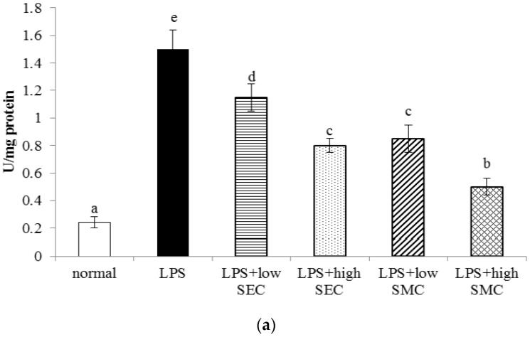
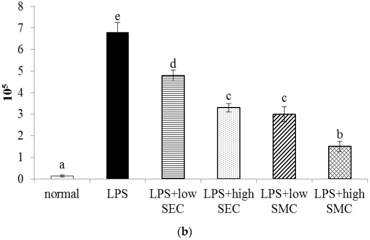
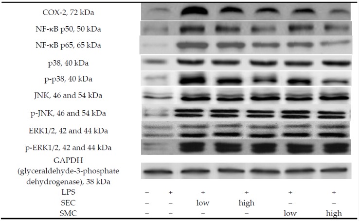
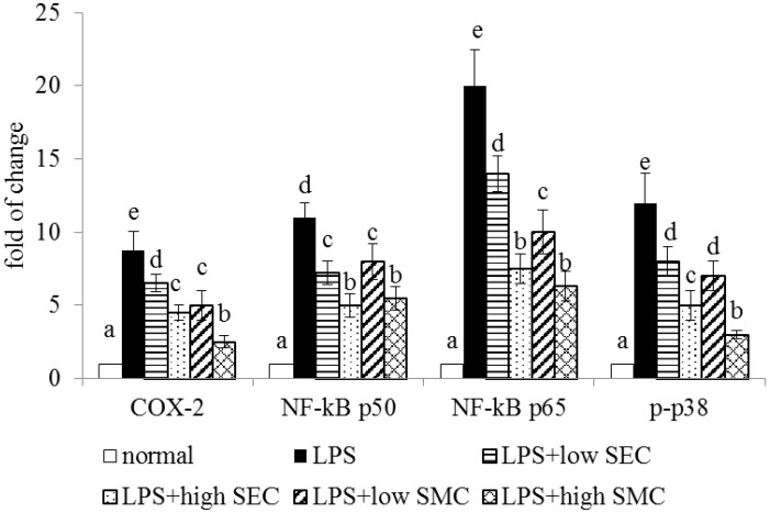
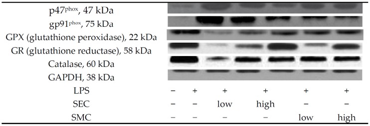
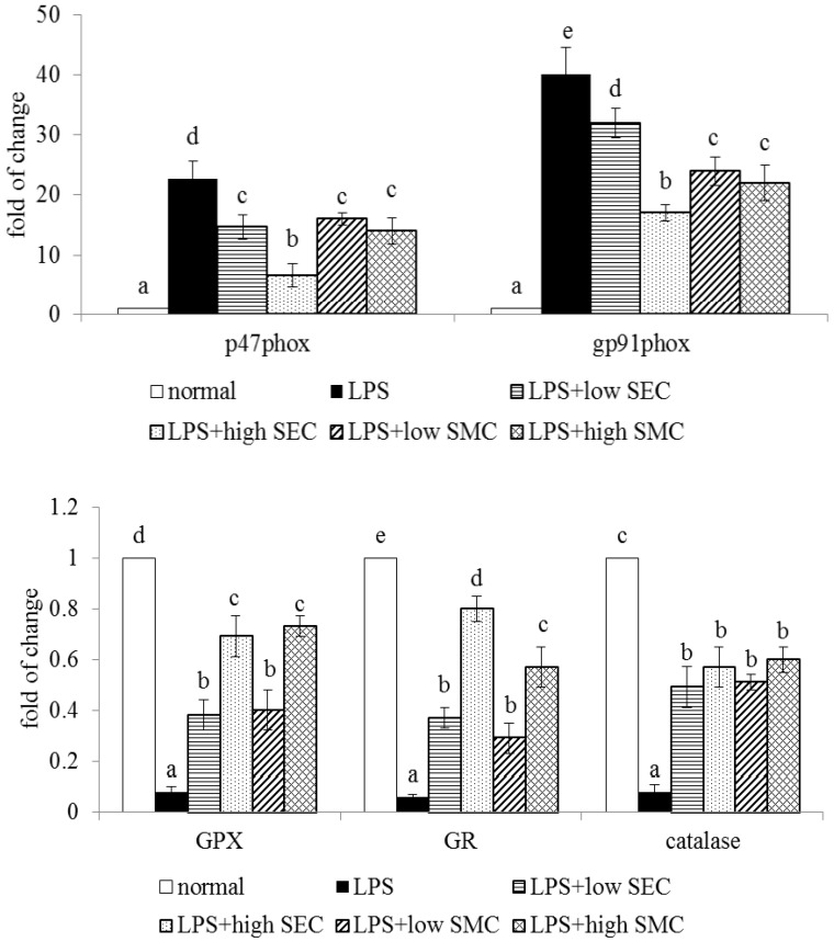
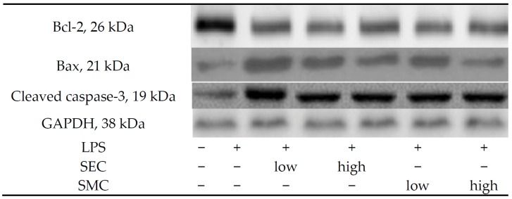
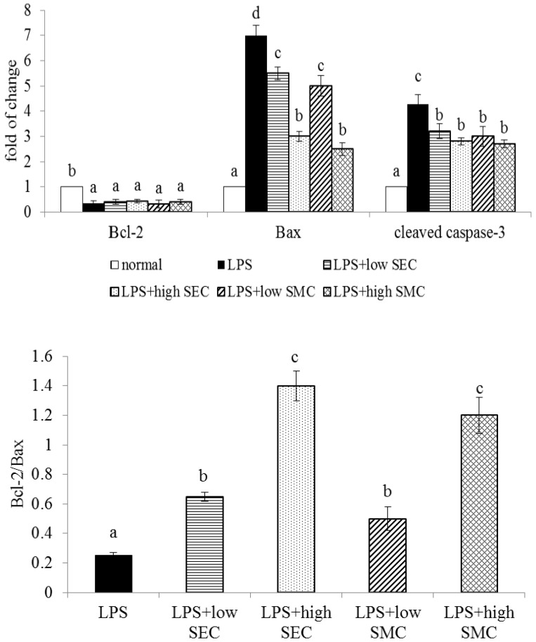
The effects of S-ethyl cysteine (SEC) and S-methyl cysteine (SMC) on lipopolysaccharide (LPS)-induced acute lung injury in mice were examined. Eight hours after LPS challenge, SEC or SMC was supplied in drinking water at 0.5% or 1% for 3 days. LPS increased lung myeloperoxidase activity, neutrophil counts and edema. SEC or SMC post-intake attenuated these events. SEC or SMC suppressed LPS-induced lung expression of cyclooxygenase-2, nuclear factor-κB and mitogen-activated protein kinase, and lowered the generation of tumor necrosis factor-alpha, monocyte chemoattractant protein-1 and prostaglandin E₂. LPS enhanced the expression of p47(phox), gp91(phox), Bax and cleaved caspase-3, and increased the production of reactive oxygen species in the lung. SEC or SMC post-intake reversed these alterations. These findings suggest that these agents could protect the lung through their anti-inflammatory, anti-oxidative and anti-apoptotic activities.

Keywords: LPS; S-ethyl cysteine; S-methyl cysteine; inflammation; lung.


Figures

Figure 1
Figure 1
Lung MPO (myeloperoxidase) activity (a); and neutrophil count (b) of mice in normal group, LPS (lipopolysaccharide) group, LPS + low SEC (S-ethyl cysteine) group, LPS + high SEC group, LPS + low SMC (S-methyl cysteine) group and LPS + high SMC group after 3-day treatments. Data are mean ± SD (standard deviation), n = 10. a–e Means among bars without a common letter differ, p < 0.05.
Figure 1
Figure 1
Lung MPO (myeloperoxidase) activity (a); and neutrophil count (b) of mice in normal group, LPS (lipopolysaccharide) group, LPS + low SEC (S-ethyl cysteine) group, LPS + high SEC group, LPS + low SMC (S-methyl cysteine) group and LPS + high SMC group after 3-day treatments. Data are mean ± SD (standard deviation), n = 10. a–e Means among bars without a common letter differ, p < 0.05.
Figure 2
Figure 2
Lung expression of COX-2, NF-κB and MAPK of mice in normal group, LPS group, LPS + low SEC group, LPS + high SEC group, LPS + low SMC group and LPS + high SMC group after 3-day treatments. Data are mean ± SD, n = 10; a−e Means among bars without a common letter differ, p < 0.05.
Figure 2
Figure 2
Lung expression of COX-2, NF-κB and MAPK of mice in normal group, LPS group, LPS + low SEC group, LPS + high SEC group, LPS + low SMC group and LPS + high SMC group after 3-day treatments. Data are mean ± SD, n = 10; a−e Means among bars without a common letter differ, p < 0.05.
Figure 3
Figure 3
Lung expression of p47phox, gp91phox, GPX, GR and catalase of mice in normal group, LPS group, LPS + low SEC group, LPS + high SEC group, LPS + low SMC group and LPS + high SMC group after 3-day treatments. Data are mean ± SD, n = 10; a−e Means among bars without a common letter differ, p < 0.05.
Figure 3
Figure 3
Lung expression of p47phox, gp91phox, GPX, GR and catalase of mice in normal group, LPS group, LPS + low SEC group, LPS + high SEC group, LPS + low SMC group and LPS + high SMC group after 3-day treatments. Data are mean ± SD, n = 10; a−e Means among bars without a common letter differ, p < 0.05.
Figure 4
Figure 4
Lung expression of Bcl-2, Bax and cleaved caspase-3, and ratio of Bcl-2/Bax of mice in normal group, LPS group, LPS + low SEC group, LPS + high SEC group, LPS + low SMC group and LPS + high SMC group after 3-day treatments. Data are mean ± SD, n = 10; a−d Means among bars without a common letter differ, p < 0.05.
Figure 4
Figure 4
Lung expression of Bcl-2, Bax and cleaved caspase-3, and ratio of Bcl-2/Bax of mice in normal group, LPS group, LPS + low SEC group, LPS + high SEC group, LPS + low SMC group and LPS + high SMC group after 3-day treatments. Data are mean ± SD, n = 10; a−d Means among bars without a common letter differ, p < 0.05.
Figure 5
Figure 5
Schematic diagram for lung protein of SEC or SMC against LPS.

Similar articles

Cited by

References

    1. Johnson E.R., Matthay M.A. Acute lung injury: Epidemiology, pathogenesis, and treatment. J. Aerosol. Med. Pulm. Drug Deliv. 2010;23:243–252. doi: 10.1089/jamp.2009.0775. - DOI - PMC - PubMed
    1. Puneet P., Moochhala S., Bhatia M. Chemokines in acute respiratory distress syndrome. Am. J. Physiol. Lung Cell. Mol. Physiol. 2005;288:3–15. doi: 10.1152/ajplung.00405.2003. - DOI - PMC - PubMed
    1. Chi G., Wei M., Xie X., Soromou L.W., Liu F., Zhao S. Suppression of MAPK and NF-κB pathways by limonene contributes to attenuation of lipopolysaccharide-induced inflammatory responses in acute lung injury. Inflammation. 2013;36:501–511. doi: 10.1007/s10753-012-9571-1. - DOI - PubMed
    1. Thangavel J., Samanta S., Rajasingh S., Barani B., Xuan Y.T., Dawn B., Rajasingh J. Epigenetic modifiers reduce inflammation and modulate macrophage phenotype during endotoxemia-induced acute lung injury. J. Cell Sci. 2015;128:3094–3105. doi: 10.1242/jcs.170258. - DOI - PMC - PubMed
    1. Ang S.F., Sio S.W., Moochhala S.M., MacAry P.A., Bhatia M. Hydrogen sulfide upregulates cyclooxygenase-2 and prostaglandin E metabolite in sepsis-evoked acute lung injury via transient receptor potential vanilloid type 1 channel activation. J. Immunol. 2011;187:4778–4787. doi: 10.4049/jimmunol.1101559. - DOI - PubMed

Publication types

LinkOut - more resources