

Leukocyte Beta-Catenin Expression Is Disturbed in Systemic Lupus Erythematosus
- PMID: 27548498
- PMCID: PMC4993388
- DOI: 10.1371/journal.pone.0161682
Leukocyte Beta-Catenin Expression Is Disturbed in Systemic Lupus Erythematosus
Abstract
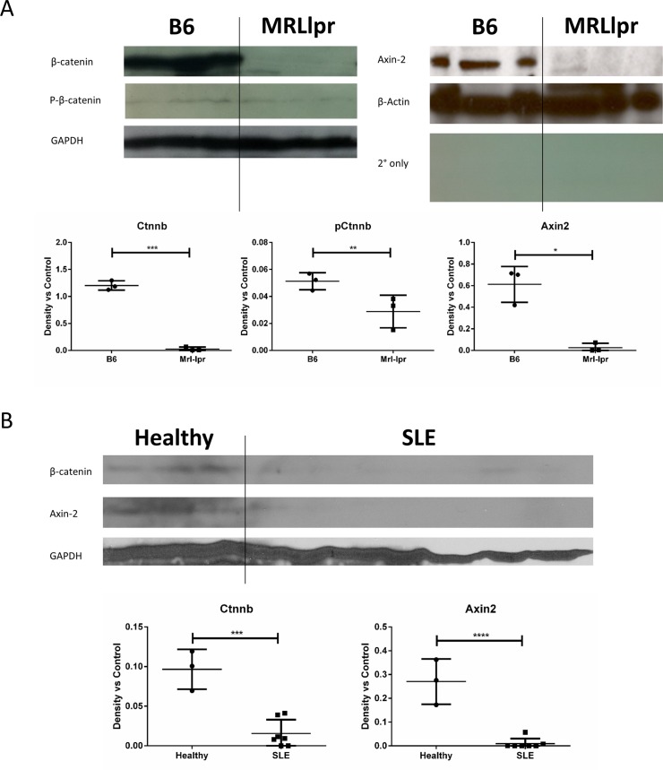
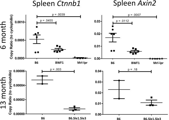
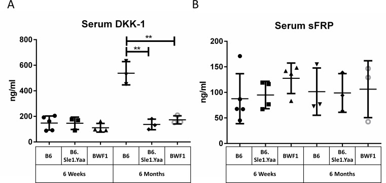
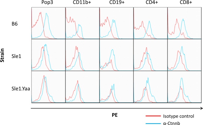
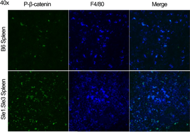
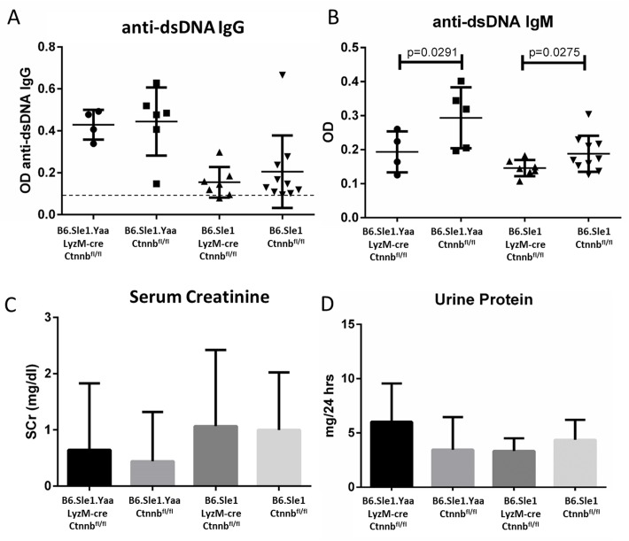
Wnt/β-catenin signaling is relatively understudied in immunity and autoimmunity. β-catenin blocks inflammatory mediators and favors tolerogenic dendritic cell (DC) phenotypes. We show here that leukocytes from lupus-prone mice and SLE patients express diminished β-catenin transcriptional activity, particularly in myeloid cells, although other leukocytes revealed similar trends. Serum levels of DKK-1, an inhibitor under transcriptional control of Wnt/β-catenin, were also decreased in lupus-prone mice. Surprisingly, however, preemptive deletion of β-catenin from macrophages appears to have no effect on lupus development, even in mice with varying genetic loads for lupus. Although myeloid-specific loss of β-catenin does not seem to be important for lupus development, the potential role of this transcription factor in other leukocytes and renal cells remain to be elucidated.
Conflict of interest statement
The authors have declared that no competing interests exist.
Figures
Similar articles
-
Aberrant activation of the WNT/β-catenin signaling pathway in lupus nephritis.PLoS One. 2014 Jan 21;9(1):e84852. doi: 10.1371/journal.pone.0084852. eCollection 2014. PLoS One. 2014. PMID: 24465439 Free PMC article.
-
Emerging Role and Therapeutic Implication of Wnt Signaling Pathways in Autoimmune Diseases.J Immunol Res. 2016;2016:9392132. doi: 10.1155/2016/9392132. Epub 2016 Mar 27. J Immunol Res. 2016. PMID: 27110577 Free PMC article. Review.
-
WNT/β-catenin pathway is modulated in asthma patients and LPS-stimulated RAW264.7 macrophage cell line.Immunopharmacol Immunotoxicol. 2012 Feb;34(1):56-65. doi: 10.3109/08923973.2011.574704. Epub 2011 Jun 24. Immunopharmacol Immunotoxicol. 2012. PMID: 21699440 Clinical Trial.
-
Axin2 marks quiescent hair follicle bulge stem cells that are maintained by autocrine Wnt/β-catenin signaling.Proc Natl Acad Sci U S A. 2016 Mar 15;113(11):E1498-505. doi: 10.1073/pnas.1601599113. Epub 2016 Feb 22. Proc Natl Acad Sci U S A. 2016. PMID: 26903625 Free PMC article.
-
Breakdown of Immune Tolerance in Systemic Lupus Erythematosus by Dendritic Cells.J Immunol Res. 2016;2016:6269157. doi: 10.1155/2016/6269157. Epub 2016 Feb 29. J Immunol Res. 2016. PMID: 27034965 Free PMC article. Review.
Cited by
-
Pertussis toxin-induced inhibition of Wnt/β-catenin signaling in dendritic cells promotes an autoimmune response in experimental autoimmune uveitis.J Neuroinflammation. 2023 Feb 4;20(1):24. doi: 10.1186/s12974-023-02707-y. J Neuroinflammation. 2023. PMID: 36739434 Free PMC article.
-
Molecular and Cellular Mediators of Renal Fibrosis in Lupus Nephritis.Int J Mol Sci. 2025 Mar 14;26(6):2621. doi: 10.3390/ijms26062621. Int J Mol Sci. 2025. PMID: 40141260 Free PMC article. Review.
-
Wnt Signaling Is Deranged in Asthmatic Bronchial Epithelium and Fibroblasts.Front Cell Dev Biol. 2021 Mar 15;9:641404. doi: 10.3389/fcell.2021.641404. eCollection 2021. Front Cell Dev Biol. 2021. PMID: 33791298 Free PMC article.
-
Dickkopf-1 Is a Biomarker for Systemic Lupus Erythematosus and Active Lupus Nephritis.J Immunol Res. 2017;2017:6861575. doi: 10.1155/2017/6861575. Epub 2017 Mar 8. J Immunol Res. 2017. PMID: 28373995 Free PMC article.
-
A ceRNA regulatory network in systemic lupus erythematosus and its molecular interplay with cancer.Ann Transl Med. 2022 May;10(10):563. doi: 10.21037/atm-22-1533. Ann Transl Med. 2022. PMID: 35722400 Free PMC article.
References
-
- Kemler R. From cadherins to catenins: cytoplasmic protein interactions and regulation of cell adhesion. Trends Genet. 1993;9(9):317–21. - PubMed
-
- McCREA PD, Turck CW, Gumbiner B. A homolog of the armadillo protein in Drosophila (plakoglobin) associated with E-cadherin. Science. 1991;254(5036):1359–61. - PubMed
-
- Behrens J, von Kries JP, Kühl M, Bruhn L, Wedlich D, Grosschedl R, et al. Functional interaction of β-catenin with the transcription factor LEF-1. Nature. 1996;382(6592):638–42. - PubMed
MeSH terms
Substances
Grants and funding
LinkOut - more resources
Full Text Sources
Other Literature Sources
Medical
Molecular Biology Databases
