Review
. 2016 Sep;8(13):1589-607.
doi: 10.4155/fmc-2016-0071. Epub 2016 Aug 22.

H3K36 methyltransferases as cancer drug targets: rationale and perspectives for inhibitor development

Affiliations
Review

H3K36 methyltransferases as cancer drug targets: rationale and perspectives for inhibitor development

David S Rogawski et al. Future Med Chem. 2016 Sep.

Abstract

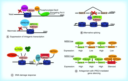
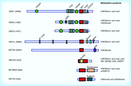
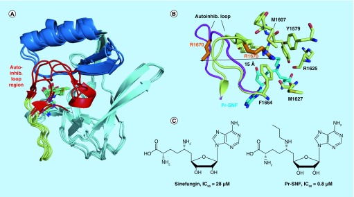
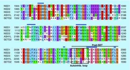
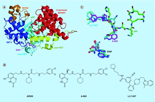
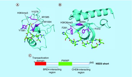
Methylation at histone 3, lysine 36 (H3K36) is a conserved epigenetic mark regulating gene transcription, alternative splicing and DNA repair. Genes encoding H3K36 methyltransferases (KMTases) are commonly overexpressed, mutated or involved in chromosomal translocations in cancer. Molecular biology studies have demonstrated that H3K36 KMTases regulate oncogenic transcriptional programs. Structural studies of the catalytic SET domain of H3K36 KMTases have revealed intriguing opportunities for design of small molecule inhibitors. Nevertheless, potent inhibitors for most H3K36 KMTases have not yet been developed, underlining the challenges associated with this target class. As we now have strong evidence linking H3K36 KMTases to cancer, drug development efforts are predicted to yield novel compounds in the near future.

Keywords: cancer; chromosomal translocation; epigenetics; gene expression; genome integrity; histone methyltransferase; oncogenes; protein–protein interactions; structure-based drug design.


Conflict of interest statement

Financial & competing interests disclosure This research has been supported by the Leukemia and Lymphoma Society TRP grants 6111-14 to T Cierpicki and 6485-16 to J Grembecka, NIH 1R01CA160467 to J Grembecka and 1R01CA181185 to T Cierpicki. J Grembecka and T Cierpicki are Leukemia and Lymphoma Society Scholars (grants 1215-14 and 1340-17). DS Rogawski acknowledges training grant support from the University of Michigan Chemistry–Biology Interface (CBI) training program (NIH Grant 5T32GM008597) and from the University of Michigan Medical Scientist Training Program (NIH Grant 5T32GM007863). J Grembecka and T Cierpicki receive research support from Kura Oncology. They are also receiving compensation as members of the scientific advisory board of Kura Oncology, and they have an equity ownership in the company. The authors have no other relevant affiliations or financial involvement with any organization or entity with a financial interest in or financial conflict with the subject matter or materials discussed in the manuscript apart from those disclosed. No writing assistance was utilized in the production of this manuscript.

Figures

<b>Figure 1.</b>
Figure 1.. The functions of H3K36 methylation in biology.
(A) H3K36 methylation plays a conserved role in suppressing intragenic transcription. Methylated H3K36 recruits histone deacetylases (yeast) or histone demethylases (humans) to maintain a suppressive transcriptional environment within gene bodies. (B) Splicing regulators including pyrimidine tract binding protein are recruited to loci by H3K36 methylation. (C) Methyl marks installed by the SETMAR and SETD2 KMTases are important for recruiting mediators of the DNA damage response including Ku70/Ku80, MRE11-RAD50-NBS1 (MRN) and CtIP. (D) Methylated H3K36 antagonizes polycomb-repressive complex 2 mediated H3K27 methylation and gene silencing. In NSD2-low cells, active genes are marked by H3K36 methylation, whereas lower expressed genes are marked by H3K27 methylation. In NSD2-high cells (as in t(4;14) multiple myeloma), overexpression of NSD2 increases genome-wide levels of H3K36 methylation, forcing accumulation of H3K27 methylation at a subset of silenced loci. Adapted with permission from [18].
<b>Figure 2.</b>
Figure 2.. Domain organization of human H3K36 methyltransferases.
SET domain is shown in red.
<b>Figure 3.</b>
Figure 3.. Structures of H3K36-specific S-adenosyl methionine domains and SAM-competitive inhibitors.
(A) Overlay of the core SET (pale cyan), SET-I (sky blue) and post-SET (pale yellow) subdomains of NSD1 (PDB code 3OOI), NSD3 (4YZ8), ASH1L (4YNM) and SETD2 (4H12). The structurally variable autoinhibitory loop region is colored in red. (B) Binding of the N-propyl sinefungin (Pr-SNF) inhibitor to SETD2 causes opening of the autoinhibitory loop. Autoinhibitory loop conformation is shown with S-adenosyl homocysteine bound (pale yellow, 4H12) and with Pr-SNF bound (magenta, 4FMU). Steric clash between the propyl moiety of Pr-SNF (cyan) and the Arg1670 sidechain (orange) causes Arg1670 to flip out a distance of 15 Å. Residues stabilizing Arg1670 in the putative substrate lysine-binding channel in the S-adenosyl homocysteine bound form of SETD2 are shown in pale yellow sticks. (C) Chemical structures of SETD2 inhibitors sinefungin and Pr-SNF. In vitro IC50 values for SETD2 are listed.
<b>Figure 4.</b>
Figure 4.. Sequence alignment of core SET and post-SET regions of the related NSD, ASH1L and SETD2 KMTases.
Blue lines indicate conserved contacts to S-adenosyl methionine cofactor. Black box indicates the autoinhibitory loop, black line indicates post-SET subdomain.
<b>Figure 5.</b>
Figure 5.. Peptide substrate-competitive inhibitors of the SMYD2 KMTase.
(A) Crystal structure of SMYD2 bound to p53 peptide (PDB code 3TG5). (B) Chemical structures of SMYD2 inhibitors. (C) Overlay of binding modes of SMYD2 inhibitors LLY-507 (4WUY) and A-893 (4YND) and p53 peptide. The weaker compound AZ505 binds to the same site but has been omitted for clarity. S-adenosyl methionine position included for reference. SAM: S-adenosyl methionine.
<b>Figure 6.</b>
Figure 6.. Targeting protein–protein interacting domains of H3K36 KMTases as an alternative to the SET domain.
Examples of PHD and PWWP domains: (A) Third PHD domain of MLL1 (cyan) bound to H3K4me3 peptide (green) (PDB code 3LQJ) and (B) PWWP domain of ZMYND11 (cyan) bound to H3.3K36me3 peptide (green) (4N4I). Residues on the protein–protein interacting domain that form the aromatic cage are colored magenta. (C) Protein–protein interacting regions in the N-terminus of NSD3 that are required for leukemogenesis. Adapted with permission from [147].

References

    1. Dawson MA, Kouzarides T. Cancer epigenetics: from mechanism to therapy. Cell. 2012;150(1):12–27. - PubMed
    1. Daigle SR, Olhava EJ, Therkelsen CA, et al. Selective killing of mixed lineage leukemia cells by a potent small-molecule DOT1L inhibitor. Cancer Cell. 2011;20(1):53–65. - PMC - PubMed
    1. Kuo AJ, Cheung P, Chen K, et al. NSD2 links dimethylation of histone H3 at lysine 36 to oncogenic programming. Mol. Cell. 2011;44(4):609–620. - PMC - PubMed
    2. •• Demonstrates that NSD2 KMTase activity is required for oncogenesis in t(4;14) multiple myeloma (MM).

    1. McCabe MT, Creasy CL. EZH2 as a potential target in cancer therapy. Epigenomics. 2014;6(3):341–351. - PubMed
    1. Waters NJ, Smith SA, Olhava EJ, et al. Metabolism and disposition of the DOT1L inhibitor, pinometostat (EPZ-5676), in rat, dog and human. Cancer Chemother. Pharmacol. 2016;77(1):43–62. - PubMed

Publication types

MeSH terms