. 2016 Oct 1;138(10):1010061-10100611.
doi: 10.1115/1.4034490.

Fiber Network Models Predict Enhanced Cell Mechanosensing on Fibrous Gels

Fiber Network Models Predict Enhanced Cell Mechanosensing on Fibrous Gels

Maziar Aghvami et al. J Biomech Eng. .

Abstract

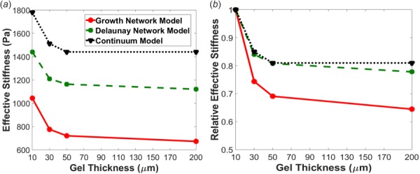
The propagation of mechanical signals through nonlinear fibrous tissues is much more extensive than through continuous synthetic hydrogels. Results from recent studies indicate that increased mechanical propagation arises from the fibrous nature of the material rather than the strain-stiffening property. The relative importance of different parameters of the fibrous network structure to this propagation, however, remains unclear. In this work, we directly compared the mechanical response of substrates of varying thickness subjected to a constant cell traction force using either a nonfibrous strain-stiffening continuum-based model or a volume-averaged fiber network model consisting of two different types of fiber network structures: one with low fiber connectivity (growth networks) and one with high fiber connectivity (Delaunay networks). The growth network fiber models predicted a greater propagation of substrate displacements through the model and a greater sensitivity to gel thickness compared to the more connected Delaunay networks and the nonlinear continuum model. Detailed analysis of the results indicates that rotational freedom of the fibers in a network with low fiber connectivity is critically important for enhanced, long-range mechanosensing. Our findings demonstrate the utility of multiscale models in predicting cells mechanosensing on fibrous gels, and they provide a more complete understanding of how cell traction forces propagate through fibrous tissues, which has implications for the design of engineered tissues and the stem cell niche.


Figures

Fig. 1
Fig. 1
Schematic of the FE model. Cell traction forces are applied to the FE nodes (red) associated with the focal adhesion area on the FE top surface. For the fiber model, 3D growth or Delaunay fiber networks similar to the microstructures found in fibrous gels were compared to the nonlinear continuum model.
Fig. 2
Fig. 2
Shear simulation results fitted to experimental data for a 2 mg/mL acellular fibrin gel from Ref. [33]. Model parameters were selected to match the nonlinear increase in stress with strain.
Fig. 3
Fig. 3
Maximum principal stress for 10 μm, 30 μm, and 50 μm thick gels in response to 6 nN of traction force. Stress contours for the (a) continuum model, the (b) growth network model, and the (c) Delaunay network model. Black nodes correspond to the inner and outer nodes of the focal adhesion area where the traction forces were applied.
Fig. 4
Fig. 4
Displacement fields for 10 μm, 30 μm, and 50 μm thick gels in response to 6 nN of traction force for the (a) continuum model, the (b) growth network model, and the (c) Delaunay network model. Black nodes correspond to the inner and outer nodes of the focal adhesion area where the traction forces were applied.
Fig. 5
Fig. 5
Magnitude of displacement along the length of the FE domain for nodes at the top surface of (a) 10 μm, (b) 30 μm, and (c) 50 μm thick gels for the growth network, Delaunay network, and continuum models. Shaded regions indicate which FE nodes were associated with the focal adhesion area.
Fig. 6
Fig. 6
Comparison of (a) effective stiffness and (b) relative effective stiffness normalized to the 10 μm thick gel for each model. The plot of relative effective stiffness demonstrates that there was a larger change in stiffness with thickness in the growth network, indicating this type of network is more sensitive to mechanical transmission.
Fig. 7
Fig. 7
(a) Growth network model and (b) Delaunay network model fiber network realignments for 10 μm, 30 μm, and 50 μm thick gels in response to 6 nN of traction force. The color map indicates the change in the degree of fiber alignment (Δα) from the initial, nominally isotropic, traction-free configuration. Also, depicted is the principal direction of fiber alignment (white lines). For clarity, principal directions are only shown for those elements with Δα  > 0.1.
Fig. 8
Fig. 8
Growth fiber network behavior at selected locations. The direction of fiber alignment is shown on the 10 μm case displacement field for Δα  > 0.1, the orange circles indicate the inner and outer boundaries of the focal adhesion area, and the lettered white circles indicate the locations of networks A and B shown below. Network organization, fiber forces, and fiber anisotropy index are shown for the initial, middle, and final steps of the simulation. Fiber force histograms are also shown for the middle and final step of the simulation.
Fig. 9
Fig. 9
Delaunay fiber network behavior at selected locations. The direction of fiber alignment is shown on the 10 μm case displacement field for Δα  > 0.1, the orange circles indicate the inner and outer boundaries of the focal adhesion area, and the lettered white circles indicate the locations of networks A and B. Network organization, fiber forces, and fiber anisotropy index are shown for the initial, middle, and final steps of the simulation. Fiber force histograms are also shown for the middle and final step.

References

    1. Yip, C. Y. Y. , Chen, J. H. , Zhao, R. G. , and Simmons, C. A. , 2009, “ Calcification by Valve Interstitial Cells is Regulated by the Stiffness of the Extracellular Matrix,” Arterioscler., Thromb., Vasc. Biol., 29(6), pp. 936–942.10.1161/ATVBAHA.108.182394 - DOI - PubMed
    1. Saha, K. , Keung, A. J. , Irwin, E. F. , Li, Y. , Little, L. , Schaffer, D. V. , and Healy, K. E. , 2008, “ Substrate Modulus Directs Neural Stem Cell Behavior,” Biophys. J., 95(9), pp. 4426–4438.10.1529/biophysj.108.132217 - DOI - PMC - PubMed
    1. Discher, D. E. , Janmey, P. , and Wang, Y. L. , 2005, “ Tissue Cells Feel and Respond to the Stiffness of Their Substrate,” Science, 310(5751), pp. 1139–1143.10.1126/science.1116995 - DOI - PubMed
    1. Lutolf, M. P. , Gilbert, P. M. , and Blau, H. M. , 2009, “ Designing Materials to Direct Stem-Cell Fate,” Nature, 462(7272), pp. 433–441.10.1038/nature08602 - DOI - PMC - PubMed
    1. Reilly, G. C. , and Engler, A. J. , 2010, “ Intrinsic Extracellular Matrix Properties Regulate Stem Cell Differentiation,” J. Biomech., 43(1), pp. 55–62.10.1016/j.jbiomech.2009.09.009 - DOI - PubMed

Publication types

MeSH terms

LinkOut - more resources