. 2016 Aug 23:6:32084.
doi: 10.1038/srep32084.

Indoxyl sulfate potentiates skeletal muscle atrophy by inducing the oxidative stress-mediated expression of myostatin and atrogin-1

Affiliations

Indoxyl sulfate potentiates skeletal muscle atrophy by inducing the oxidative stress-mediated expression of myostatin and atrogin-1

Yuki Enoki et al. Sci Rep. .

Abstract

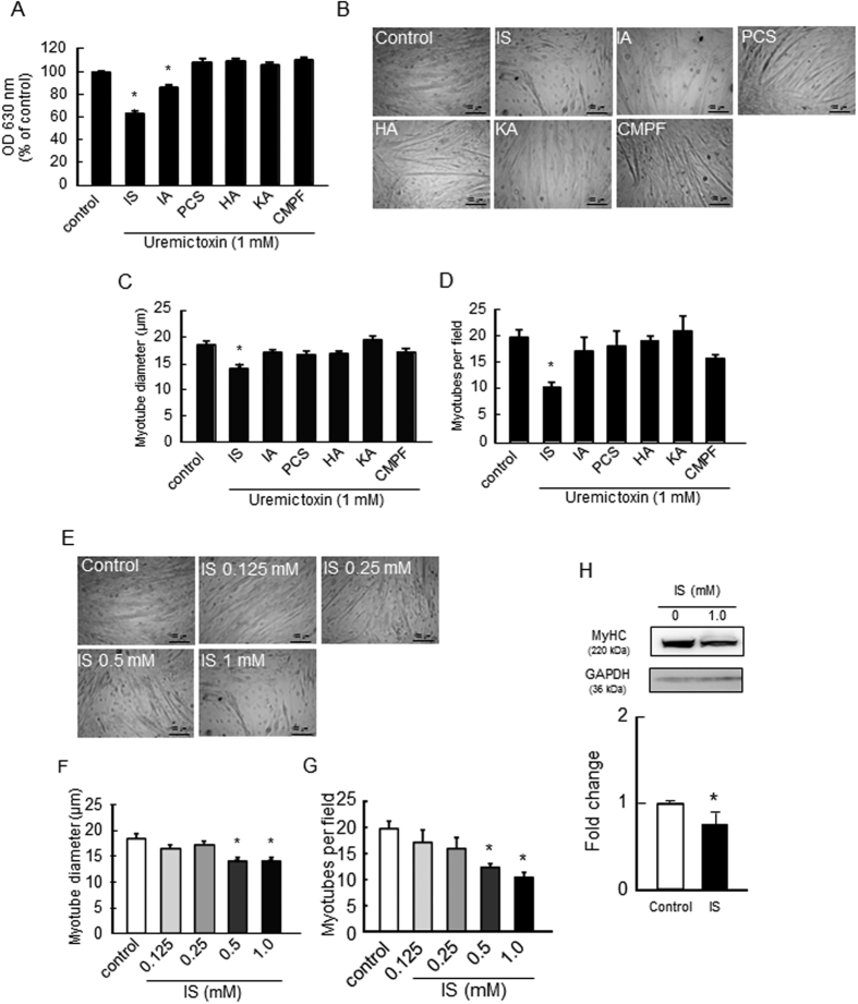
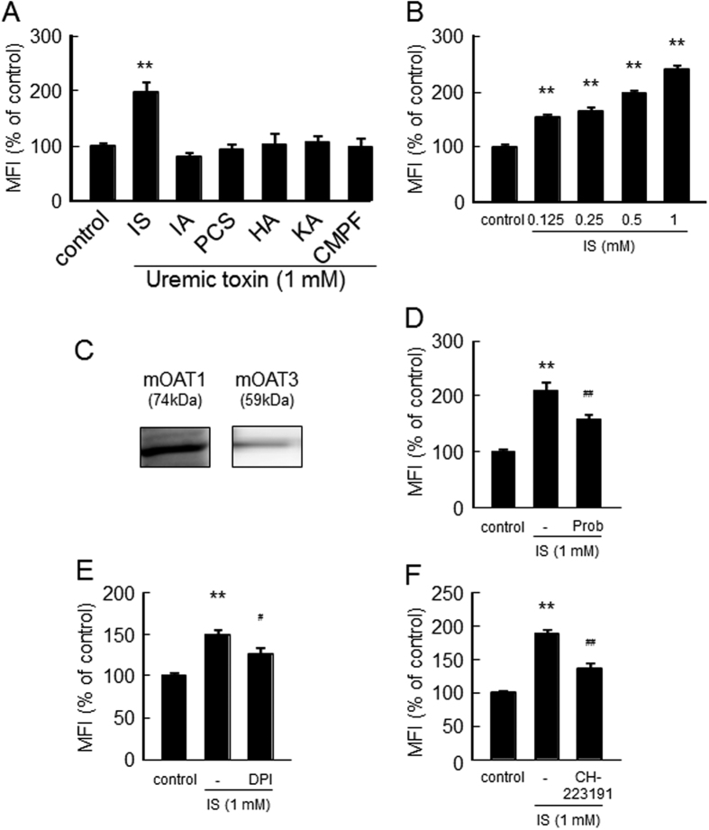
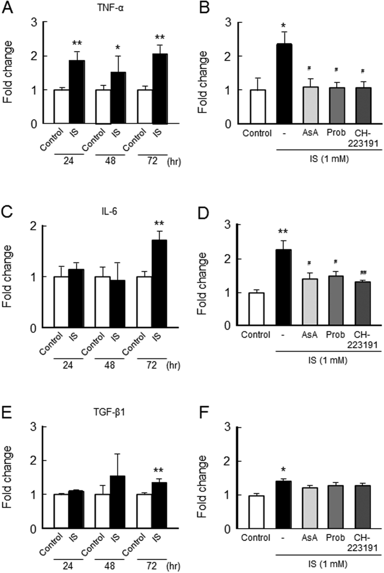
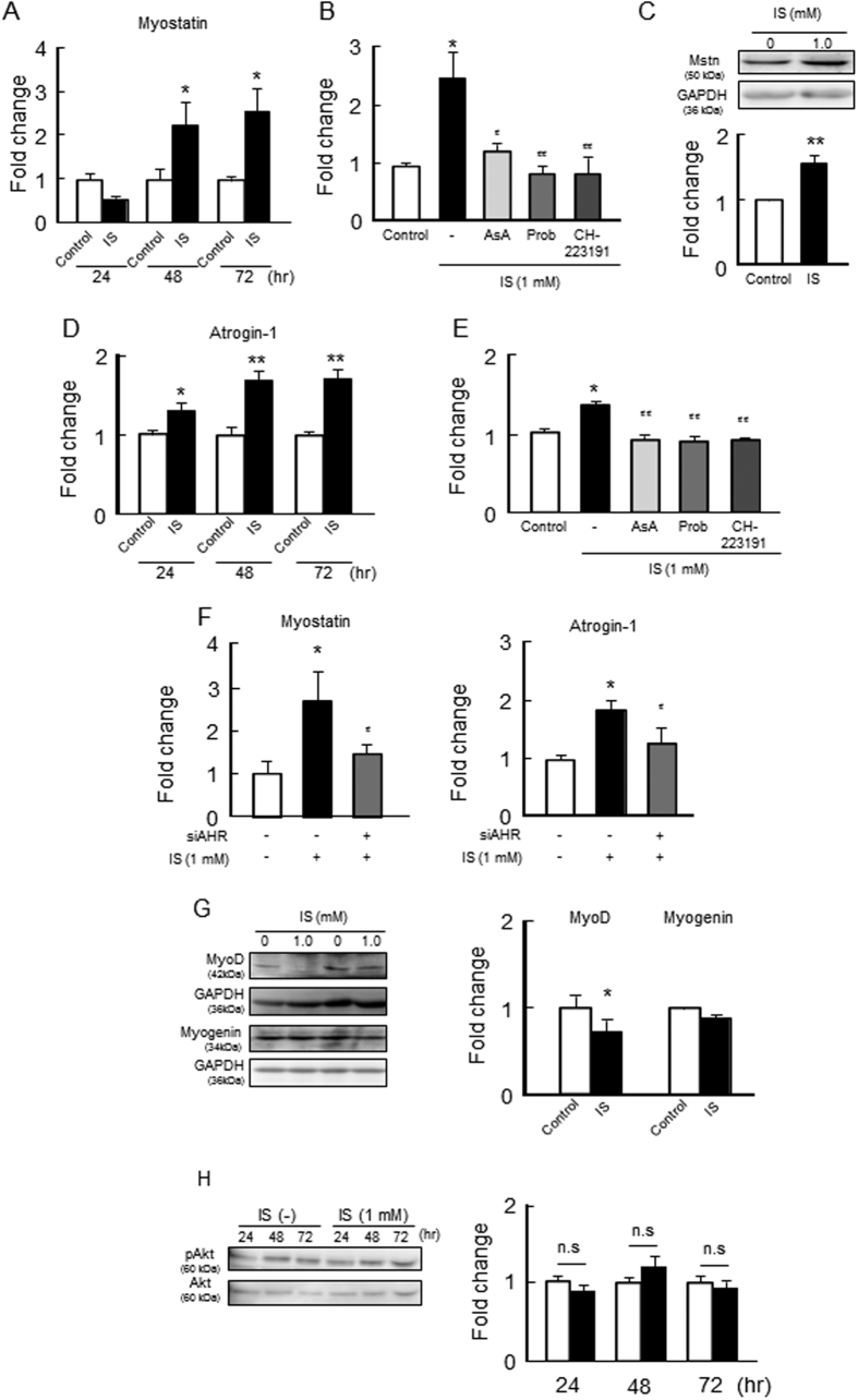
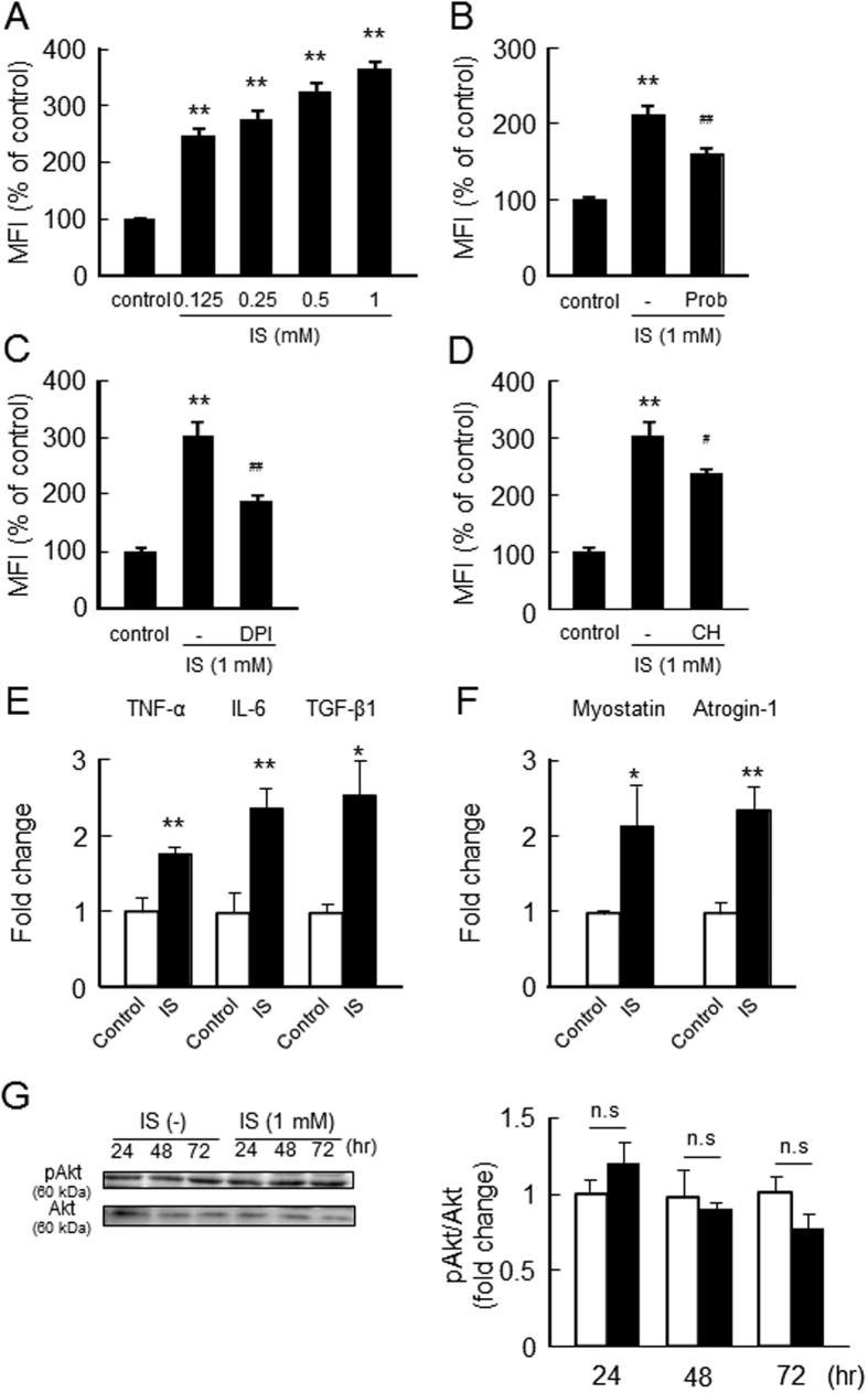
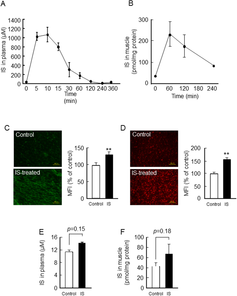
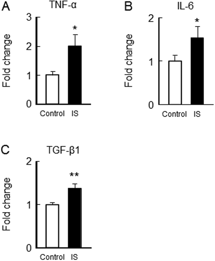
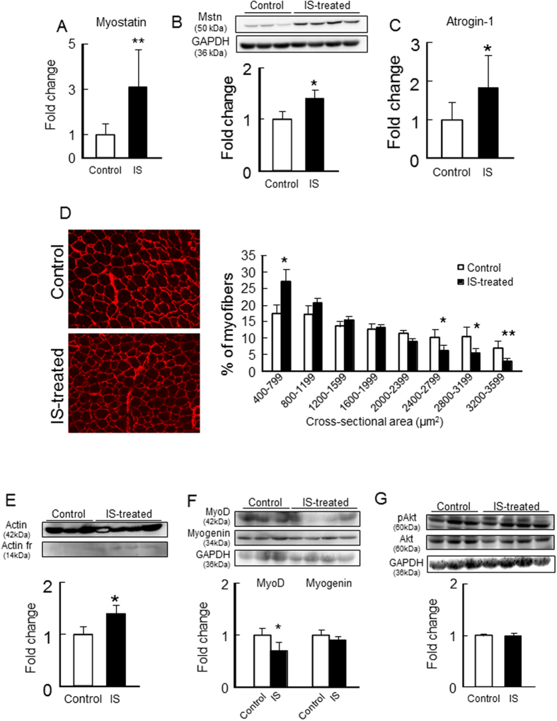
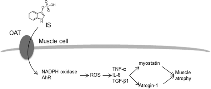
Skeletal muscle atrophy, referred to as sarcopenia, is often observed in chronic kidney disease (CKD) patients, especially in patients who are undergoing hemodialysis. The purpose of this study was to determine whether uremic toxins are involved in CKD-related skeletal muscle atrophy. Among six protein-bound uremic toxins, indole containing compounds, indoxyl sulfate (IS) significantly inhibited proliferation and myotube formation in C2C12 myoblast cells. IS increased the factors related to skeletal muscle breakdown, such as reactive oxygen species (ROS) and inflammatory cytokines (TNF-α, IL-6 and TGF-β1) in C2C12 cells. IS also enhanced the production of muscle atrophy-related genes, myostatin and atrogin-1. These effects induced by IS were suppressed in the presence of an antioxidant or inhibitors of the organic anion transporter and aryl hydrocarbon receptor. The administered IS was distributed to skeletal muscle and induced superoxide production in half-nephrectomized (1/2 Nx) mice. The chronic administration of IS significantly reduced the body weights accompanied by skeletal muscle weight loss. Similar to the in vitro data, IS induced the expression of myostatin and atrogin-1 in addition to increasing the production of inflammatory cytokines by enhancing oxidative stress in skeletal muscle. These data suggest that IS has the potential to accelerate skeletal muscle atrophy by inducing oxidative stress-mediated myostatin and atrogin-1 expression.


Figures

Figure 1
Figure 1. Effect of uremic toxins on the proliferation and myotube formation of C2C12 myoblast cells.
(A) Effect of uremic toxins on C2C12 myoblast cell proliferation was determined by a methylene blue assay. C2C12 myoblast cells were seeded on a 96-well plate and cultured overnight. After adhesion, cells were treated with uremic toxins (1 mM) and incubated for 72 hr. Adherent cells were fixed and stained with methylene blue. The OD630 nm was then measured. Effect of uremic toxins on cell differentiation was determined by C2C12 myoblast cell tubular formation. C2C12 myoblast cells were seeded on a 12-well plate and cultured overnight. After cell adhesion, cultured medium was changed to differentiation medium containing with uremic toxins and cultured for 7 days. The cell density in each treatment group was the same before changing the culture medium to differentiation medium. (B) Cell morphology was observed by microscopy, and (C) average diameters of at least 50 myotubes were determined to be 40 μm along the length of the myotube. (D) The number of the myotubes per field was measured 5 fields per sample. Myotubes were counted, and diameter was assessed on Image J. Dose-dependent effect of IS on (E) cell morphology, (F) myotube diameter, and (G) the number of myotubes were shown. Scale bar = 100 μm. (H) Myotube formation was also evaluated using second marker, namely, the myosin heavy chain (MyHC) by Western blot. Data are expressed the means ± SEM (n = 3~6). *p < 0.05, **p < 0.01 compared with control.
Figure 2
Figure 2. Effect of uremic toxins on ROS production in C2C12 myoblast cells.
(A) Effect of uremic toxins on ROS production was determined using CM-H2DCFDA. C2C12 myoblast cells were starved with serum free medium for 2 hr, and then treated with CM-H2DCFDA in D-PBS for 30 min. After removal of the D-PBS, the cells were treated with uremic toxins and incubated for 2 hr. Fluorescence intensity was measured at an excitation wavelength of 485 nm and at an emission wavelength of 535 nm. (B) Dose-dependent effect of IS on ROS production was measured in C2C12 myoblast cells (C) Expression of mouse Oat1 and Oat3 in C2C12 cells was detected by Western blotting. To determine the effect of inhibitors of Oats, NADPH oxidase or AHR on the IS-induced ROS production, cells were incubated with CM-H2DCFDA for 30 min followed by incubation with (D) Oats inhibitors (probenecid (0.5 mM)), (E) NADPH oxidase inhibitor (diphenylene iodonium: DPI (50 μM)) or (F) AHR inhibitor (CH223191 (10 μM)) for 30 min. The cells were then incubated with IS (1 mM) for 1 hr with Oats inhibitors or 2 hr with others. Data are expressed the means ± SEM (n = 6). **p < 0.01 compared with control. #p < 0.05, ##p < 0.01 compared with IS only.
Figure 3
Figure 3. Effect of IS on inflammatory cytokine expression on C2C12 myoblast cells.
Effect of IS on inflammatory cytokines expression was determined by real time RT-PCR. C2C12 myoblast cells were starved with serum free medium for 2 hr, and then treated with IS for 24, 48 or 72 hr. After the incubation, total RNA was collected and the mRNA expression of (A) TNF-α, (C) IL-6 or (E) TGF-β1 in C2C12 myoblast cells were determined. To determine the effect of inhibitors, cells were co-incubated in the presence or absence of ascorbic acid (AsA, a ROS scavenger), probenecid (Prob, Oat inhibitor) and CH-223191 (AHR inhibitor) for 72 hr, and the mRNA expression of (B) TNF-α (D) IL-6 or (F) TGF-β1 in C2C12 myoblast cells were determined. Data are expressed the means ± SEM (n = 3~5). *p < 0.05, **p < 0.01 compared with control. #p < 0.05, ##p < 0.01 compared with IS only.
Figure 4
Figure 4. Effect of IS on the expression of myostatin, skeletal muscle atrophy- or myogenic-related genes and Akt phosphorylation in C2C12 myoblast cells.
Effect of IS on (A,B) mRNA and (C) protein expression of myostatin, mRNA expression of (D,E) atrogin-1 were determined by real time RT-PCR. (F) Effect of AHR RNAi on IS-induced myostatin or atrogin-1 expression was determined by real time RT-PCR. (G) Protein expression of MyoD and myogenin in C2C12 myoblast cells were determined by Western blots. (H) Phosphorylation of Akt was detected by Western blots. C2C12 myoblast cells were starved with serum free medium for 2 hr, and then treated with IS for 24, 48 or 72 hr. After the incubation, total RNA or a whole cell lysate was collected and mRNA or protein expressions in the C2C12 myoblast cells were determined. To determine the effect of inhibitors, the cells were co-incubated in the presence or absence of ascorbic acid (AsA, a ROS scavenger), probenecid (Prob, Oat inhibitor) and CH-223191 (AHR inhibitor) for 72 hr, and the mRNA expression of (B) myostatin or (E) atrogin-1 in C2C12 myoblast cells were determined. Data are expressed the means ± SEM (n = 3~4). *p < 0.05, **p < 0.01 compared with control. #p < 0.05, ##p < 0.01 compared with IS only.
Figure 5
Figure 5. Effect of IS on the ROS production and expression of inflammatory cytokines, myostatin and atrogin-1 in C2C12 myotubes.
C2C12 myotubes were differentiated for 4 days in differentiated medium. (A) To determine the ROS production, C2C12 myotubes were starved with serum free medium for 2 hr, and then treated with CM-H2DCFDA in D-PBS for 30 min. After removal of the D-PBS, the cells were treated with IS and incubated for 2 hr. Fluorescence intensity was measured at an excitation wavelength of 485 nm and an emission wavelength of 535 nm. To determine the effect of inhibitors of Oats, NADPH oxidase or AHR, C2C12 myotubes were incubated with CM-H2DCFDA for 30 min followed by incubation with (B) probenecid (Prob: 0.5 mM), (C) diphenylene iodonium (DPI: 50 μM), or (D) CH223191 (10 μM) for 30 min. The cells were then incubated with IS (1 mM) for 1 hr with an Oats inhibitor or 2 hr with others. C2C12 myotubes were starved with serum free medium for 2 hr, and then treated with IS for 72 hr. After incubation, total RNA was collected and the mRNA expression of (E) IL-6, TNF-α, TGF-β1, (F) myostatin and atrogin-1 were determined. (G) Akt phosphorylation was determined after 72 hr incubation with IS. Data are expressed the means ± SEM (n = 3~6). *p < 0.05, **p < 0.01 compared with control. #p < 0.05, ##p < 0.01 compared with IS only.
Figure 6
Figure 6. Plasma and skeletal muscle concentration of IS in IS-administered 1/2 Nx mice.
(A) Plasma concentration-time profile of IS after intraperitoneal administration of IS (100 mg/kg) into 1/2 Nx mice was determined by HPLC methods. (B) The gastrocnemius concentration of IS was measured after IS administration. (C) Immunofluorostaining image of IS using anti-IS antibody on gastrocnemius was performed. (D) DHE staining of gastrocnemius was performed, and significant increasing of superoxide production was observed 1 hr after IS administration. (E) Plasma and (F) gastrocnemius muscle concentration of IS were measured at the trough level after 12 weeks IS administration. Scale bar = 100 μm. Data are expressed the means ± SEM (n = 3~8). **p < 0.01 compared with control.
Figure 7
Figure 7. Effect of IS administration on inflammatory cytokines expression in the skeletal muscle of 1/2 Nx mice.
After IS administration for 12 weeks, the mRNA expression of (A) TNF-α, (B) IL-6 and (C) TGF-β1 in gastrocnemius were determined by real time RT-PCR. Data are expressed the means ± SEM (n = 6~8). *p < 0.05, **p < 0.01 compared with control.
Figure 8
Figure 8. Effect of IS administration on myostatin expression and muscle atrophy- or myogenic-related genes expression or Akt phosphorylation in the skeletal muscle of 1/2 Nx mice.
After IS administration for 12 weeks, (A) mRNA and (B) protein expressions of myostatin in gastrocnemius were determined by real time RT-PCR and Western blots. mRNA expression of (C) atrogin-1 in gastrocnemius were determined by real time RT-PCR. (D) Cryosections of tibialis anterior muscles were immunostained with anti-laminin to assess myofiber size. (E) Muscle degradation marker, 14 kDa actin fragment was determined by Western blot. (F) MyoD and myogenin expression in gastrocnemius were determined by Western blots. (G) Akt phosphorylation in gastrocnemius was determined by Western blots. Relative intensity of pAkt/Akt was quantified using the ImageJ software. Data are expressed the means ± SEM (n = 6~8). *p < 0.05, **p < 0.01 compared with control.
Figure 9
Figure 9. Proposed mechanism of IS-induced muscle atrophy.
IS accumulates in muscle cells via Oat where IS activates NADPH oxidase and the AHR pathway to cause increased ROS production. The enhanced ROS production, in turn, triggers inflammatory cytokines (TNF-α, IL-6 and TGF-β1) induce myostatin and atrogin-1 expression that are involved in muscle wasting.

References

    1. Wang A. Y. et al.. Resting energy expenditure and subsequent mortality risk in peritoneal dialysis patients. J Am Soc Nephrol 15, 3134–3143, doi: 10.1097/01.ASN.0000144206.29951.B2 (2004). - DOI - PubMed
    1. Kalantar-Zadeh K. et al.. Mortality prediction by surrogates of body composition: an examination of the obesity paradox in hemodialysis patients using composite ranking score analysis. Am J Epidemiol 175, 793–803, doi: 10.1093/aje/kwr384 (2012). - DOI - PMC - PubMed
    1. Foletta V. C., White L. J., Larsen A. E., Léger B. & Russell A. P. The role and regulation of MAFbx/atrogin-1 and MuRF1 in skeletal muscle atrophy. Pflugers Arch 461, 325–335, doi: 10.1007/s00424-010-0919-9 (2011). - DOI - PubMed
    1. Rodriguez J. et al.. Myostatin and the skeletal muscle atrophy and hypertrophy signaling pathways. Cell Mol Life Sci, doi: 10.1007/s00018-014-1689-x (2014). - DOI - PMC - PubMed
    1. Bodine S. C. & Baehr L. M. Skeletal muscle atrophy and the E3 ubiquitin ligases MuRF1 and MAFbx/atrogin-1. Am J Physiol Endocrinol Metab 307, E469–E484, doi: 10.1152/ajpendo.00204.2014 (2014). - DOI - PMC - PubMed

Publication types

MeSH terms