. 2016 Nov;92(11):fiw176.
doi: 10.1093/femsec/fiw176. Epub 2016 Aug 21.

RNA-Seq reveals virus-virus and virus-plant interactions in nature

Affiliations

RNA-Seq reveals virus-virus and virus-plant interactions in nature

Mari Kamitani et al. FEMS Microbiol Ecol. 2016 Nov.

Abstract

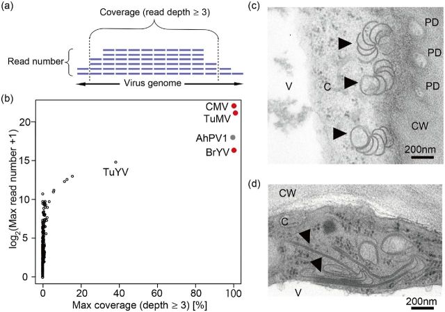
As research on plant viruses has focused mainly on crop diseases, little is known about these viruses in natural environments. To understand the ecology of viruses in natural systems, comprehensive information on virus-virus and virus-host interactions is required. We applied RNA-Seq to plants from a natural population of Arabidopsis halleri subsp. gemmifera to simultaneously determine the presence/absence of all sequence-reported viruses, identify novel viruses and quantify the host transcriptome. By introducing the criteria of read number and genome coverage, we detected infections by Turnip mosaic virus (TuMV), Cucumber mosaic virus and Brassica yellows virus Active TuMV replication was observed by ultramicroscopy. De novo assembly further identified a novel partitivirus, Arabidopsis halleri partitivirus 1 Interestingly, virus reads reached a maximum level that was equivalent to that of the host's total mRNA, although asymptomatic infection was common. AhgAGO2, a key gene in host defence systems, was upregulated in TuMV-infected plants. Multiple infection was frequent in TuMV-infected leaves, suggesting that TuMV facilitates multiple infection, probably by suppressing host RNA silencing. Revealing hidden plant-virus interactions in nature can enhance our understanding of biological interactions and may have agricultural applications.

Keywords: Arabidopsis halleri; Arabidopsis halleri partitivirus 1; Argonaute2; RNA-Seq; Turnip mosaic virus; multiple infection.


Figures

Figure 1.
Figure 1.
Detection of TuMV, CMV and BrYV in a natural population of A. halleri. (a) Definitions of the read number and the coverage for a particular virus genome used in this study. Reads obtained from RNA-Seq were mapped onto each virus genome. Read depth refers to the number of reads covering the same sequence. The coverage indicates the percentage of the genome area covered with more than three reads. (b) Determination of infection by sequence-reported viruses. Maximum values of the log2 of the read number and the genome coverage for the 3981 sequence reported viruses (NCBI database) are shown. Each point indicates a single virus. Red-filled points represent infecting viruses. A novel virus identified by the de novo assembly, AhPV1, is shown by a grey-filled point after re-mapping. (c and d) TEM images of an infected leaf. Filled arrowheads show the pinwheel (c) and rod-shaped (d) structures of TuMV. V, C and CW indicate the vacuole, cytoplasm and cell wall, respectively. PD indicates the secondary plasmodesma.
Figure 2.
Figure 2.
AhPV1, a novel partitivirus identified by the de novo assembly. (a) Schematic diagrams of AhPV1-RdRP and AhPV1-CP fragments. The total nucleotide lengths of the fragments are shown in parentheses. Coding regions of RdRP and CP are shown by boxes with amino acid lengths in parentheses. Numbers under the diagrams indicate the starting and terminative positions of the fragment and coding region. (b) Amplifications of RdRP- and CP-specific fragments for five putative infected (shown by asterisks) and five non-infected leaves. M and nc represent the size marker (400 and 500 bp) and no-template negative control, respectively. (c) Phylogenetic locations of AhPV1 (boxed) based on RdRP AA sequences. Corresponding viruses shown by abbreviations are listed in Table S3. Colours correspond to the four genera of partitivirus. Cryspovirus was used as an outgroup. Underlining indicates related Alphapartitivirus for which the CP sequence is unknown. Numbers represent bootstrap values (percentages).
Figure 3.
Figure 3.
Spatial distributions of A. halleri plants infected by different combinations of viruses, and chlorosis phenotypes in the study site. Spatial pattern in samples collected along the creek (a) and in the 20 m × 25 m rectangular plot (b). Each small circle represents a single plant. Presence/absence of leaf chlorosis is shown by filled/open circles, respectively. Combinations of infecting virus species are shown by coloured shadings. Circle, square, hexagon and diamond shadings represent single, double, triple and quadruple infections, respectively. The grey area indicates a stream that flows from the top to the bottom of diagrams. White bars crossing the stream indicate the positions of erosion-control dams. (c) Typical chlorosis of A. halleri leaves.
Figure 4.
Figure 4.
Multiple infections and host response. (a) Frequency of multiple infection at the Omoide River site. Numbers of plants infected by the viruses are shown in a Venn diagram (n = 68). The overlapping areas represent multiple infections from corresponding viruses. (b) Test of coinfection of virus pairs. The results of Fisher's exact test (P-value) between all virus pairs are shown. (c) Differentially expressed genes in the comparison between TuMV-infected and non-infected host transcriptomes. In the volcano plot, statistical significance (–log2P-value) is plotted against the log ratio of the average expression (rpm, infection/non-infection) for each host gene. Red dots represent the genes with FDR-adjusted P-values of <0.05. (d) Difference in AhgAGO2 gene expression between TuMV-non-infected and TuMV-infected plants (black and red circles, respectively). The vertical axis is the read number of the AhgAGO2 gene obtained from RNA-Seq. The box and lines denote the median, first quartile, and third quartile values of each sample group. (e) The hypothesised mechanism of facilitation in multiple infection by TuMV. TuMV-infected plants recognise the infection and respond with RNA silencing. TuMV inhibits the host RNA silencing with HC-Pro protein. A plant whose defence is suppressed becomes susceptible to infection by other viruses.

References

    1. Aikawa S, Kobayashi MJ, Satake A et al. . Robust control of the seasonal expression of the Arabidopsis FLC gene in a fluctuating environment. P Natl Acad Sci USA 2010;107:11632–7 - PMC - PubMed
    1. Alexander HM, Mauck KE, Whitfield AE et al. . Plant-virus interactions and the agro-ecological interface. Eur J Plant Pathol 2014;138:529–47
    1. Alvarado VY, Scholthof HB.. AGO2: a new argonaute compromising plant virus accumulation. Front Plant Sci 2011;2:112. - PMC - PubMed
    1. Anandalakshmi R, Pruss GJ, Ge X et al. . A viral suppressor of gene silencing in plants. P Natl Acad Sci USA 1998;95:13079–84 - PMC - PubMed
    1. Barba M, Czosnek H, Hadidi A.. Historical perspective, development and applications of next-generation sequencing in plant virology. Viruses 2014;6:106–36 - PMC - PubMed