. 2016 Oct;4(10):869-880.
doi: 10.1158/2326-6066.CIR-15-0230. Epub 2016 Aug 22.

Reduction of MDSCs with All-trans Retinoic Acid Improves CAR Therapy Efficacy for Sarcomas

Affiliations

Reduction of MDSCs with All-trans Retinoic Acid Improves CAR Therapy Efficacy for Sarcomas

Adrienne H Long et al. Cancer Immunol Res. 2016 Oct.

Abstract

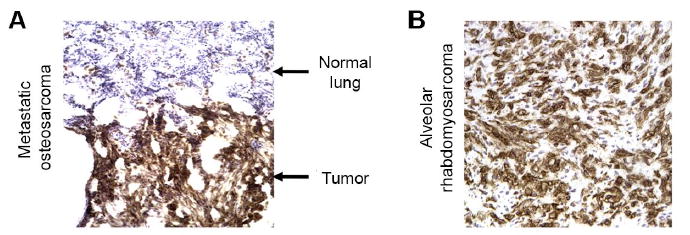
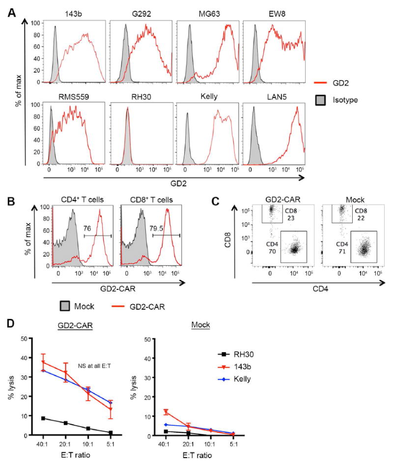
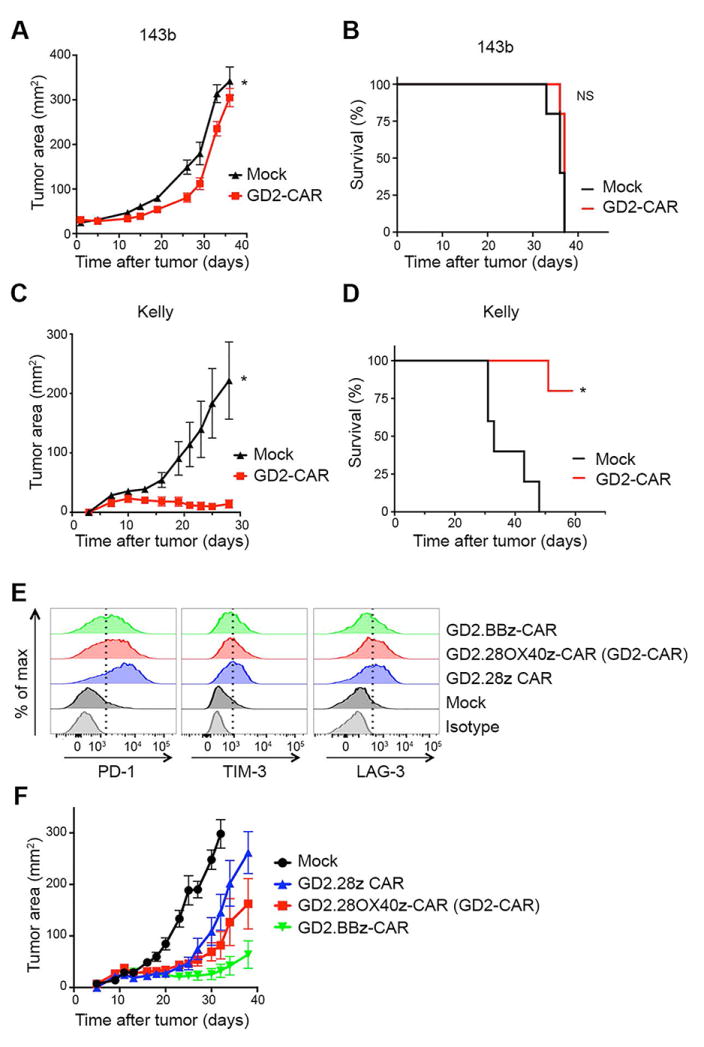
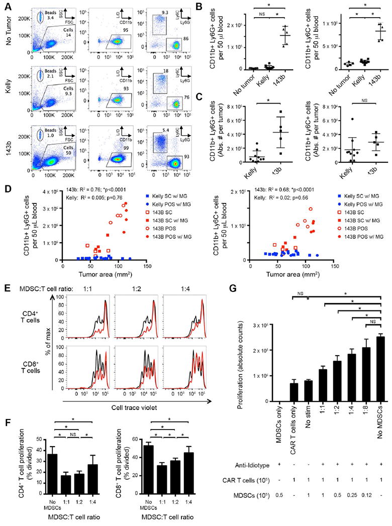
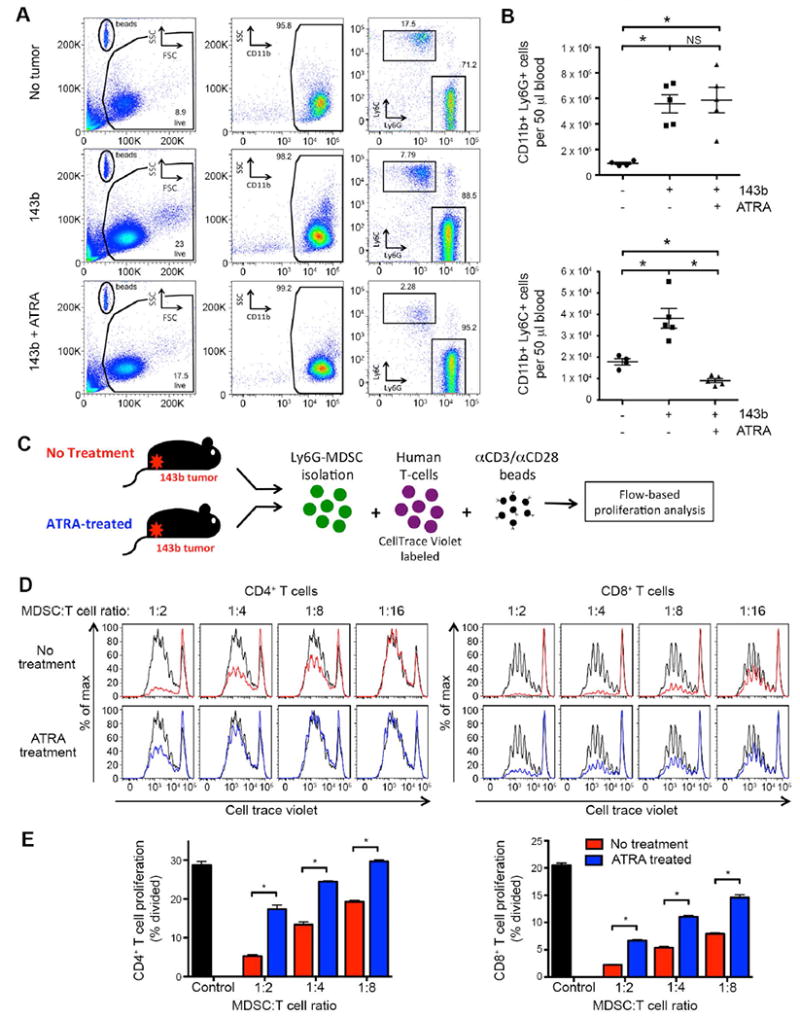
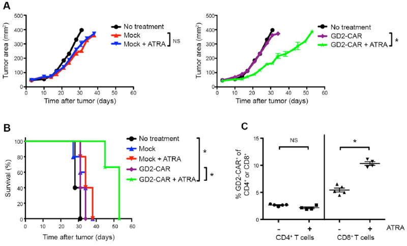
Genetically engineered T cells expressing CD19-specific chimeric antigen receptors (CAR) have shown impressive activity against B-cell malignancies, and preliminary results suggest that T cells expressing a first-generation disialoganglioside (GD2)-specific CAR can also provide clinical benefit in patients with neuroblastoma. We sought to assess the potential of GD2-CAR therapies to treat pediatric sarcomas. We observed that 18 of 18 (100%) of osteosarcomas, 2 of 15 (13%) of rhabdomyosarcomas, and 7 of 35 (20%) of Ewing sarcomas expressed GD2. T cells engineered to express a third-generation GD2-CAR incorporating the 14g2a-scFv with the CD28, OX40, and CD3ζ signaling domains (14g2a.CD28.OX40.ζ) mediated efficient and comparable lysis of both GD2+ sarcoma and neuroblastoma cell lines in vitro However, in xenograft models, GD2-CAR T cells had no antitumor effect against GD2+ sarcoma, despite effectively controlling GD2+ neuroblastoma. We observed that pediatric sarcoma xenografts, but not neuroblastoma xenografts, induced large populations of monocytic and granulocytic murine myeloid-derived suppressor cells (MDSC) that inhibited human CAR T-cell responses in vitro Treatment of sarcoma-bearing mice with all-trans retinoic acid (ATRA) largely eradicated monocytic MDSCs and diminished the suppressive capacity of granulocytic MDSCs. Combined therapy using GD2-CAR T cells plus ATRA significantly improved antitumor efficacy against sarcoma xenografts. We conclude that retinoids provide a clinically accessible class of agents capable of diminishing the suppressive effects of MDSCs, and that co-administration of retinoids may enhance the efficacy of CAR therapies targeting solid tumors. Cancer Immunol Res; 4(10); 869-80. ©2016 AACR.


Conflict of interest statement

The authors report no conflict of interest exists.

Figures

Figure 1
Figure 1. GD2 expression on sarcoma patient samples by immunohistochemistry
Representative immunohistochemical staining for GD2 on sarcoma patient samples, demonstrating high expression of GD2 on (A) metastatic osteosarcoma compared to adjacent lung tissue and on (B) alveolar rhabdomyosarcoma.
Figure 2
Figure 2. GD2-CAR T cells effectively lyse GD2+ sarcoma cell lines in vitro
(A) Flow cytometric analysis of GD2 expression on human osteosarcoma (143b, G292, MG63), Ewing sarcoma (EW8), rhabdomyosarcoma (RMS559, RH30) and neuroblastoma (Kelly, LAN5) cell lines grown in vitro. Numerous sarcoma cell lines demonstrated strong GD2 surface expression. (B) Representative transduction efficiency of GD2-CAR T cells, evaluated on day 7 following initial activation. Transduction efficiency measured by staining with clone 1A7, an anti-idiotype antibody specific for the 14g2a antibody. (C) Representative CD4:CD8 ratios of GD2-CAR and Mock T-cell products, evaluated on day 7 following initial activation. (D) Lysis of target cells measured by 51Cr release. GD2-CAR T cells equivalently lyse the GD2+ 143b and Kelly cells lines, but do not lyse the GD2 negative cell line RH30.
Figure 3
Figure 3. GD2-CAR has poor in vivo efficacy against the 143b osteosarcoma cell line in a xenograft model that is not fully attributable to T-cell exhaustion
(A) Tumor growth curves and (B) survival of NSG mice inoculated with 106 143b cells periosteally on day 0, followed by adoptive transfer of 107 untransduced-Mock or GD2-CAR T cells on day 3. GD2-CAR T cells have no antitumor effect against 143b. n=5 mice/group. (C) Tumor growth curves and (D) survival of NSG mice inoculated with 106 Kelly cells subcutaneously with Matrigel on day 0, followed by adoptive transfer of 107 untransduced-Mock or GD2-CAR T cells on day 3. GD2-CAR T cells prevent outgrowth of Kelly tumors leading to enhanced overall survival. n=5 mice/group. (E) Exhaustion marker expression on T cells transduced with GD2-specific CARs incorporating different costimulatory domains (GD2-CAR, a third generation CAR with CD28 and OX40; a second generation CAR with CD28; and a second generation CAR with 4-1BB) 9 days following initial activation. The third generation GD2-CAR T cells have an intermediate expression of exhaustion markers compared to the second generation CD28 CAR and the 4-1BB CAR. (F) Tumor growth curves of NSG mice inoculated with 106 Kelly cells subcutaneously with Matrigel on day 0, followed by adoptive transfer of 107 untransduced-Mock or GD2-specific CAR T cells on day 3. The third generation GD2-CAR T cells had an intermediate efficacy compared to the second generation CD28 CAR and the 4-1BB CAR. n=10 mice/group.
Figure 4
Figure 4. Human sarcoma implanted in NSG mice induces expansion of host MDSCs
(A) Representative flow cytometry plots of peripheral blood, evaluating presence of CD11b+Ly6G+ or CD11b+Ly6C+ MDSCs in mice inoculated with 106 143b osteosarcoma periosteally or 106 Kelly cells subcutaneously with Matrigel. Evaluated day 28 after tumor inoculation. L/D = live/dead stain. (B) Cumulative data from A, quantifying absolute number of CD11b+Ly6G+ (left) and CD11b+Ly6C+ (right) cells per 50 μl of blood. 143b tumors induce significant expansion of both CD11b+Ly6C+ and CD11b+Ly6G+ populations, compared to non-tumor controls or Kelly tumor-bearing mice with similarly sized tumors. (C) Absolute number of CD11b+Ly6G+ (left) and CD11b+Ly6C+ (right) cells per tumor. 143b tumors contain significant CD11b+Ly6G+ populations, compared to Kelly tumors. (D) Impact of tumor location and Matrigel on induction of CD11b+Ly6G+ (left) and CD11b+Ly6C+ (right) cells in blood. NSG mice were inoculated with 106 143b (red) or Kelly (blue) tumor cells. MDSCs were quantified in the blood 20 days post tumor injection and plotted as a function of tumor size. Tumors were placed subcutaneously (SC, square) or periosteally (POS, circle), with (filled) or without (open) Matrigel (MG). R2 and p value statistics calculated from linear regression of pooled 143b and Kelly data. (E) Cell trace violet dilution of human T cells following co-incubation with murine CD11b+ MDSCs in vitro. Human αCD3/αCD28 beads were added as a proliferative stimulus. Flow cytometry was performed 4 days later. αCD3/αCD28 bead-induced proliferation without MDSCs in co-culture shown in black. CD11b+ MDSCs isolated from 143b tumor-bearing mice suppress human T-cell proliferation in a dose dependent manner (red). (F) Quantification of T-cell proliferation in E, measured as percentage of T cells divided. (G) Absolute cell counts of GD2-CAR T cells (9 days after initial activation) co-incubated with murine CD11b+ MDSCs derived from 143b tumor bearing mice. GD2-CAR T cells were stimulated via the CAR with the anti-idiotype 1A7 antibody (0.5 μg/mL) and a cross-linking anti-mouse F(ab’)2 (2.5 μg/mL). Cell counts from cultures were performed 5 days after CAR stimulation. CD11b+ MDSCs isolated from 143b tumor-bearing mice suppress human CAR T-cell proliferation in a dose dependent manner.
Figure 5
Figure 5. ATRA treatment reduces number and suppressive capacity of murine MDSCs induced by sarcoma in NSG mice
(A) Representative flow cytometry plots of peripheral blood, evaluating presence of CD11b+Ly6G+ or CD11b+Ly6C+ MDSCs in mice inoculated with 106 143b osteosarcoma periosteally on day 0, treated with or without ATRA sustained-release subcutaneous pellets on day -1. Evaluated day 15 after tumor inoculation. (B) Cumulative data from A, showing the absolute number of CD11b+Ly6C+ (top) and CD11b+Ly6C+ (bottom) cells per 50 μl of blood. ATRA treatment leads to a significant reduction in CD11b+Ly6C+ cells in 143b tumor-bearing mice. The number of CD11b+Ly6G+ MDSCs observed following ATRA treatment was not consistently decreased. (C) Schematic of MDSC T-cell suppression assay. Splenic Ly6G+ cells from 143b tumor-bearing mice (± ATRA treatment on day -1) were magnetically isolated and mixed at increasing ratios with CellTrace Violet-labeled human T cells. Flow cytometry was performed 4 days later. Human αCD3/αCD28 beads were added as a proliferative stimulus. (D) CellTrace Violet dilution of CD4+ and CD8+ human T cells following co-incubation with murine MDSCs in vitro. αCD3/αCD28 bead induced proliferation without MDSCs in co-culture shown in black. CD11b+Ly6G+ MDSCs isolated from 143b tumor-bearing mice not treated with ATRA suppress human T-cell proliferation in a dose dependent manner (red). CD11b+Ly6G+ MDSC isolated from 143b mice treated with ATRA have decreased ability to suppress T-cell proliferation (blue). (E) Quantification of T-cell proliferation in D, measured as percentage of CD4+ (left) or CD8+ (right) T cells dividing. n=6/group.
Figure 6
Figure 6. Co-administration of ATRA with GD2-CAR T cells leads to enhanced antitumor efficacy against 143b osteosarcoma tumors in NSG mice
(A) Tumor growth curves and (B) survival of NSG mice inoculated with 5 × 10⁵ 143b cells on day 0, followed by adoptive transfer of 30 × 106 untransduced-Mock or GD2-CAR T cells on day 3 and 15 × 106 on day 5. Where indicated, mice received ATRA pellets subcutaneously or sham surgeries day -1 before tumor injection. GD2-CAR T cells have enhanced antitumor effect and prolonged survival against 143b tumors in the presence of ATRA. n=3-5 mice/group. (C) Frequency of GD2-CAR+ expression within CD4+ (left) and CD8+ (right) T cells populations in the peripheral blood 15 days after adoptive transfer, assessed by flow cytometry. Adoptively transferred T cells distinguished from host murine cells by hCD45+. CAR+ identified by 1A7 anti-idiotype.

References

    1. Lee DW, Barrett DM, Mackall C, Orentas R, Grupp SA. The Future Is Now: Chimeric Antigen Receptors as New Targeted Therapies for Childhood Cancer. Clinical Cancer Research. 2012;18(10):2780–90. - PMC - PubMed
    1. Sadelain M, Brentjens R, Rivière I. The Basic Principles of Chimeric Antigen Receptor Design. Cancer Discovery. 2013;3(4):388–98. - PMC - PubMed
    1. Lee DW, Kochenderfer JN, Stetler-Stevenson M, Cui YK, Delbrook C, Feldman SA, et al. T cells expressing CD19 chimeric antigen receptors for acute lymphoblastic leukaemia in children and young adults: a phase 1 dose-escalation trial. The Lancet. 2014 - PMC - PubMed
    1. Maude SL, Frey N, Shaw PA, Aplenc R, Barrett DM, Bunin NJ, et al. Chimeric Antigen Receptor T Cells for Sustained Remissions in Leukemia. New England Journal of Medicine. 2014;371(16):1507–17. - PMC - PubMed
    1. Kochenderfer JN, Dudley ME, Feldman SA, Wilson WH, Spaner DE, Maric I, et al. B-cell depletion and remissions of malignancy along with cytokine-associated toxicity in a clinical trial of anti-CD19 chimeric-antigen-receptor–transduced T cells. Blood. 2012;119(12):2709–20. - PMC - PubMed

MeSH terms