

The potential impact of bone tissue engineering in the clinic
- PMID: 27549369
- PMCID: PMC5007661
- DOI: 10.2217/rme-2016-0042
The potential impact of bone tissue engineering in the clinic
Abstract
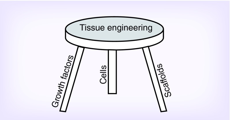
Bone tissue engineering (BTE) intends to restore structural support for movement and mineral homeostasis, and assist in hematopoiesis and the protective functions of bone in traumatic, degenerative, cancer, or congenital malformation. While much effort has been put into BTE, very little of this research has been translated to the clinic. In this review, we discuss current regenerative medicine and restorative strategies that utilize tissue engineering approaches to address bone defects within a clinical setting. These approaches involve the primary components of tissue engineering: cells, growth factors and biomaterials discussed briefly in light of their clinical relevance. This review also presents upcoming advanced approaches for BTE applications and suggests a probable workpath for translation from the laboratory to the clinic.
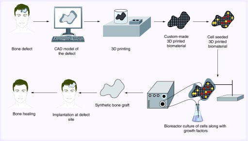
Keywords: 3D printing; additive manufacturing; biomaterials; bioreactor; bone tissue engineering; cell-based therapy.
Conflict of interest statement
Financial & competing interests disclosure The authors acknowledge partial support from NIH grant R01-DE013740, NIH grant R01-AR061460, the Army, Navy, NIH, Air Force, VA, and Health Affairs to support the AFIRM II effort under award No. W81XWH-14-2-0004. The US Army Medical Research Acquisition Activity is the awarding and administering acquisition office for award No. W81XWH-14-2-0004. Some of the implant design and fabrication technology discussed in this review have been patented and assigned or licensed to Osteoplastics, LLC (Shaker Heights, OH, USA). Other technology discussed in this manuscript is the subject of sponsored research agreements with 3DBioResins (Pepper Pike, OH, USA) and 3DServicePros, LLC (Pepper Pike, OH, USA). D Dean is a co-founder and has an ownership stake in all three companies. D Dean has received compensation from Osteoplastics, LLC. The authors have no other relevant affiliations or financial involvement with any organization or entity with a financial interest in or financial conflict with the subject matter or materials discussed in the manuscript apart from those disclosed. No writing assistance was utilized in the production of this manuscript.
Figures

References
-
- Bao CLM, Teo EY, Chong MSK, Liu Y, Choolani M, Chan JKY. Advances in bone tissue engineering. In: Andrades JA, editor. Regenerative Medicine and Tissue Engineering. InTech; Rijeka, Croatia: 2013. pp. 599–614.
-
- Van Der Stok J, Van Lieshout EM, El-Massoudi Y, Van Kralingen GH, Patka P. Bone substitutes in The Netherlands – a systematic literature review. Acta Biomater. 2011;7(2):739–750. - PubMed
-
- Global Industry Analysts I. US Bone Grafts Market to Reach US$2.3 Billion by 2017. 2011. www.prweb.com/releases/bone_grafts/standard_bone_allografts/prweb8953883...
-
- Burg KJ, Porter S, Kellam JF. Biomaterial developments for bone tissue engineering. Biomaterials. 2000;21(23):2347–2359. - PubMed
Publication types
MeSH terms
Grants and funding
LinkOut - more resources
Full Text Sources
Other Literature Sources
Medical