. 2016 Aug 23;13(1):46.
doi: 10.1186/s12989-016-0158-0.

Mesothelioma response to carbon nanotubes is associated with an early and selective accumulation of immunosuppressive monocytic cells

Affiliations

Mesothelioma response to carbon nanotubes is associated with an early and selective accumulation of immunosuppressive monocytic cells

François Huaux et al. Part Fibre Toxicol. .

Abstract

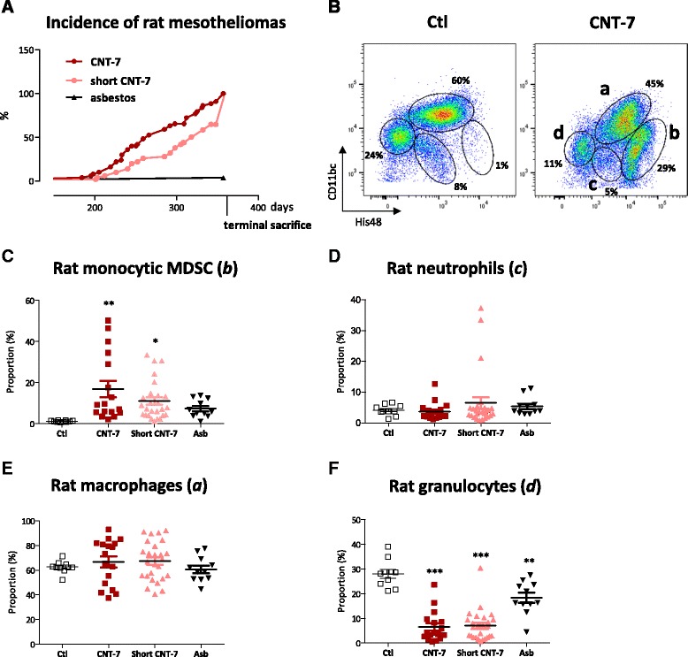
Background: The asbestos-like toxicity of some engineered carbon nanotubes (CNT), notably their capacity to induce mesothelioma, is a serious cause of concern for public health. Here we show that carcinogenic CNT induce an early and sustained immunosuppressive response characterized by the accumulation of monocytic Myeloid Derived Suppressor Cells (M-MDSC) that counteract effective immune surveillance of tumor cells.

Methods: Wistar rats and C57BL/6 mice were intraperitoneally injected with carcinogenic multi-walled Mitsui-7 CNT (CNT-7) or crocidolite asbestos. Peritoneal mesothelioma development and immune cell accumulation were assessed until 12 months. Leukocyte sub-populations were identified by recording expression of CD11b/c and His48 by flow cytometry. The immunosuppressive activity on T lymphocytes of purified peritoneal leukocytes was assessed in a co-culture assay with activated spleen cells.

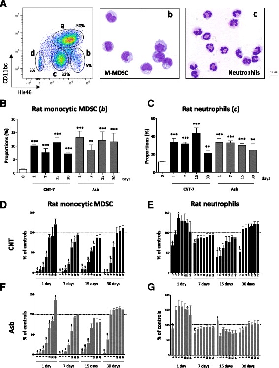
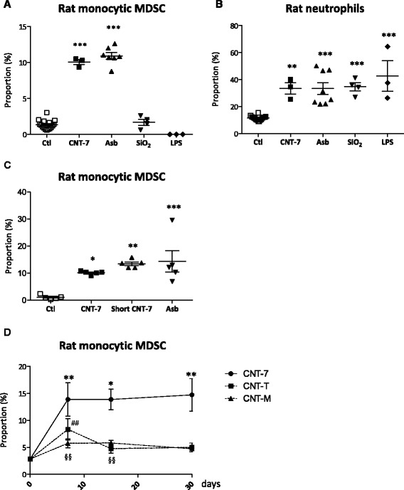
Results: We demonstrate that long and short mesotheliomagenic CNT-7 injected in the peritoneal cavity of rats induced, like asbestos, an early and selective accumulation of monocytic cells (CD11b/c(int) and His48(hi)) which possess the ability to suppress polyclonal activation of T lymphocytes and correspond to M-MDSC. Peritoneal M-MDSC persisted during the development of peritoneal mesothelioma in CNT-7-treated rats but were only transiently recruited after non-carcinogenic CNT (CNT-M, CNT-T) injection. Peritoneal M-MDSC did not accumulate in mice which are resistant to mesothelioma development.

Conclusions: Our data provide new insights into the initial pathogenic events induced by CNT, adding a new component to the adverse outcome pathway leading to mesothelioma development. The specificity of the M-MDSC response after carcinogenic CNT exposure highlights the interest of this response for detecting the ability of new nanomaterials to cause cancer.

Keywords: Asbestos; Carbon nanotubes; Crocidolite; Fibers; Immunosuppression; Inflammation; Mesothelioma; Myeloid cells; Rats and mice.


Figures

Fig. 1
Fig. 1
M-MDSC are specifically accumulated in the peritoneal cavity of rats bearing mesotheliomas after carcinogenic CNT-7 exposure. Incidence of peritoneal mesotheliomas (a). Flow cytometry dot plots show the proportions of peritoneal leukocyte sub-populations (b) present in the peritoneal cavity of Wistar rats bearing or not mesotheliomas after carcinogenic CNT-7 (6 mg) or saline (Ctl) treatments and identified by using CD11b/c and His48 markers (a = macrophages, b = M-MDSC, c = neutrophils, d = granulocytes, i.e. eosinophils and mast cells, as indicated in CNT-7 square). Proportion of (c) monocytic CD11b/cint and His48high Myeloid Derived Suppressor Cells (M-MDSC) (see b in B), (d) CD11b/cint and His48int neutrophils (see c in B), (e) CD11b/chigh and His48int macrophages (see a in B), and (f) CD11b/cint and His48low granulocytic cells (see d in B, eosinophils-mast cells) in peritoneal fluid after saline, CNT-7, short CNT-7 (6 mg) or asbestos (Asb, 2 mg) intra-peritoneally injected in rats. Each bar represents mean ± SEM. The results were statistically analyzed using the Dunnett’s Multiple Comparison test. ** = p < 0.01, *** = p < 0.001 indicate a statistically significant difference with saline-treated rats (Ctl)
Fig. 2
Fig. 2
An early accumulation of immunosuppressive M-MDSC is specifically associated with the peritoneal response to carcinogenic CNT-7 in Wistar rats. a Flow cytometry dot plots show the proportions of leukocyte sub-populations present in peritoneal lavages obtained from CNT-7-treated rats (2 mg) at day 7 and identified by using CD11b/c and His48 markers (a = macrophages, b = M-MDSC, c = neutrophils, d = granulocytes, i.e. eosinophils and mast cells). Microscopic views (Diff-Quick staining, 400x) of FACS-sorted monocytic CD11b/cint and His48hi Myeloid Derived Suppressor Cells (M- MDSC) (b) and CD11b/cint and His48int neutrophils (c) freshly obtained from peritoneal cells of CNT-7-treated rats at day 7. Proportion of (b) M-MDSC and (c) neutrophils in the peritoneal lavages performed on CNT-7- or crocidolite asbestos- (Asb) treated Wistar rats at different time points (from 1 to 30 days) after ip injection of 2 mg particles. Each bar represents mean ± SEM of 4–5 observations. The results were statistically analyzed using the Dunnett’s Multiple Comparison test. ** = p < 0.01, *** = p < 0.001 indicate a statistically significant difference with saline-treated rats (day 0). In vitro anti-proliferative activity of different numbers of M-MDSC (d-f) or neutrophils (e-g) purified at different time points (from 1 to 30 days) from CNT-7- (d-e) or asbestos- (f-g) treated rats (2 mg) on spleen T lymphocytes activated with anti-CD3/CD28 antibodies. Graphs represent the average level of 3H-thymidine incorporation expressed as percent of controls (activated T lymphocytes alone) for each M-MDSC or neutrophils/splenocytes ratio. Each bar represents mean ± SEM. The results were statistically analyzed using the Dunnett’s Multiple Comparison test. * = p < 0.05, $ = p < 0.01, # = p < 0.001 indicate a statistically significant difference with activated splenocytes cultured alone
Fig. 3
Fig. 3
The early peritoneal M-MDSC response is specific of mesotheliomagenic CNT. Proportion of (a) monocytic CD11b/cint and His48hi Myeloid Derived Suppressor Cells (M-MDSC) and (b) inflammatory CD11b/cint and His48int neutrophils in the peritoneal lavages performed on saline-, CNT-7- (2 mg), crocidolite asbestos- (Asb, 2 mg), DQ12 silica- (SiO2, 2 mg) or LPS- (E. coli, 5 μg) treated Wistar rats 1 day after ip injection. SiO2 and LPS are not mesotheliomagenic. c Proportion of peritoneal M-MDSC after ip injection of 2 mg CNT-7, short CNT-7 or asbestos at day 1. Each bar represents mean ± SEM of 4–5 observations. The results were statistically analyzed using the Dunnett’s Multiple Comparison test. * = p < 0.05, ** = p < 0.01, *** = p < 0.001 indicate a statistically significant difference with saline-treated rats (Ctl). d Proportion of peritoneal M-MDSC in the peritoneal cavity of rats after carcinogenic CNT-7 or non-carcinogenic CNT-M or CNT-T treatment (2 mg) at days 7, 15 and 30. The results were statistically analyzed using the Dunnett’s Multiple Comparison test. * = p < 0.05, ** = p < 0.01, *** = p < 0.001 indicate a statistically significant difference between saline-treated (day 0) and CNT-7-treated animals. ## = p < 0.01 indicates a statistically significant difference between saline-treated (day 0) and CNT-T-treated animals. $$ = p < 0.01 indicates a statistically significant difference between saline-treated (day 0) and CNT-M-treated animals

References

    1. Jaurand MC, Renier A, Daubriac J. Mesothelioma: Do asbestos and carbon nanotubes pose the same health risk? Part Fibre Toxicol. 2009;6:16. doi: 10.1186/1743-8977-6-16. - DOI - PMC - PubMed
    1. Grosse Y, Loomis D, Guyton KZ, Lauby-Secretan B, El Ghissassi F, Bouvard V, Benbrahim-Tallaa L, Guha N, Scoccianti C, Mattock H, et al. Carcinogenicity of fluoro-edenite, silicon carbide fibres and whiskers, and carbon nanotubes. Lancet Oncol. 2014;15:1427–1428. doi: 10.1016/S1470-2045(14)71109-X. - DOI - PubMed
    1. Donaldson K, Poland CA, Murphy FA, MacFarlane M, Chernova T, Schinwald A. Pulmonary toxicity of carbon nanotubes and asbestos - similarities and differences. Adv Drug Deliv Rev. 2013;65:2078–2086. doi: 10.1016/j.addr.2013.07.014. - DOI - PubMed
    1. Poland CA, Duffin R, Kinloch I, Maynard A, Wallace WA, Seaton A, Stone V, Brown S, Macnee W, Donaldson K. Carbon nanotubes introduced into the abdominal cavity of mice show asbestos-like pathogenicity in a pilot study. Nat Nanotechnol. 2008;3:423–428. doi: 10.1038/nnano.2008.111. - DOI - PubMed
    1. Joyce JA, Fearon DT. T cell exclusion, immune privilege, and the tumor microenvironment. Science. 2015;348:74–80. doi: 10.1126/science.aaa6204. - DOI - PubMed

Publication types