. 2016 Oct 7;291(41):21474-21484.
doi: 10.1074/jbc.M116.729806. Epub 2016 Aug 22.

Composition and Activity of the Non-canonical Gram-positive SecY2 Complex

Affiliations

Composition and Activity of the Non-canonical Gram-positive SecY2 Complex

Mikaila Bandara et al. J Biol Chem. .

Abstract

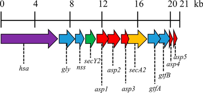
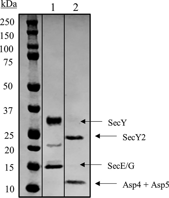
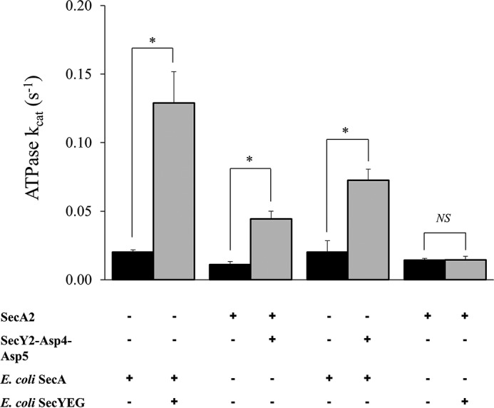
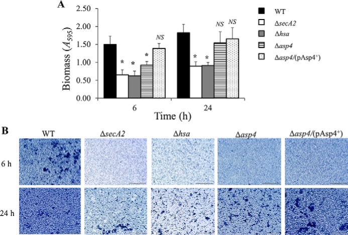
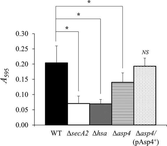
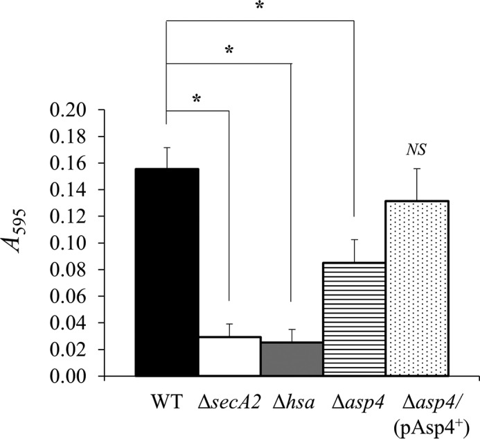
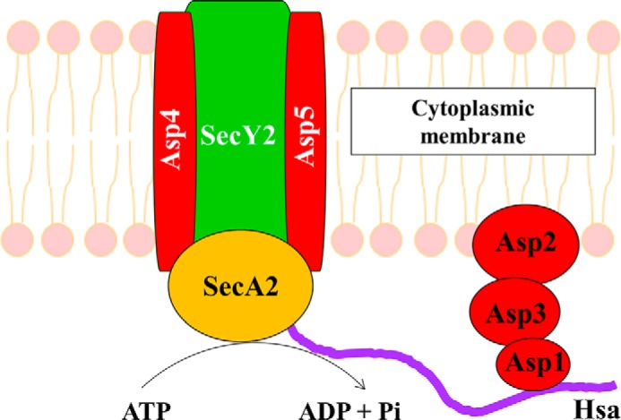
The accessory Sec system in Streptococcus gordonii DL1 is a specialized export system that transports a large serine-rich repeat protein, Hsa, to the bacterial surface. The system is composed of core proteins SecA2 and SecY2 and accessory Sec proteins Asp1-Asp5. Similar to canonical SecYEG, SecY2 forms a channel for translocation of the Hsa adhesin across the cytoplasmic membrane. Accessory Sec proteins Asp4 and Asp5 have been suggested to work alongside SecY2 to form the translocon, similar to the associated SecY, SecE, and SecG of the canonical system (SecYEG). To test this theory, S. gordonii secY2, asp4, and asp5 were co-expressed in Escherichia coli The resultant complex was subsequently purified, and its composition was confirmed by mass spectrometry to be SecY2-Asp4-Asp5. Like SecYEG, the non-canonical complex activates the ATPase activity of the SecA motor (SecA2). This study also shows that Asp4 and Asp5 are necessary for optimal adhesion of S. gordonii to glycoproteins gp340 and fibronectin, known Hsa binding partners, as well as for early stage biofilm formation. This work opens new avenues for understanding the structure and function of the accessory Sec system.

Keywords: ATPase; SecY2; Streptococcus gordonii; accessory Sec system; complex; membrane; secretion; translocon; transport.


Figures

FIGURE 1.
FIGURE 1.
Accessory sec locus of S. gordonii DL1. Schematic representation of the ∼20.5-kb accessory sec locus (derived from the genome sequence of S. gordonii CH1: GenBankTM accession number CP000725.1). Surface protein, hsa (SGO_0966); glycosyltransferases, gly (SGO_0968), nss (SGO_0969), gtfA (SGO_0975), and gtfB (SGO_0976); accessory secretion proteins, asp1 (SGO_0971), asp2 (SGO_0972), asp3 (SGO_0973), asp4 (SGO_0977), and asp5 (SGO_0978); secY2 (SGO_0970), secA2 (SGO_0974).
FIGURE 2.
FIGURE 2.
Gel filtration chromatography of SecY2-Asp4-Asp5. Trace from size exclusion chromatography of SecY2-Asp4-Asp5. Fractions volumes for SecY2-Asp4-Asp5 (186–201 ml) that were collected are represented by red dashed lines. The column was run in TSG130 buffer containing 0.02% DDM. mAU, milli-absorbance units.
FIGURE 3.
FIGURE 3.
SDS-PAGE analysis of purified SecY2-Asp4-Asp5 complex. Coomassie Blue-stained gel of purified components. Lane 1, E. coli SecYEG (4 μg of protein), with bands for SecY and SecE/G indicated. Note that at ∼22 kDa, a SecY breakdown product is often present. Lane 2, S. gordonii SecY2-Asp4-Asp5 (4 μg protein), with bands at 25 and 10 kDa. These were subjected to LC-MS, where the 25-kDa band corresponded to SecY2, whereas Asp4 and Asp5 co-migrated at a molecular mass of 10 kDa.
FIGURE 4.
FIGURE 4.
Mass spectrometry peptides identified within the sequences of SecY2-Asp4-Asp5. Isolated SecY2-Asp4-Asp5 samples were subjected to LC-MS analysis to identify the components present. The amino acid sequences of the proteins are shown above: A, SecY2; B, Asp4; and C, Asp5. Yellow highlights are the identified peptides within each protein sequence. Green highlights predicted post-translational modification.
FIGURE 5.
FIGURE 5.
SDS-PAGE analysis of purified SecA2. Coomassie Blue-stained gel of purified S. gordonii SecA2 (2 μg of protein) with a band between 75 and 100 kDa (the predicted mass is 92 kDa). Protein bands between 37- and 50-kDa markers were presumed to be degradation products due to proteolysis.
FIGURE 6.
FIGURE 6.
ATPase activities of S. gordonii SecA2 with and without SecY2-Asp4-Asp5. Steady-state ATPase activity of 0.3 μm S. gordonii SecA2 or E. coli SecA in the presence of 0.46 μm S. gordonii SecY2-Asp4-Asp5 or E. coli SecYEG, reconstituted in proteoliposomes. Statistical significance is indicated by an asterisk (*, p < 0.05, t test). No statistical significance (NS) is also indicated. Error bars are ± S.E. (n = 5).
FIGURE 7.
FIGURE 7.
Molecular dynamics simulation of SecY2-Asp4-Asp5-SecA2 model. A, homology model of SecY2-Asp4-Asp5-SecA2 was created using Modeler and aligned to the crystal structure of SecYEG-SecA from T. maritima (shown in yellow), Protein Data Bank code 3DIN. Green, SecY2; blue, Asp4; pink, Asp5; beige, SecA2. r.m.s.d. of the alignment of the backbone atoms of the 2 structures is 0.95 Å. B, r.m.s.d. analysis of C-α atoms over the course of a 100-ns molecular dynamics simulation. The system stabilized within 10 ns, at an r.m.s.d. of ∼0.7 nm from its original position. C, pre- and post-simulation images of S. gordonii SecY2-Asp4-Asp5-SecA2 after 100 ns.
FIGURE 8.
FIGURE 8.
WGA dot blot analysis of S. gordonii WT and mutants. 2-Fold dilutions of intact cells of S. gordonii WT, ΔsecA2, Δhsa, and Δasp4 were dot blotted with WGA, which has primary sugar specificity to N-acetylglucosamine, a major component of Hsa.
FIGURE 9.
FIGURE 9.
Biofilm formation by S. gordonii WT, mutants as indicated, and asp4 complemented strain. A, S. gordonii monospecies biofilms were grown on saliva-coated coverslips for 6 or 24 h. Total biomass was quantified by crystal violet staining and measuring A595 following release of stain with acetic acid. Statistical significance to the wild type is indicated by an asterisk (*, p < 0.05, t test). No statistical significance to the wild type (NS) is also indicated. Error bars ± S.D. (n = 2). B, representative light micrographs of S. gordonii biofilms grown for 6 or 24 h and stained with crystal violet. Scale bar, 50 μm.
FIGURE 10.
FIGURE 10.
gp340 binding by S. gordonii WT, mutants as indicated, and asp4 complement strain. gp340 (50 ng) was immobilized on the surface of MTP wells and incubated in the presence of bacteria. Bound bacterial cells were quantified by staining with crystal violet. Statistical significance to the wild type is indicated by an asterisk (*, p < 0.05, t test). No statistical significance to the wild type (NS) is also indicated. Error bars are ± S.D. (n = 5).
FIGURE 11.
FIGURE 11.
Cellular fibronectin binding by S. gordonii WT, mutants as indicated, and asp4 complement strain. Cellular fibronectin (1 μg) was immobilized on the surface of MTP wells and incubated in the presence of bacteria. Bound bacterial cells were quantified by staining with crystal violet. Statistical significance to the wild type is indicated by an asterisk (*, p < 0.05, t test). No statistical significance to the wild type (NS) is also indicated. Error bars are ± S.D. (n = 4).
FIGURE 12.
FIGURE 12.
Model of SecA2 bound to SecY2-Asp4-Asp5 translocon in S. gordonii. SecY2, Asp4, and Asp5 form the cytoplasmic membrane translocon of the accessory Sec system, where SecA2 associates with the complex working as an ATPase to provide energy for Hsa substrate translocation.

References

    1. Garnier F., Gerbaud G., Courvalin P., and Galimand M. (1997) Identification of clinically relevant viridans group streptococci to the species level by PCR. J. Clin. Microbiol. 35, 2337–2341 - PMC - PubMed
    1. Nyvad B., and Kilian M. (1990) Comparison of the initial streptococcal microflora on dental enamel in caries-active and in caries-inactive individuals. Caries Res. 24, 267–272 - PubMed
    1. Whittaker C. J., Klier C. M., and Kolenbrander P. E. (1996) Mechanisms of adhesion by oral bacteria. Annu. Rev. Microbiol. 50, 513–552 - PubMed
    1. Douglas C. W., Heath J., Hampton K. K., and Preston F. E. (1993) Identity of viridans streptococci isolated from cases of infective endocarditis. J. Med. Microbiol. 39, 179–182 - PubMed
    1. Jakubovics N. S., Kerrigan S. W., Nobbs A. H., Strömberg N., van Dolleweerd C. J., Cox D. M., Kelly C. G., and Jenkinson H. F. (2005) Functions of cell surface-A anchored antigen I/II family and Hsa polypeptides in interactions of Streptococcus gordonii with host receptors. Infect. Immun. 73, 6629–6638 - PMC - PubMed

MeSH terms

Associated data

LinkOut - more resources