. 2016 Oct 7;291(41):21729-21739.
doi: 10.1074/jbc.M116.744011. Epub 2016 Aug 22.

Severe Molecular Defects Exhibited by the R179H Mutation in Human Vascular Smooth Muscle α-Actin

Affiliations

Severe Molecular Defects Exhibited by the R179H Mutation in Human Vascular Smooth Muscle α-Actin

Hailong Lu et al. J Biol Chem. .

Abstract

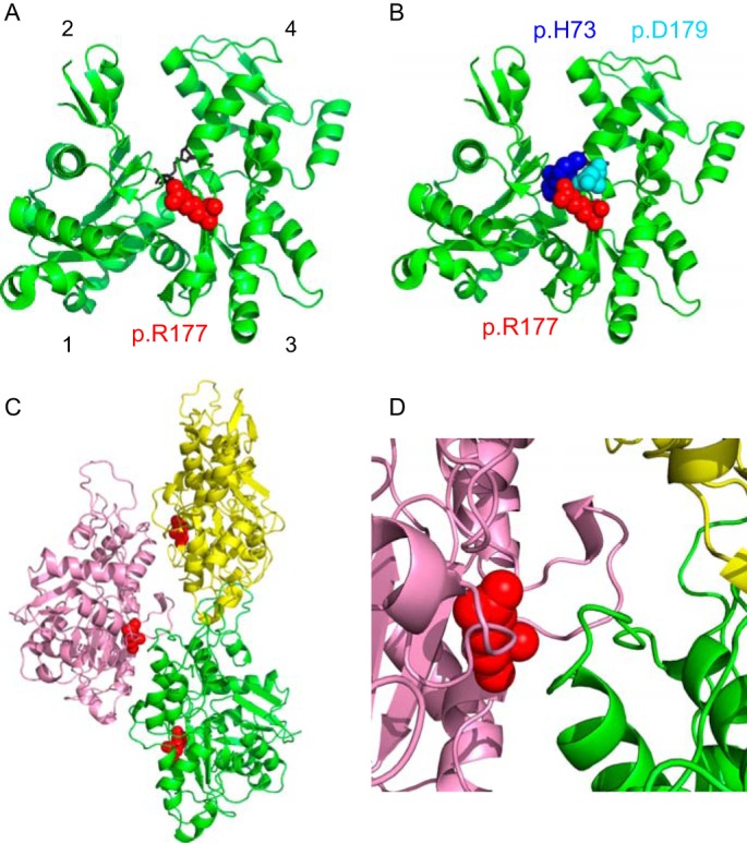
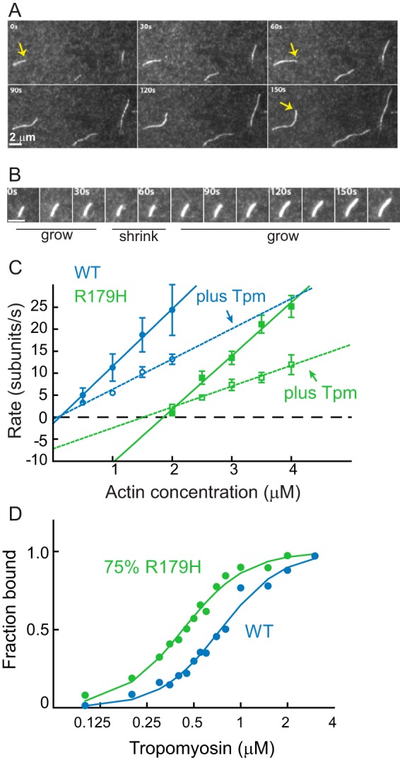
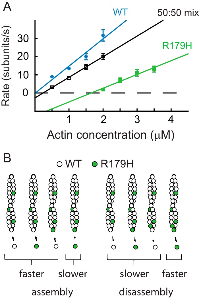
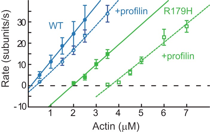
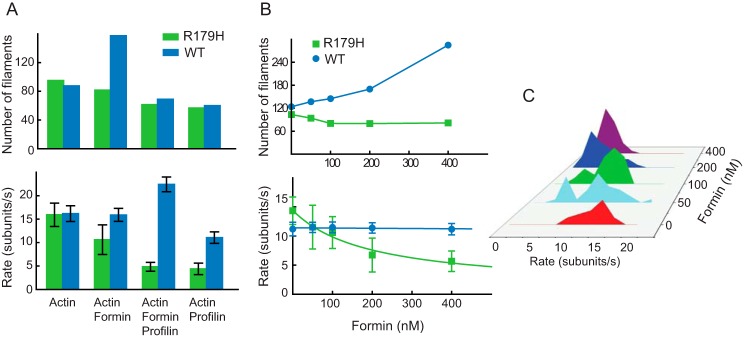
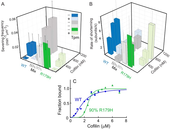
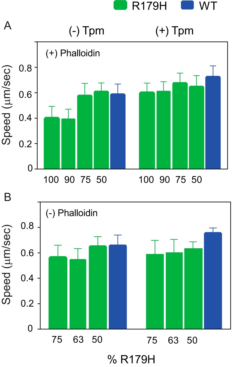
Mutations in vascular smooth muscle α-actin (SM α-actin), encoded by ACTA2, are the most common cause of familial thoracic aortic aneurysms that lead to dissection (TAAD). The R179H mutation has a poor patient prognosis and is unique in causing multisystemic smooth muscle dysfunction (Milewicz, D. M., Østergaard, J. R., Ala-Kokko, L. M., Khan, N., Grange, D. K., Mendoza-Londono, R., Bradley, T. J., Olney, A. H., Ades, L., Maher, J. F., Guo, D., Buja, L. M., Kim, D., Hyland, J. C., and Regalado, E. S. (2010) Am. J. Med. Genet. A 152A, 2437-2443). Here, we characterize this mutation in expressed human SM α-actin. R179H actin shows severe polymerization defects, with a 40-fold higher critical concentration for assembly than WT SM α-actin, driven by a high disassembly rate. The mutant filaments are more readily severed by cofilin. Both defects are attenuated by copolymerization with WT. The R179H monomer binds more tightly to profilin, and formin binding suppresses nucleation and slows polymerization rates. Linear filaments will thus not be readily formed, and cells expressing R179H actin will likely have increased levels of monomeric G-actin. The cotranscription factor myocardin-related transcription factor-A, which affects cellular phenotype, binds R179H actin with less cooperativity than WT actin. Smooth muscle myosin moves R179H filaments more slowly than WT, even when copolymerized with equimolar amounts of WT. The marked decrease in the ability to form filaments may contribute to the poor patient prognosis and explain why R179H disrupts even visceral smooth muscle cell function where the SM α-actin isoform is present in low amounts. The R179H mutation has the potential to affect actin structure and function in both the contractile domain of the cell and the more dynamic cytoskeletal pool of actin, both of which are required for contraction.

Keywords: actin; cofilin; contractile protein; formin; myosin; profilin; vascular smooth muscle cells.


Figures

FIGURE 1.
FIGURE 1.
Location of protein residue Arg-177 in the G- and F-actin structure. A, ribbon representation of G-actin (Protein Data Bank code 1NWK). The four subdomains of actin are indicated. Arg-179, which is residue p.R177 in the processed protein, is shown in red space-filling spheres. It is located in a short β-strand in the “backside” of subdomain 3. ATP is shown in black sticks. B, nitrogen on the side chain of residue p.R177 is ∼5 Å from the carbonyl oxygen of p.H73 (blue) in subdomain 1, and ∼3 Å from the negatively charged side chain of p.D179. Neither interaction would occur in the mutant p.H177. C, ribbon representation of three protomers (yellow, pink, and green) from a model of filamentous actin (Protein Data Bank code 3J8A). p.R177 is shown in red spheres at the inter-strand interface. D, close-up of the filament inter-strand interface. Protein residue p.R177 is adjacent to an uncharged loop in subdomain 4 of an adjacent protomer (green) in the filament (protein residues 197–202; Gly-Tyr-Ser-Phe-Val-Thr).
FIGURE 2.
FIGURE 2.
Quantification of R179H actin filament growth observed by TIRF microscopy and tropomyosin binding affinity. A, growth of filaments from 4 μm R179H G-actin as a function of time. Each panel is separated by 30 s. The yellow arrow marks the growth of one filament. Bar in bottom left corner is 2 μm. B, time series images showing the growing and shrinking of R179H filaments at 2.5 μm actin. The white bar in the lower left corner is 2 μm long. C, rate of filament growth as a function of actin concentration for WT SM α-actin (blue) and R179H (green) actin, in the absence (solid line) or presence (dashed line) of smooth muscle tropomyosin. Error bars are S.E. Parameters obtained from these data (assembly rate, disassembly rate, and critical concentration) with multiple independent preparations are given in Table 1. D, affinity of WT (blue) and R179H (green, filaments formed from 25%WT and 75% R179H actin) F-actin for smooth muscle tropomyosin determined by an actin pelleting assay. Maximal binding was normalized to 1. Data were fit to the Hill equation. Kapp for WT is 1.3 × 10⁶ m−1 and for R179H is 2.3 × 10⁶ m−1. The Hill coefficient is 2.2 for WT and 2.1 for R179H.
FIGURE 3.
FIGURE 3.
Copolymerization of equal amounts of WT and R179H actin. A, rate of filament growth versus actin concentration graph for WT SM α-actin (blue), R179H (green), and an equimolar mixture of WT and R179H actin (black). Error bars are S.E. B, schematic of a simple model to explain the copolymerization data (see “Experimental Procedures” for details). The slower assembly rate of the mutant actin occurs only when R179H adds onto a filament with a mutant protomer at the end. Disassembly only occurs at the fast mutant rate if both end protomers are R179H actin. Table 2 tabulates the polymerization parameters and values calculated from the model.
FIGURE 4.
FIGURE 4.
R179H actin binds more strongly to profilin than WT SM α-actin. Rate of polymerization of WT SM α-actin (blue) or R179H actin (green) in the absence (solid line) or presence (dashed line) of profilin. Error bars are S.E. Profilin concentration was 1 μm for R179H actin and 3 μm for WT. The dashed lines are fits to the data as described under “Experimental Procedures.” Profilin binds to R179H actin ∼4-fold tighter than to WT actin (see Table 3).
FIGURE 5.
FIGURE 5.
R179H actin has a defect in formin-mediated polymerization. A, number of filaments (in 54 × 54 μm2 area after 2 min) and the polymerization rate for WT SM α-actin (blue bars) and R179H actin (green bars) in the presence of 50 nm formin (FH1-FH2 fragment of mDia1) and/or 1 μm profilin. B, number of filaments and polymerization rate versus formin concentration for WT and R179H actin. The curve was fit to r = (r2r1)a + r1, in which r1 is the assembly rate of formin-free actin filaments (14.4 subunits/s); r2 is the assembly rate of formin-capped filaments (1 subunit/s), and a is the percentage of filaments that are formin-capped. C, polymerization rate histogram of R179H in the presence of increasing amounts of formin. Two distinct rates were observed; the slower rate is due to polymerization with formin bound to the growing barbed end, and the faster rate is polymerization onto a formin-free barbed end.
FIGURE 6.
FIGURE 6.
R179H filaments are more susceptible to cofilin-induced shortening and severing than WT SM α-actin, even in the presence of tropomyosin. A, frequency of severing actin filaments by cofilin for WT SM α-actin (blue bar), R179H (green bar), and mixtures of WT and mutant actin (50% R179H, darker gray bars; 90% R179H, lighter gray bars). Lighter shaded blue and green bars are in the presence of smooth muscle tropomyosin. Frequency is reported as the number of events per 1 min per 1 μm of actin filament. Error bars are S.E. Equimolar copolymers of R179H and WT are more resistant to cofilin severing than R179H homopolymers. B, rate of actin filament shortening induced by cofilin. Same color scheme as A. Error bars are S.E. Equimolar copolymers of R179H and WT are more resistant to cofilin shortening than R179H homopolymers. C, affinity of WT (blue) and R179H (green, formed from 90% R179H actin and 10% WT) filaments for cofilin determined by an actin pelleting assay. Maximal binding was normalized to 1. Data were fit to the Hill equation. Kapp for WT filaments is 0.6 × 10⁶ m−1 and 0.4 × 10⁶ m−1 for R179H filaments. The Hill coefficient is 2.2 for WT and 6.9 for R179H.
FIGURE 7.
FIGURE 7.
R179H actin binds less cooperatively to the transcription factor MRTF-A than WT SM α-actin. A, rate of polymerization of WT SM α-actin (blue) or R179H actin (green) in the absence (solid line) or presence (dashed line) of MRTF-A (3 μm for WT, 1 μm for R179H). Error bars are S.E. The dashed curves are fits to the Hill equation as described under “Experimental Procedures.” The fits yield a Kd of 1.8 μm for WT and 1.1 μm for R179H. Hill coefficients are 3 for WT and 1.5 for R179H. B, rate of polymerization of WT SM α-actin (blue, repeated from A) or a 50:50 mixture of WT and R179H actin (gray) in the presence (dashed line) of 3 μm MRTF-A. Error bars are S.E. The dashed curves are fits to the Hill equation as described under “Experimental Procedures.” The fits yield a Kd of 1.7 μm for the 50:50 mix of WT and R179H, and a Hill coefficient of 1.7.
FIGURE 8.
FIGURE 8.
In vitro motility performed with R179H or WT SM α-actin in the absence or presence of tropomyosin. A, speed at which phosphorylated human smooth muscle myosin moves phalloidin-stabilized actin filaments. Filaments were polymerized from mixtures containing the indicated percentage of R179H actin. All pairs showed a statistically significant decrease compared with WT, except for the 50 and 75% R179H mixtures in the absence of tropomyosin. With bare actin, speeds were as follows: 0.585 ± 0.083 (n = 100) for WT; 0.606 ± 0.071 (n = 41) for 50% R179H; 0.574 ± 0.100 (n = 41) for 75% R179H; 0.392 ± 0.079 (n = 41) for 90% R179H; and 0.397 ± 0.096 (n = 58) for 100% R179H. In the presence of Tpm1.4, speeds were as follows: 0.722 ± 0.090 (n = 100) for WT; 0.645 ± 0.090 (n = 40) for 50% R179H; 0.673 ± 0.082 (n = 40) for 75% R179H; 0.610 ± 0.077 (n = 40) for 90% R179H; and 0.597 ± 0.079 (n = 40) for 100% R179H. B, speed at which phosphorylated human smooth muscle myosin moves actin filaments that were not stabilized with phalloidin. Filaments were polymerized from mixtures containing the indicated percentage of R179H actin. All pairs showed a statistically significant decrease compared with WT, except for the 50% R179H mixtures with bare actin. Bare actin speeds were as follows: 0.616 ± 0.084 (n = 40) for WT; 0.591 ± 0.089 (n = 40) for 50% R179H; 0.490 ± 0.106 (n = 40) for 63% R179H; and 0.494 ± 0.111 (n = 40) for 75% R179H. In the presence of Tpm1.4, speeds were as follows: 0.732 ± 0.052 (n = 40) for WT; 0.613 ± 0.048 (n = 40) for 50% R179H; 0.584 ± 0.087 (n = 40) for 63% R179H; and 0.571 ± 0.092 (n = 40) for 75% R179H. Data were obtained using three independent protein preparations each of WT and R179H.

References

    1. Milewicz D. M., Østergaard J. R., Ala-Kokko L. M., Khan N., Grange D. K., Mendoza-Londono R., Bradley T. J., Olney A. H., Ades L., Maher J. F., Guo D., Buja L. M., Kim D., Hyland J. C., and Regalado E. S. (2010) De novo ACTA2 mutation causes a novel syndrome of multisystemic smooth muscle dysfunction. Am. J. Med. Genet. A 152A, 2437–2443 - PMC - PubMed
    1. Guo D. C., Pannu H., Tran-Fadulu V., Papke C. L., Yu R. K., Avidan N., Bourgeois S., Estrera A. L., Safi H. J., Sparks E., Amor D., Ades L., McConnell V., Willoughby C. E., Abuelo D., et al. (2007) Mutations in smooth muscle α-actin (ACTA2) lead to thoracic aortic aneurysms and dissections. Nat. Genet. 39, 1488–1493 - PubMed
    1. Guo D. C., Papke C. L., Tran-Fadulu V., Regalado E. S., Avidan N., Johnson R. J., Kim D. H., Pannu H., Willing M. C., Sparks E., Pyeritz R. E., Singh M. N., Dalman R. L., Grotta J. C., Marian A. J., et al. (2009) Mutations in smooth muscle α-actin (ACTA2) cause coronary artery disease, stroke, and Moyamoya disease, along with thoracic aortic disease. Am. J. Hum. Genet. 84, 617–627 - PMC - PubMed
    1. Morisaki H., Akutsu K., Ogino H., Kondo N., Yamanaka I., Tsutsumi Y., Yoshimuta T., Okajima T., Matsuda H., Minatoya K., Sasaki H., Tanaka H., Ishibashi-Ueda H., and Morisaki T. (2009) Mutation of ACTA2 gene as an important cause of familial and nonfamilial nonsyndromatic thoracic aortic aneurysm and/or dissection (TAAD). Hum Mutat 30, 1406–1411 - PubMed
    1. Regalado E. S., Guo D. C., Prakash S., Bensend T. A., Flynn K., Estrera A., Safi H., Liang D., Hyland J., Child A., Arno G., Boileau C., Jondeau G., Braverman A., Moran R., et al. (2015) Aortic disease presentation and outcome associated with ACTA2 mutations. Circ. Cardiovasc. Genet. 8, 457–464 - PMC - PubMed

LinkOut - more resources