. 2016 Sep 6;113(36):10168-73.
doi: 10.1073/pnas.1602425113. Epub 2016 Aug 22.

Structural basis for the CsrA-dependent modulation of translation initiation by an ancient regulatory protein

Affiliations

Structural basis for the CsrA-dependent modulation of translation initiation by an ancient regulatory protein

Florian Altegoer et al. Proc Natl Acad Sci U S A. .

Abstract

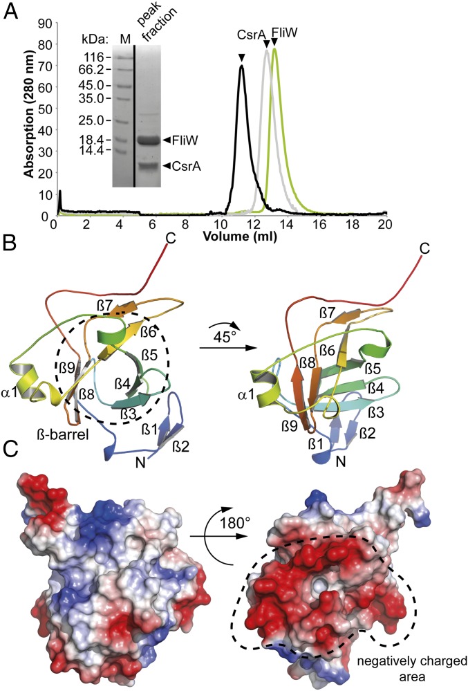
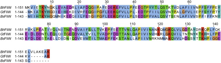
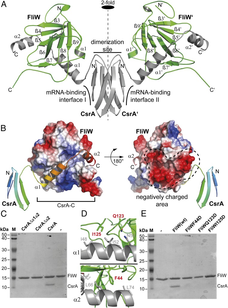
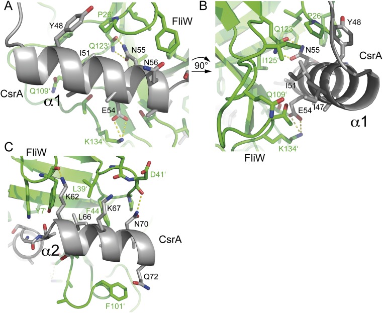
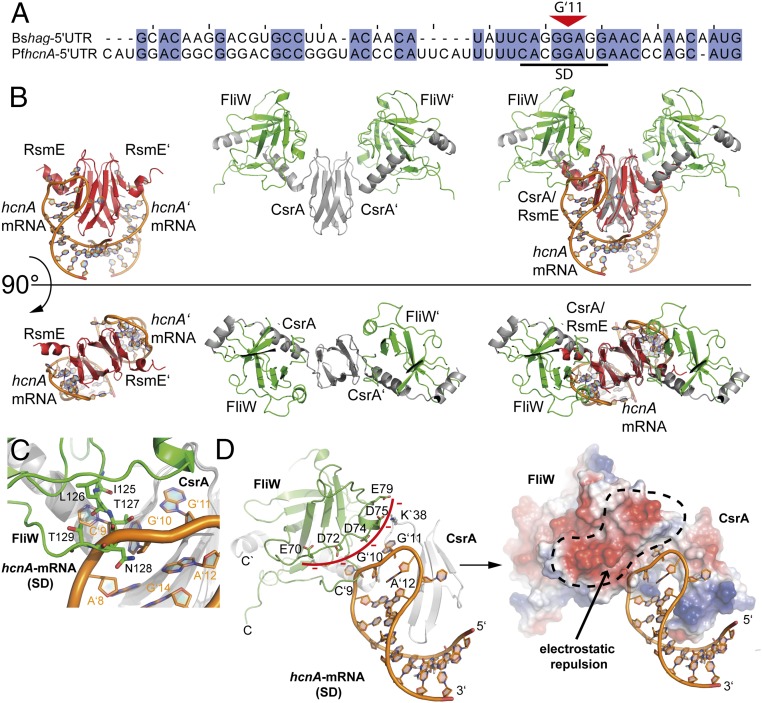
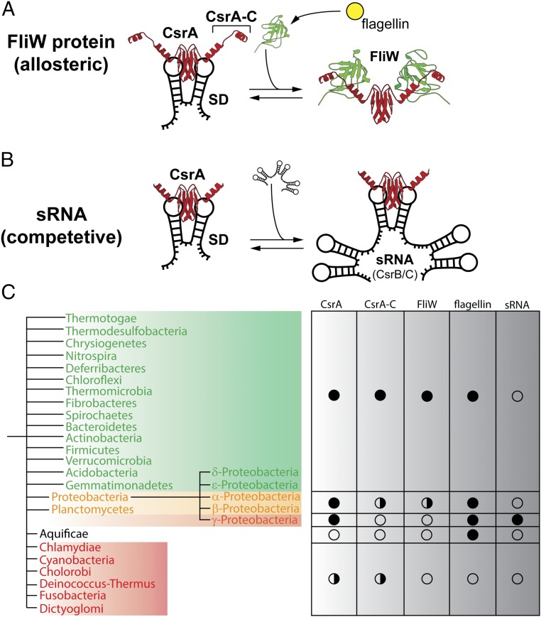
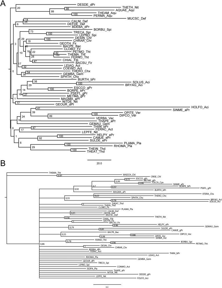
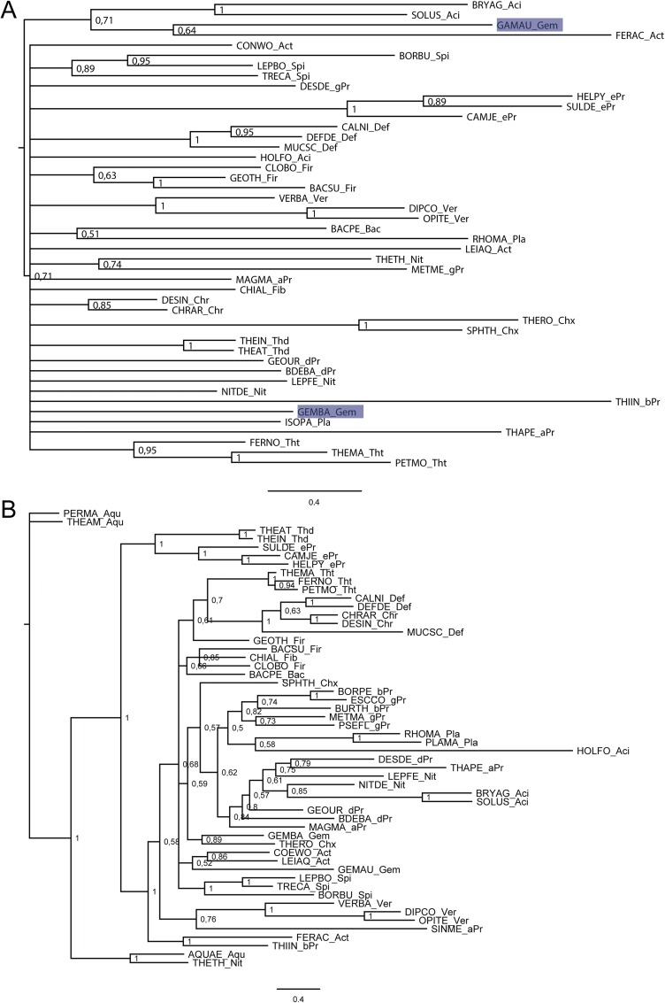
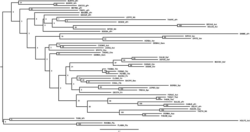
Regulation of translation is critical for maintaining cellular protein levels, and thus protein homeostasis. The conserved RNA-binding protein CsrA (also called RsmA; for carbon storage regulator and regulator of secondary metabolism, respectively; hereafter called CsrA) represents a well-characterized example of regulation at the level of translation initiation in bacteria. Binding of a CsrA homodimer to the 5'UTR of an mRNA occludes the Shine-Dalgarno sequence, blocking ribosome access for translation. Small noncoding RNAs (sRNAs) can competitively antagonize CsrA activity by a well-understood mechanism. However, the regulation of CsrA by the protein FliW is just emerging. FliW antagonizes the CsrA-dependent repression of translation of the flagellar filament protein, flagellin. Crystal structures of the FliW monomer reveal a novel, minimal β-barrel-like fold. Structural analysis of the CsrA/FliW heterotetramer shows that FliW interacts with a C-terminal extension of CsrA. In contrast to the competitive regulation of CsrA by sRNAs, FliW allosterically antagonizes CsrA in a noncompetitive manner by excluding the 5'UTR from the CsrA-RNA binding site. Our phylogenetic analysis shows that the FliW-mediated regulation of CsrA regulation is the ancestral state in flagellated bacteria. We thus demonstrate fundamental mechanistic differences in the regulation of CsrA by sRNA in comparison with an ancient regulatory protein.

Keywords: CsrA; FliW; crystallography; flagellum; translation.


Conflict of interest statement

The authors declare no conflict of interest.

Figures

Fig. 1.
Fig. 1.
Crystal structure of FliW. (A) SEC chromatograms of GtFliW (green), GtCsrA (gray), and the GtCsrA–FliW complex (black) (Right). The Inset shows a Coomassie-stained SDS/PAGE of the GtCsrA–FliW peak fraction. (B) The crystal structure of GtFliW is shown in rainbow colors from the N to the C terminus indicated by “N” and “C,” respectively. (C) The electrostatic surface representation of FliW shows that one side of the molecule is highly negatively charged, as indicated by red colors.
Fig. S1.
Fig. S1.
Amino acid sequence alignment of FliW primary sequences from B. halodurans, B. subtilis, and Geobacillus thermodenitrificans.
Fig. S2.
Fig. S2.
Structures of GtFliW were obtained from two crystal forms. (A) Superimposition of the two crystal structures in two orientations. The P22121-form is shown in green and the P21 structure is colored in blue. The overall rmsd of the Cα-atoms is 0.766, showing no significant differences between the two structures. (B) Crystallographic symmetry of the two GtFliW structures. The main difference between the two crystal lattices is the amount of molecules within the asymmetric unit. The P22121 form contains only one molecule, whereas the P21 contains two molecules in the asymmetric unit.
Fig. 2.
Fig. 2.
Crystal structure of CsrA–FliW. (A) Cartoon representation of the GtCsrA–FliW crystal structure shows that the CsrA homodimer (gray) binds two FliW molecules (green). “N” and “C” indicate N and C termini, respectively. The dashed line indicates the twofold symmetry axis. (B) Electrostatic surface representation of FliW and cartoon representation of a CsrA monomer rainbow-colored from the N to the C terminus. Helices α1 and α2 of CsrA-C bind into an extended groove of FliW (Left) located opposite to the negatively charged area of FliW (Right) (compare also to Fig. 1C). (C) Coomassie-stained SDS/PAGE of an in vitro pulldown assay using GtFliW as bait and GtCsrA, GtCsrAΔα2, and GtCsrAΔα1/α2 as prey. (D) Detailed view of the interaction of FliW with α1 (Upper) and α2 (Lower) of CsrA-C. Residues in FliW that were tested in the in vitro pull-down assay are shown in red (compare with E). (E) Coomassie-stained SDS/PAGE of an in vitro pulldown assay using wild-type GtFliW and its variants as bait and GtCsrA as prey.
Fig. S3.
Fig. S3.
Detailed interaction network of CsrA and FliW. (A) The helix α1 of CsrA is mainly coordinated by polar and electrostatic interactions. Asn55 of FliW and Gln123 of FliW establish a hydrogen bond; Lys134 of FliW coordinates Glu54. (B) Changing the orientation of helix α1 of CsrA by 90° allows a closer look on the residues of FliW that coordinate CsrA. Ile51 is part of a hydrophobic patch. (C) Interaction surface of helix α2 with FliW. Lys62 establishes a hydrogen bonding to the backbone of Tyr7 FliW; Asn70 is coordinated by the backbone of Asp41 and Leu66 of CsrA is part of a hydrophobic patch at FliW.
Fig. 3.
Fig. 3.
FliW and RNA interaction with CsrA. (A) Sequence alignment of the 5′UTR regions of the hag and hcnA mRNAs from B. subtilis (Bs) and P. fluorescens (Ps), respectively. A black bar indicates the Shine–Dalgarno (SD) sequence. The red triangle indicates the central base G11. (B) Side-by-side views of the structures of CsrA (red) bound to hcnA (orange) (Left), CsrA (gray) bound to FliW (green) (Center) and the superimposition of both structures (Right). The Upper and Lower panels show two orientations of the same that are rotated by 90 ° relative to each other. (C) Detailed view on the overlapping binding sites of FliW (green) and hcnA (orange) at CsrA (gray). (D) Detailed view of how FliW (green) hinders interaction of CsrA (gray) with hcnA (orange) in a steric (Left) and electrostatic manner (Right).
Fig. 4.
Fig. 4.
Regulation of CsrA by sRNA and the protein FliW. (A) The different modes of CsrA (red) regulation by FliW (green, Right) and (B) sRNA (black, Right) are depicted. Both FliW and sRNA antagonize the CsrA-dependent repression of translation initiation (Left). (C) Schematic summary of the phylogenetic analysis of CsrA and FliW. Flagellated species containing FliW and CsrA are shown in green; a full circle marks the general presence of the genes in the respective clades. The flagellin protein serves as a marker for flagella-mediated motility. Some members of the Proteobacteria and Planctomycetes contain FliW, whereas some lack FliW (yellow), which is also indicated by the split circle (Right). sRNAs have only been experimentally confirmed within the class of γ-proteobacteria. The Aquificae only contain flagellin, but lack FliW and CsrA. Species at the bottom (red, half circle) lack all of the proteins with the exception of Chlamydiae and Cyanobacteria, in which some species possess a CsrA (Table S1).
Fig. S4.
Fig. S4.
Sequence alignment of CsrA/RsmA homologs from representatives of different phyla sorted by length. The abbreviations are: BACSU_Fir: Bacillus subtilis (Firmicutes); BORPE_bPr: Bordetella petrii (β-proteobacteria); CAMJE_gPr: Campylobacter jejuni (ε-proteobacteria); ESCCO_gPr: Escherichia coli (γ-proteobacteria); GEMAU_Gem: Gemmatimonas aurantiaca (Gemmatimonadetes); HELPY_ePr: Helicobacter pylori (ε-proteobacteria); LEIAQ_Act: Leifsonia aquatic (Actinobacteria); NITDE_Nit: Candidatus Nitrospira defluvii (Nitrospira); PLAMA_Pla: Planctomyces maris (Planctomycetes); THAPE_aPr: Thalassospira permensis (γ-proteobacteria); VERBA_Ver: Verrucomicrobiae bacterium (Verrucomicrobia).
Fig. S5.
Fig. S5.
(A) Midpoint-rooted Bayesian inference tree of CsrA, the numbers at the nodes represent posterior probabilities. (B) Midpoint-rooted Bayesian inference tree of FliW, the numbers at the nodes represent posterior probabilities.
Fig. S6.
Fig. S6.
(A) Midpoint-rooted Bayesian inference tree of flagellin, the numbers at the nodes represent posterior probabilities. (B) Midpoint-rooted neighbor-joining tree of CsrA, the numbers at the nodes represent bootstrap values.
Fig. S7.
Fig. S7.
(A) Midpoint-rooted neighbor-joining tree of FliW, the numbers at the nodes represent bootstrap values. (B) Midpoint-rooted neighbor-joining tree of flagellin, the numbers at the nodes represent bootstrap values.
Fig. S8.
Fig. S8.
(A) Midpoint-rooted ML tree of CsrA, the numbers at the nodes represent bootstrap values. (B) Midpoint-rooted ML tree of FliW, the numbers at the nodes represent bootstrap values.
Fig. S9.
Fig. S9.
Midpoint-rooted ML tree of flagellin; the numbers at the nodes represent bootstrap values.

References

    1. Steitz JA, Jakes K. How ribosomes select initiator regions in mRNA: Base pair formation between the 3′ terminus of 16S rRNA and the mRNA during initiation of protein synthesis in Escherichia coli. Proc Natl Acad Sci USA. 1975;72(12):4734–4738. - PMC - PubMed
    1. Shine J, Dalgarno L. Determinant of cistron specificity in bacterial ribosomes. Nature. 1975;254(5495):34–38. - PubMed
    1. Vakulskas CA, Potts AH, Babitzke P, Ahmer BMM, Romeo T. Regulation of bacterial virulence by Csr (Rsm) systems. Microbiol Mol Biol Rev. 2015;79(2):193–224. - PMC - PubMed
    1. Romeo T. Global regulation by the small RNA-binding protein CsrA and the non-coding RNA molecule CsrB. Mol Microbiol. 1998;29(6):1321–1330. - PubMed
    1. Heroven AK, Böhme K, Dersch P. The Csr/Rsm system of Yersinia and related pathogens: A post-transcriptional strategy for managing virulence. RNA Biol. 2012;9(4):379–391. - PubMed

Publication types

MeSH terms

LinkOut - more resources